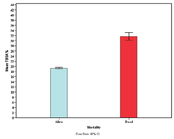
Figure 1: Mortality according to mean TBSA

Habibullah Shah1* Huma Gul2 M Mukhtar Khan3 Rashid Khan3
1Habib Burn Centre, Zia Medical Complex, Peshawar, Pakistan*Corresponding author: Habibullah Shah, Habib Burn Centre, Zia Medical Complex, Peshawar, KPK, Pakistan, Tel: 0092 334 88 00 33 9; E-mail: drhabibulllahshah@gmail.com
Objective: Specialised burn wound management is the need of the hour. We conducted this study to present our 3-year experience of burn wound management and its outcome from a specialist burn centre in Peshawar Pakistan.
Methods: This is a retrospective analysis of prospectively collected data about burn patients who were managed at Habib Burn Centre Peshawar between January 2013 and December 2015 (3 years). Data was collected prospectively about patient demographics, burn types, burn thickness, total burn surface area (TBSA) and the body sites involvement. The outcome was measured in terms of rates of wound healing, the length of stay, early and late complications and mortality. Data was analysed to observe associations between patient and burn characteristics to the outcome parameters.
Results: Mean TBSA was 20.83 ± 10.81 SD, mean total healing time was 18.15 days ± 8.50 SD while mean LOS was 9.51 days ± 5.45 SD. On Chi-square analysis for mortality versus burn thickness, no statistical significance was noted (p=0.53, OR: 0.93, 95% CI, 0.73 to 1.18) while mode of treatment (dressing versus grafting) was significantly affected by the thickness of the burn (p<0.001, OR: 0.69, 95% CI, 0.58 to 0.81). TBSA above 40% was strongly associated with mortality as well as with the presence of inhalational injury. TBSA was also associated with prolonged healing times and increased the length of stay (R2=0.48 and 0.73 respectively).
Conclusion: Good outcome can be achieved with antibiotic and silver sulphadiazine dressing technique for partial thickness wounds while those with full thickness or deep partial thickness wounds will ultimately require split skin grafting.
Burn management; Antibiotic coated dressing; Split skin grafting; Outcome
World Health Organization (WHO) estimates show global burn deaths over 310,000 persons every year with a staggering majority (up to 95%) of these deaths occurring in the low and middle-income countries (LMIC) [1,2]. Such a high mortality in the developing nations is an eye opener for the acute care medical community as well as the governments to enhance burn care on an emergent basis. Even more concern in these statistics is a majority of children and women [3]. In light of the immense impact of burn injury on patient lives, WHO has called upon the healthcare professionals to improve data collection and reporting as well as utilisation of resources for this subset of patients [1].
Burns covering more than 20% of total body surface area (TBSA) are the top-ranked long- and short-term disability causing trauma types and they are estimated with a prevalence of more than 100 million cases worldwide. Of the total burn loads, Asia holds more than half of their proportion due to its dense population demographics [3,4]. These global statistics emphasize the need for specialised wound management in burn patients in order to reduce a load of chronic disability and suffering worldwide.
Careful plastic surgical management of burn wounds have been shown to improve outcomes for burn patients in terms of rapid wound healing, good cosmesis and high patient satisfaction [5-7]. Good dressing technique and wound debridement are paramount in achieving rapid healing of partial thickness wounds [8]. Similarly, in full thickness or deep burns, the utilisation of various techniques of skin grafting or utilisation of skin substitutes has been shown to improve outcomes [9]. The treatment of a burn patient does not end at the healing of the skin wound, rather, the effects of psychological injury linger on for far too long, especially if the burns are inflicted in by others as a tool of violence [10-12]. Despite the scientific insight that we have obtained so far about burns and their impact, studies have shown that specialised plastic surgical services are scarce in LMICs, as compared to the high rates of burn patients requiring advanced care [2].
We, therefore, aim to present our experience with advanced management of the burn patients at our centre in Peshawar, Pakistan over a period of three years. By this, we want to show our way of management for improving outcomes in terms of wound healing rates, lowering complications with very simple cost-effective approaches.
The study was conducted at Habib Burn Centre Peshawar between January 2013 and December 2015. The data collected was analysed in a retrospective manner. Patients irrespective of age, gender and burn type were included after taking their informed consent. Severe comorbidities and (American Society of Anaesthesiologists) ASA class V and VI. Patients who presented with burn management complications or those who were initially managed elsewhere were also excluded from the study.
All partial thickness wounds were initially treated with wound cleaning and dressing. All full thickness wounds were treated with split-thickness grafts. In the case of compartment syndromes, fasciotomies/escharotomies were initially performed on an emergent basis.
All burn patients received the initial burn management according to international emergency burn management protocols and the Advanced Burn Life Support guidelines [13]. Analgesia and antibiotic prophylaxis were provided and the wound was dressed according to our special procedures.
After adequate analgesia with combined opioids and NSAIDs (intravenous ketorolac 30 mg TDS plus tramadol 50 mg TDS), the patient was sedated with intravenous midazolam infusion (total not more than 5 mg IV). Cleaning of the wound was done with an adequate wash with normal saline. A paste of antibiotic ointment (Polymyxin B 10000 IU/g +Bacitracin 500 IU/g (Polyfax®)) with a mixture of Silver Sulphadiazine (1% w/w, Quench®) ointment was prepared in a ratio of 2/3rd of the antibacterial and 1/3rd of the silver ointment. A coat of at least 3 mm thickness was applied over the burnt surface. Once the coat was completely applied, we covered the area with an antibiotic impregnated mesh dressing.
Grafts were harvest from unaffected skin areas. Intermediate thickness split skin grafts were prepared and were meshed in a 1.5-3:1. Grafts were applied over the burnt area and were fixed with staples. The non-adherent dressing was done after grafting and the patients were advised bed rest so as to minimise displacement of the graft.
Dressings for both procedures were opened at 5-day intervals. Wounds were assessed for epithelialisation and graft take. The graft take percentage was recorded and if necessary additional dressings were applied. All subsequent dressings were opened at 5 days thereof and wounds were reassessed.
Data was collected about patient demographics, wound characteristics (TBSA, thickness), burn type, inhalational injury, body site(s) involved, treatment applied, healing times, the length of stay, complications and mortality.
Data was analysed using IBM SPSS Statistics, version 22.0. Frequencies and percentages were presented in tables and graphs. Data was stratified according to age groups, burn type, thickness, treatment applied and mortality. Independent samples t-tests were applied to see significance for healing rates and length of stay. Mortality rates association with burn surface area and an inhalational component of burn trauma were assessed. Correlations between various continuous variables were assessed using Spearman’s rank correlation.
A total of 3223 patients were treated for burns during the 3-year period. Out of these 1633 (50.7%) were male and 1590 (49.3%) were female patients. There were 2325 (72.1%) cases of partial thickness and 898 (27.9%) cases of full-thickness burns. Scalds were the most common (n=2253, 69.9%) type of burns which was followed by flame burns in 25.4% (820) and 100 (3.1%) cases of electric burns. The majority (n=1636, 50.8%) of burns involved upper limbs while lower limbs were involved in 25.7% (n=827) and head, face or neck regions in 14.7% (n=475) patients (Table 1). The concomitant inhalational injury was present in 155 (4.8%) patients (Table 2).
Clinical Variable |
Frequency (N) |
Percentage (%) |
Gender |
|
|
Male |
1633 |
50.7% |
Female |
1590 |
49.3% |
Time to presentation |
|
|
less than 24 hours |
1133 |
35.2% |
24 to 48 hours |
902 |
28.0% |
48 to 120 hours |
521 |
16.2% |
More than 120 hours |
667 |
20.7% |
Burn Thickness |
|
|
Partial Thickness |
2325 |
72.1% |
Full Thickness |
898 |
27.9% |
Body Sites Involved |
|
|
Head, Face, Neck |
475 |
14.7% |
Upper Limbs |
1636 |
50.8% |
Lower Limbs |
827 |
25.7% |
Trunk |
285 |
8.8% |
Table 1: Clinical features and their frequencies
Mean age was 21.82 years ± 16.024 SD with the majority (58.1%) of patients in less than 20-year age group while only 2.9% patients were above the age of 60 years (Table 3).
2100 (65.2%) patients were treated with dressing and debridement only while 1123 (34.8%) patients required skin grafting. Complications were wound infection (n=341, 10.6%), graft loss (n=202, 6.3%) and scar problems (n=276, 8.6%) such as hypertrophic scars and contractures development. Mortality was 12.2% (n=392) (Table 2).
Clinical Variable |
Frequency (N) |
Percent (%) |
Type of burn |
|
|
Scalds |
2253 |
69.9% |
Flame |
820 |
25.4% |
Chemical |
50 |
1.6% |
Electric |
100 |
3.1% |
Age groups |
|
|
1 (1 to 10 years old) |
962 |
29.8% |
2 (11 to 20 years old) |
911 |
28.3% |
3 (21 to 30 years old) |
609 |
18.9% |
4 (31 to 40 years old) |
291 |
9.0% |
5 (41 to 50 years old) |
162 |
5.0% |
6 (51 to 60 years old) |
194 |
6.0% |
7 (above 60 years old) |
94 |
2.9% |
Inhalational injury |
155 |
4.8% |
Final treatment |
|
|
Dressing & Debridement |
2100 |
65.2% |
Grafting |
1123 |
34.8% |
Complications |
|
|
Infection |
341 |
10.6% |
Graft Loss |
202 |
6.3% |
Scar Problems |
276 |
8.6% |
Mortality |
392 |
12.2 |
Table 2: Clinical features and their frequencies
Mean time to presentation was 54.65 hours ± 52.63 SD while mean TBSA was 20.83 ± 10.81 SD. Similarly, mean total healing time was 18.15 days ± 8.50 SD while mean LOS was 9.51 days ± 5.45 SD (Table 3).
Statistics |
Patient Age |
Time to presentation |
TBSA % |
Total Healing Time |
Length of Stay |
Mean |
21.82 |
54.65 |
20.83 |
18.15 |
9.51 |
Median |
15.00 |
36.00 |
20.00 |
15.00 |
8.00 |
Mode |
12 |
6 |
16 |
9 |
8 |
Std. Deviation |
16.024 |
52.636 |
10.806 |
8.502 |
5.446 |
Minimum |
4 |
1 |
3 |
1 |
2 |
Maximum |
65 |
182 |
59 |
39 |
31 |
Table 3: Descriptive statistics
The independent t-test was performed for the difference in length of stay, total healing time, TBSA, time to presentation and patient age with regard to mortality. There were significant mean differences (p<0.001) for all the variables tested as is shown in table 4. On Chi-square analysis for mortality versus burn thickness, no statistical significance was noted (p=0.53, OR: 0.93, 95% CI, 0.73 to 1.18) while mode of treatment (dressing versus grafting) was significantly affected by the thickness of the burn (p<0.001, OR: 0.69, 95% CI, 0.58 to 0.81).
|
t |
(p) |
Mean Difference |
95% Confidence Interval of the Difference |
|
Lower |
Upper |
||||
Length of Stay |
-9.84 |
<0.001 |
-3.49 |
-4.18 |
-2.79 |
Total Healing Time |
-6.30 |
<0.001 |
-2.9 |
-3.8 |
-1.99 |
TBSA % |
-16.08 |
<0.001 |
-12.37 |
-13.88 |
-10.86 |
Time to |
-5.43 |
<0.001 |
-17.24 |
-23.48 |
-11.00 |
Patient Age |
-2.40 |
0.017 |
-2.39 |
-4.34 |
-0.43 |
Table 4: Independent t-test for equality of means (in terms of mortality)
t: test value
df: degrees of freedom
Sig.: significance
The Spearman rank correlation test was performed between patient age, TBSA, total healing time and length of stay. The extent of TBSA was positively correlated with total healing time and length of stay, and patient age was negatively correlated with TBSA, total healing time and length of stay. These results are shown in table 5 and figure 1.
Figure 1: Mortality according to mean TBSA
A significantly high number of deaths and chronic disabilities are associated with burn trauma in the lower or middle income countries all around the world. Disabilities due to burns not only affect the physical functioning of an individual, rather, it acts as a barrier to the mental and social functioning of an individual, especially the in young age groups and female gender [14]. Palmu R et al. [15] assessed the social and mental functioning of individuals for up to 6 months after burn injury and they concluded that great impact is conferred by mental problems such as major depressive disorders and delirium, however, the length of stay and body sites such as hand burn and burns to the face and head area are also frequently associated with social and mental functioning of individuals [15]. Murphy ME et al. [16] in a cross-sectional study interviewed burn patients 2.5 to 12 years after injury and they concluded that young adult burn survivors demonstrate increased disability scores than their nonburned counterparts [16]. They have emphasized the need for long-term psychosocial intervention in these patients, especially when associated with larger TBSA, males, school age kids and adolescents [16]. A mortality rate of 2.3 per 100,000 persons per year is also a factor to improve early burn care and specialised wound management of these patients [17].
In light of the above discussion, it is imperative to improve our level of expertise regarding burn wound management. In our study, the important demographics were a high number of children below ten years of age and a similar number of adolescents (Table 2 for age groups distribution). Since scalds are the most common mode of burn affecting young children, it emphasizes the importance of implementing good safety measures at homes and an urgent need for educating the public about proactive measures in preventing burns. Ceniceros A et al. [18] recently evaluated mortality trends and factors affecting it in burn patients with established bacteraemia. They recorded a mortality rate of nearly 25% where the most important factors were age, TBSA and bacterial infection with pseudomonas. Delayed presentation to primary or tertiary care for burn patients is highly associated with increased wound infections and high rates of prolonged morbidity and mortality. Chelidze KL et al. [19] also observed similar findings in our study where the primary mode of burn injury was scalds (85%) and 98% of injuries occurred at home with a mortality of above 7%. They concluded that increasing TBSA and younger age was strongly associated with mortality [19]. In our study, we found that young age was increasingly associated with high TBSA (R2:-0.239, p<0.001, table 5). Young children have lower overall body surface area and apparently a trivial injury may cause more damage in terms of affecting more percentage of the body surface leading to significant morbidity and mortality. We also found that TBSA above 40% was strongly associated with mortality as well as the presence of inhalational injury. TBSA was also associated with prolonged healing times and increased the length of stay (R2=0.48 and 0.73 respectively, table 5).
|
|
|
Patient Age |
TBSA |
Total Healing Time |
Length of Stay |
Spearman's rho |
Patient Age |
Correlation |
1.00 |
-0.239** |
-0.079** |
-0.145** |
Sig. (2-tailed) |
- |
<0.001 |
<0.001 |
<0.001 |
||
N |
3223 |
3223 |
3223 |
3223 |
||
TBSA % |
Correlation |
-0.239 |
1 |
0.482** |
0.739** |
|
Sig. (2-tailed) |
<0.001 |
- |
<0.001 |
<0.001 |
||
N |
3223 |
3223 |
3223 |
3223 |
||
Total Healing Time |
Correlation |
-0.079** |
0.482** |
1 |
0.345** |
|
Sig. (2-tailed) |
<0.001 |
<0.001 |
- |
<0.001 |
||
N |
3223 |
3223 |
3223 |
3223 |
||
Length of Stay |
Correlation |
-0.145** |
0.739** |
.345** |
1.00 |
|
Sig. (2-tailed) |
<0.001 |
<0.001 |
<0.001 |
- |
||
N |
3223 |
3223 |
3223 |
3223 |
Table 5: Spearman Rank Correlations
**Correlation is significant at the 0.01 level (2-tailed).
Various wound dressing techniques [20-23] have been introduced over the course of time. All of these techniques have the primary aim of accelerating wound healing rates as well as to lower cost of care and complications [24]. Silver Sulphadiazine ointment, however, have remained the standard of care to which other treatments have been compared. Hansbrough JF et al. [25] studied the healing rates of silver Sulphadiazine cream and a collagenase ointment. Though they found speedy healing rates for the collagenase ointment, the silver Sulphadiazine dressing has been comparatively effective in achieving clean wounds. Olawoye OA et al. [8] have studied the effectiveness of open burn wound dressing techniques with silver Sulphadiazine and they have concluded this dressing technique is highly effective with acceptable wound healing rates (mean 21 days) and fairly affordable costs to the people of low and middle income countries [8]. The Aquacel-silver dressing method was evaluated by Paddock HN et al. [24] and they found it significantly cost effective due to rapid healing rates and shorter hospital stay than silver sulphadiazine. Some studies have also suggested that silver sulphadiazine retards wound healing and therefore either should not be used or used only if there are no alternatives available [26,27]. We prefer antibiotic ointment mixed with silver sulphadiazine for two main reasons, one is good healing rates with nearly half of partial thickness wounds healing within 5 to 10-day period and second is its cost effectiveness. The majority of our patients are poor people from far flung areas who cannot afford the expensive modern dressings and we perform dressings according to their consent after the costs and benefits have been adequately discussed.
For full thickness burn patients, those with larger TBSA or those who fail to primary dressing technique with deep partial thickness burns split skin grafting with or without tangential wound excision has been recommended in order to achieve good overall outcomes [28,29]. However, the role of early SSG has been questioned [30] in terms of LOS or wound healing rates in the resource poor countries such as Pakistan. Others have favoured early excision with benefits ranging from shorter LOS, lower mortality and good overall wound healing and scar outcomes [31,32]. Vinita et al. [32] have concluded that early wound excision with grafting has definite applicability even the resource poor countries. Our preference in this regard is to apply silver plus antibiotic ointment coat covered by antibiotic coated mesh dressing to all partial thickness burns, especially those due to scalds. We observed excellent healing rates for these patients and nearly half of patients healed within ten days of initiating treatment. On the other hand, those patients who have full thickness burns or those who fail to respond to the dressing technique undergo wound excision and SSG. Overall healing times for the SSG might be prolonged but they have better scar outcomes, while those who respond to the dressing techniques have frequently been found to develop scar complications such as hypertrophic scars or contractures.
In summary, we recommend to go for early excision and SSG if it is a full thickness burn and to apply dressing with a mixture of antibiotics ointment and silver sulphadiazine after adequate wound healing under systemic analgesia and sedation. These techniques have proven benefits and patients in our county can really get benefit from it.
The weaknesses of our study include its observational nature, no control groups and no blinding of the investigators or doctors. Designing large randomised trials with blinding of the investigators and surgeons will provide more robust evidence which can be effectively relied upon.
Burn wounds are frequently accidental affecting the very young and adolescents. A good outcome can be achieved with antibiotic and silver sulphadiazine dressing techniques for partial thickness wounds while those with full thickness or deep partial thickness wounds will ultimately require split skin grafting. Split skin grafting has a better wound and scar outcomes, though expensive and takes longer hospital admission times and if infected may become a serious problem for patients.
Download Provisional PDF Here
Article Type: Research Article
Citation: Shah H, Gul H, Khan MM, Khan R (2017) Specialised Burn Wound Management and its Outcome: A 3-Year Perspective from a Plastic Surgery Specialist Centre. J Surg Open Access 3(1): doi http://dx.doi.org/10.16966/2470-0991.139
Copyright: © 2017 Shah H, et al. This is an open-access article distributed under the terms of the Creative Commons Attribution License, which permits unrestricted use, distribution, and reproduction in any medium, provided the original author and source are credited.
Publication history:
All Sci Forschen Journals are Open Access