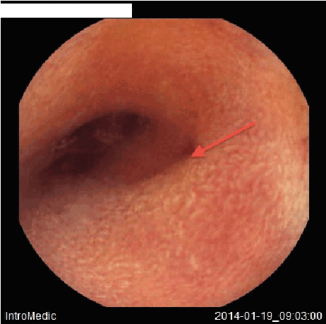
Figure 1: Small opening with minimal bleeding (arrow) and narrowing of the main lumen.

Kholoud Ayesh1 Imad Jabali Dweikat2 Thomas Ciecierega3 Mutaz Sultan1*
1Makassed Hospital-Alquds University, Medical College, Palestine*Corresponding author: Mutaz Sultan, Makassed Hospital–Alquds University, Medical College, Palestine, Tel: 00972543251282; Fax: 0097226288392; E-mail: drmutazsultan@gmail.com
Meckel’s diverticulum (MD) is the most common congenital anomaly of the gastrointestinal (GI) tract affecting about 2% of the populations. Gastrointestinal bleeding is a common presentation in pediatric and can be massive.
The diagnosis of symptomatic MD is often difficult to make. We report on a 7-year-old boy who suffered from recurrent severe GI bleeding. He had normal upper and lower endoscopy and negative Meckel scan. Meckel’s Diverticulum identification was achieved by wireless capsule endoscopy (WCE).
Meckel’s Diverticulum; Gastrointestinal bleeding; Wireless capsule endoscopy
Gastrointestinal bleeding is not uncommon in children and is often self-limited; however, bleeding may have severe consequences if left untreated and should therefore be thoroughly investigated.
The omphalomesenteric duct connects the yolk sac to the intestinal tract during early fetal life. This structure is usually obliterated by the fifth to seventh week of gestation. Failure to regress may lead to several anomalies including Meckel’s Diverticulum (MD) [1].
More than 50% of these patients present with bleeding by the age of 2 years. Individuals with a MD may develop symptoms from infancy through adulthood that include intestinal obstruction, gastrointestinal bleeding, acute intra-abdominal inflammation, Littre’s hernia (inguinal hernia with a Meckel’s diverticulum in the hernia sac), or umbilical anomalies [1].
MD represents the most common congenital anomaly of the gastrointestinal tract, with an incidence of 1%-3% in the general population [2]. It is normally located on the anti mesenteric border of the terminal ileum within 80-100 cm of the ileocecal valve and is on average 2 cm in length.
Approximately 57% of MD contains ectopic gastric mucosa [2], which actively secretes the hydrochloric acid responsible for mucosal ulcerations within the diverticulum and unprotected wall of the adjacent ileum.
Meckel scan (technetium-99 pertechnetate scan) is the current test of choice for a bleeding diverticulum [3]. Few case reports have described capsule endoscopy as a useful test in identifying a MD, but is not regularly used for this purpose.
We report the case of a 7-year-old boy who suffered from recurrent severe GI bleeding. He had normal upper and lower endoscopy. MD identification was achieved by wireless capsule endoscopy (WCE).
A 7-year-old boy was referred from outside facility for investigation of obscured recurrent GI bleeding. His story started two years ago when he passed large amount of fresh per rectum without other symptoms. His hemoglobin dropped down to 6.5 g/l and two units of packed blood were transfused and he underwent extensive evaluation. Upper and lower endoscopies were used to investigate the source of bleeding and showed normal macroscopic findings.
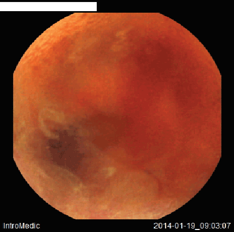
Meckel scan with 99mTc-Na-pertechnetate was negative for heterotopic gastric tissue in the small bowel area. About one year later, he had the same event with fresh bleeding per rectum associated with drop of his hemoglobin to 6 g/dl, when he was given two units of packed RBC. Again abdominal CT, upper and lower endoscopies failed to detect the source of bleeding. One month later, he had another similar attack that was treated conservatively. Two days prior to admission he had poor appetite, nausea and general weakness with profuse rectal bleeding and thus was admitted urgently to hospital. Upon admission the patient’s hemoglobin level was 8.5 g/dl. Wireless Capsule Endoscopy (WCE) was considered medically necessary in order to investigate the obscure GI bleeding suspected to be of small bowel origin. A WCE (Intromedic’s Mirocam Capsule Endoscopy System) was placed by upper endoscopy and released in the stomach, which then revealed a small opening seen in the distal ileum with active bleeding from adjacent area and mild narrowing of the lumen (Figures 1 and 2), findings can be suggestive of MD. The patient passed the capsule on the second day. The Pediatric Surgery team was consulted and the patient underwent explorative laparotomy. MD was resected as well as the appendix. Histopathological report for the diverticulum removed showed intestinal wall with ectopic gastric mucosa.
The clinical course of the patient was excellent and no adverse events occurred. During 18 months of follow up, he remained well without further bleeding episodes.

Figure 1: Small opening with minimal bleeding (arrow) and narrowing of the main lumen.

Figure 2: Active bleeding detected at the same site.
Although MD is the most prevalent congenital abnormality of the gastrointestinal tract, it is often difficult to diagnose. MD has various presentations and can be easily misdiagnosed, and hence it is necessary to maintain a high index of suspicion in the pediatric age group. Massive or intermittent bleeding may occur, such as in our case, which warrant blood transfusions. Intestinal obstruction and diverticulitis are the most frequent complications in adults. In contrast, painless rectal bleeding is a common clinical feature in childhood [2]. Our patient had manifested recurrent painless rectal bleeding before the diagnosis was confirmed.
Blevrakis et al. [4] reported that MD has various presentations and can be easily misdiagnosed. The records of 45 patients with a diagnosis of MD were retrospectively reviewed. In 25 of patients MD was an incidental finding at laparotomy because of appendicitis, the remaining 20 patients were symptomatic and presented with various clinical features. Nine patients (19.9%) had clinical features of peritonitis; of these, three had perforated MD and six had Meckel’s diverticulitis at laparotomy. Four patients were diagnosed with intestinal obstruction. Seven patients (15.5%) presented with lower gastrointestinal bleeding. Ultrasound scans revealed intussusception in three patients, requiring open reduction. The remaining four patients with bleeding per rectum underwent a Meckel’s Tc99 scan that showed a positive tracer.
Patients with upper GI bleeding often present with hematemesis or melena. The test used most often to diagnose the cause of GI bleeding is upper GI endoscopy. Colonoscopy is the test of choice in patients with rectal bleeding. Our patient suffered from fresh rectal bleeding, but unfortunately upper and lower endoscopy failed to detect the bleeding site. A lesion in the small bowel was suspected causing bleeding and MD was considered.
The most sensitive study is a Meckel radionuclide scan, which is performed after intravenous infusion of technetium-99m pertechnetate. Avid accumulation of 99m Tc-pertechnetate in gastric mucosa makes it the study of choice for identifying ectopic gastric mucosa in a MD. Properly performed scan in the appropriate clinical setting is an effective method for the detection of Meckel diverticulum containing functioning gastric mucosa, with overall sensitivity of 85%, specificity of 95%, and accuracy of 90% [5]. H2 blocker improves diagnostic accuracy by inhibiting the intra luminal release of technetium, and glucagon does so as an antiperistaltic. Meckel scan was normal in our patient and failed to show ectopic gastric mucosa in spite of premedication with H2 blockers.
Our patient had WCE study to look for the source of bleeding, which revealed a small opening seen in the distal ileum with active bleeding from adjacent area, suggestive of bleeding MD.
The fact that our patient underwent the capsule study during the bleeding episodes increased the detection rate and the sensitivity of the test.
Handful case reports have been published detecting MD using capsule endoscopy [6-9]. Xanias et al. [9] reported an 8-year-old boy who presented with GI bleeding due to MD Upper and lower endoscopy in addition to Meckel scan failed diagnose the source of bleeding. WCE of the small intestine revealed a polypoid lesion of the terminal ileum producing relative stenosis, thus delaying the capsule passage, which was possibly an MD and the diagnosis was confirmed after surgical resection of the MD.
In conclusion, WCE is a helpful test to look for obscure GI bleeding in children. It can be a helpful tool to diagnose MD specially if done during active bleeding. More controlled studies are needed to delineate the role of WCE in diagnosing symptomatic MD.
Download Provisional PDF Here
Article Type: Case Report
Citation: Ayesh K, Dweikat IJ, Ciecierega T, Sultan M (2016) Capsule Endoscopy detects Meckel’s Diverticulum in a Child with Recurrent Gastrointestinal Bleeding: Case report and Review of the Literature. J Gastric Disord Ther 2(2): doi http://dx.doi. org/10.16966/2381-8689.115
Copyright: © 2016 Ayesh K, et al. This is an open-access article distributed under the terms of the Creative Commons Attribution License, which permits unrestricted use, distribution, and reproduction in any medium, provided the original author and source are credited.
Publication history:
All Sci Forschen Journals are Open Access