Abstract
Nowadays, Endoscopic Submucosal Dissection (ESD) has been widely applied in early cancer or precancerosis of gastrointestinal tract.
With the development of endoscopic technique, the procedure advances and the operation time of ESD becomes shorter. However, there still
exist some unpredictable complications, such as gastric mucosa avulsion caused by submucosal injection during ESD procedure. And we will
report one case about it in this article.
Case
A 69-year-old man, with the chief complains of abdominal distension
and pain for one and a half years, was admitted to our hospital. Upper
endoscopy showed a 1.2 × 1.2 cm flat lesion in the greater curvature of the
antrum. It was an IIa+IIc form lesion[1]. Mucosal erosion and depression
area could be seen on the surface of the lesion. Histopathological
examination revealed a high-grade intraepithelial neoplasia lesion. Then
ESD was performed [2] (Figure 1). The procedure was generally as follows.
Indigo carmine was sprayed to define the form and range of the lesion.
Argon Plasma Coagulation (APC) was used to mark the lesion margin.
After that, mixture liquid (1 ml indigo carmine and 100 ml normal saline)
was injected into the submucosal tissue. However, when performed
submucosal injection at the anal side of the lesion, we found it with great
resistance and without doubt the mucosal didn’t lift well. So we repeated
injecting with more power, trying to lift the mucosal up. While it failed,
and we found that the gastric mucosal avulsed. We had to immediately
changed injection site, the mucosal lift sign of other areas were well.
Then we pre-cut the mucosa along the marker by Dual knife (KD-650L;
Olympus) and dissected the lesion from oral side to anal side gradually. The
anal side submucosal tissue adhered to the muscluaris propria firmly and
was difficult to be dissected. So a snare (ASM-1; Cook) was used to resect
the lesion and a white scar was seen. The anal side of the wound, where the
mucosa avulsed when performing submucosal injection, formed a cavity.
Several endo-clips (Resolution, M00522610; Boston Scientific) were used
to occlude the cavity. High-grade intraepithelial neoplasia was confirmed
by the final pathologic examination and the lesion margin and base was
both negative.
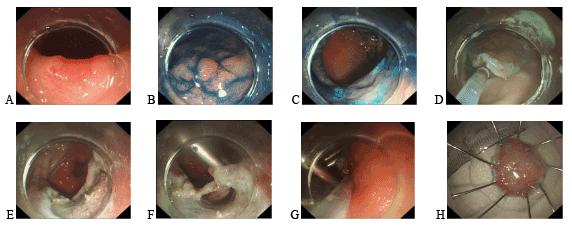
Figure 1: Mucosa avulsion during ESD procedure
A. The lesion located in greater curvature side of antrum.
B. Indigo carmine staining.
C. Marked the lesion by APC.
D. Pre-cut mucosa along the marker.
E. Lesion was resected, cicatrix was seen on the wound.
F. Mucosa avulsion and cavity forming.
G. Occlude the cavity with endo-clips.
H. Lesion specimen.
Discussion
As it is minimally invasive and with notable curative effect, ESD
has progressed rapidly and been used widely in the treatment for
gastrointestinal early cancer or precancerous lesions in recent years.
The procedure of ESD includes defining margins, marking, submucosal
injection, circumferential mucosal incision and submucosal dissection.
As we all know, in order to perform ESD safety, the submucosal space
should be expanded by injection of a lifting solution to form a safe plane
for dissection between the mucosa and muscle wall. What’s more, the
mucosa lifting sign during submucosal injection can be used to evaluate
the adhesion degree of submucosal tissue and pre-judge that whether an
en bloc and curative resection can be achieved. So submucosal injection is
an important and meaningful part of ESD.
The complication of mucosa avulsion during submucosal injection is
rare and just a minority of cases were reported in esophagus, of which the
submucosal tissue was relatively loose, while till now, none was reported in
gastro, especially in gastric antrum, as the submucosal tissue was pyknotic.
We have performed ESD successfully in several thousand patients since
we began to develop ESD procedure in our digestive endoscopy center
in 2006. And no gastric mucosa avulsion was found during submucosal
injection in ESD procedure until this case.
As for the causes, leading to gastric mucosa avulsion, are probably as
follows. Firstly, the mucosa wasn’t lifted well during submucosal injection.
As the histopathological examination revealed a high-grade intraepithelial
neoplasia lesion, which belongs to precancerosis, so adhesion caused
by malignant tumor infiltration could be excluded. The primary reason
might due to the multi-block biopsy before ESD, which can easily resulted
in mucosa and submucosal tissues cicatrization and adhesion, making
the submucosal injection more difficult, and it could be confirmed by
the white scar observed during the ESD. Secondly, the needle tip usually
directs to the anal side of the lesion when performing submucosal
injection. Due to the submucosal adhesion, the mucosa lifting sign was
poor, and the pressure of pushing mixture liquid had to be increased.
The higher the pressure was the more easily the mixture liquid spread
to the anal side quickly, bypassing the submucosal adhesion which
could not be lifted well, and as a result the anal side gastric mucosa
avulsed instantly under the high liquid pressure and flow rate. Thirdly,
individual difference also plays an important role in mucosa avulsion.
Of the several thousand cases of ESD, in our digestive endoscopy
center, in some patients submucosal adhesion reported, but not
avulsion, because there is no long history of glucocorticoids intake,
which would make the submucosal tissue around the adhesion much
looser, as the case reported in this article.
When gastric mucosa avulsion occurred, we’d better continue the ESD
and resect the lesion as procedure. Then we should confirm the avulsion
and occlude the submucosal cavity with endo-clips (Resolution, Boston
Scientific). If not, the cavity is sure to be exposed to the acidic conditions
and postoperative bleeding might occur.
Download Provisional PDF Here
Article Information
Aritcle Type: Case Report
Citation: Xiong Y, Zhang X, Chen Q, Hu H, Linghu E
(2016) Gastric Mucosa Avulsion during Endoscopic
Submucosal Dissection. J Gastric Disord Ther 2 (1):
doi http://dx.doi.org/10.16966/2381-8689.112
Copyright: © 2016 Xiong Y, et al. This is an
open-access article distributed under the terms
of the Creative Commons Attribution License,
which permits unrestricted use, distribution, and
reproduction in any medium, provided the original
author and source are credited.
Publication history:
Received date: 27 Nov 2015
Accepted date: 18
Jan 2016
Published date: 22 Jan 2016