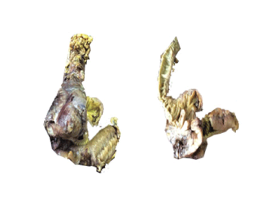
Figure 1: Segment of the right hemicolectomy specimen of a 40-year old man with fibrosis and perforation involving the base of the caecum.

Der EM1,2* Osman L1 Armah R3
1Department of Pathology, School of Biomedical Sciences, Korle-Bu Teaching Hospital, Accra, Ghana*Corresponding author: Der Muonir Edmund, Department of Pathology, School of Biomedical Sciences, Korle-Bu Teaching Hospital, Accra, Ghana, Tel: 2322444444; E-mail: maadelle@yahoo.com
Background: Caecal perforation commonly occurs in elderly patients with caecal neoplasms or distal obstructions.
Case presentation: We report a case of caecal schistosomiasis with spontaneous perforation in a 40 year old taxi driver in Accra Ghana, who presented with symptoms of acute abdomen. He had emergency laparotomy and right hemicolectomy. Findings at surgery were caecal tumour with perforation at the base and faecal peritonitis. Pathological examination revealed chronic granulomatous response to schistosoma ova with bowel wall necrosis and perforation.
Conclusions: Right iliac fossa pain is a common symptom of acute appendicitis, especially in young patients. However, in rare cases it can reflect other pathology which is easy to miss-diagnose as in this case.
Caecum; Schistosomiasis; Acute abdomen; Granulomatous reaction
Caecal perforation in a healthy individual is uncommon and if it does, it is commonly associated with distal luminal obstruction, malignancy, unrelieved volvulus of the caecum, trauma to the right side of the abdomen, or iatrogenic [1]. Spontaneous perforation of the caecum in the absence of any of the above factors is exceedingly rare. In the 1990s, cases of spontaneous caecal perforations were reported. Low and Fairley [2] in 1934 described a case of perforation occurring in a patient suffering from tropical sprue. Robertson et al. [3] in 1958 described a case following natural childbirth. Other inflammatory conditions mimicking spontaneous caecal perforations have been be reported [4,5]. Again, in 1961 Hirsch also described another case following Caesarean section [6].
Caecal perforation associated with abdominal tuberculosis [7,8] in developing countries has been reported in the literature. Similarly, intestinal schistosomiasis has been reported in Singapore, Ghana [9,10] and other countries in the tropics [11,12]. Spontaneous large bowel perforation associated with intestinal schistosomiasis has been reported in Africa [13]. However, spontaneous caecal perforation resulting from intestinal schistosomiasis in Ghana has not been published from our literature searched. This case report presents rare cause of spontaneous caecal perforation associated with intestinal schistosomiasis in a 40 year old taxi driver, a native of the Eastern Region of Ghana
Mr SF, a 40 year old male driver from Awoshie in the greater Accra Region of Ghana, presented to the Korle-Bu Teaching Hospital (KBTH) surgical medical emergency with a 3 day history of sudden onset of right iliac fossa pain associated with anorexia, fever and chills. He is a native of the Eastern Region of Ghana, where he lived until age 20-years when he migrated to Accra to learn his current profession. At the time of presentation, he was ill-looking, dehydrated and warm to touch. The pulse rate was 102 bpm. He had marked right iliac fossa tenderness and rebounce tenderness, but no guarding. The bowel sounds were present but infrequent.
The haemoglobulin level was 14.1 g/dl, WBC was 17.05 × 10⁹/L (high) and platelet count was 190 × 109/L. The Renal function test (BUE&Cr) was Sodium (Na) 135 mmol/L, Potassium (K) 4.1 mmol/L, Urea (SI), 3.1 mmol/L (Low), Creatinine (SI), 3102 µmol/L.
Pre-operative diagnosis: Peritonitis 20 perforated appendix
Mr SF had exploratory laparotomy and right hemicolectomy. The findings at surgery were: gangrenous appendix, caecal tumour with perforation at the base and faecal peritonitis. There were 11 enlarged pericolic lymph nodes.
Post-operative diagnosis: 1. Perforated Caecal tumour 2. Gangrenous Appendicitis
He had intravenous antibiotics (flagyl and ciprofloxacin), intramuscular pethedine and suppository paracetamol. He had nasogastric tube and urethral catheter passed. He had input-output fluid chart.
We received a right hemicolectomy specimen consisting of the terminal ileum (11.0 cm long), caecum (8.0 cm long) and the ascending colon (14.0 cm long). There was a perforation (3 × 2 cm) that was posteriorly located and at the base. Opening the bowel revealed a thickened and fibrotic caecal wall associated with necrosis and perforation (Figure 1). The appendix was grossly normal except moderate thickening and fibrosis at the base closed to the perforation. The rest of the specimen was normal grossly. No polyp nor and tumour seen. There were 11 lymph nodes retrieved from the pericolic fat.
Figure 1: Segment of the right hemicolectomy specimen of a 40-year old man with fibrosis and perforation involving the base of the caecum.
Microscopy examination of sections of representative portions from the caecal wall showed large areas of fibrosis, necrosis, haemorrhage and oedema. There were viable and calcified Schistosoma haematobium ova within the necrotic area and the submucosa region of the viable bowel wall. No malignancy seen. The 11 lymph nodes retrieved show reactive changes (Figure 2).
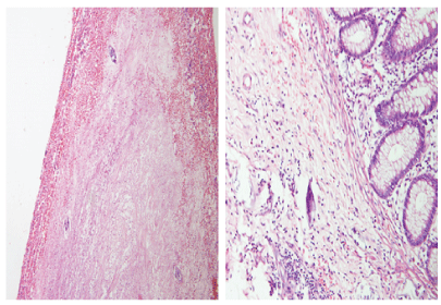
Figure 2: Histological section of the caecal wall of 40-year old man showing large areas of necrosis and oedema. There are Schistosoma haematobium ova within the necrotic and viable portions of the wall.
Caecal schistosomiasis with perforation.
Mr SF was put on a stat dose of praziquantil (1.2 G) based on the histopathology report and discharged on the post-operative day 10. He was found on his first review to be doing well.
Mr. SF presented to the KBTH surgical medical emergency with sudden onset of colicky right iliac fossa pain. He had right hemicolectomy and the final histological diagnosis was caecal schistosomiasis with perforation. The clinical picture of Mr SF mimics most acute abdominal conditions such as acute appendicitis [3,4] that requires immediate surgical management as in this case. The age of Mr SF and the clinical presentation of his condition all put together may have accounted for the pre-operative diagnosis of peritonitis 20 perforated appendicitis by the surgeons. For instance, appendicitis and spontaneous caecal perforation are common conditions within the age (Younger) group of Mr SF [5].
Caecal perforation associated with luminal obstruction by tumours or trauma has been reported in the literature, [1,6] but these were absent in this case. Infectious causes of spontaneous large bowel perforation such as tuberculosis and schistosomiasis in healthy individuals both rare complications have been reported in the developed and developing countries across the globe [7-13]. The diagnosis of spontaneous caecal perforation due to schistosomiasis in Mr. SF therefore supports previous studies.
Infections by schistosome species are commoner in Asia and SubSaharan Africa [14,15] with fresh water bodies and farming as the major occupation [16]. In Ghana, schistosomiasis have major economic and public health implications on residents living in endemic rural and periurban areas, [17,18] where people, especially children are predispose to schistosomiasis and its complications decays ago [19-21]. Mr. SF is a native of the East Region of Ghana, a farming community with fresh water bodies (rivers), where he lived up to age 20 years before moving to Accra and thus may have acquired the infection during his childhood. The chronic inflammatory response to the ova resulted in caecal wall fibrosis, ischaemic necrosis and perforation that presented as acute abdomen in this case. The commonly involved species in farming communities of Ghana [10] and other endemic regions in Africa such as Egypt [22,23] decays ago has been identified as S. haematobium, and this supported the species identified in this current case report.
The presence of the ova in the vessels and tissues evokes chronic inflammatory response with mark fibrosis and inflammatory mass formation. This inflammatory mass may grossly mimic a neoplasm as in the current case. For instance, post operatively, two of the findings were caecal mass and 11 enlarged lymph nodes, mimicking a neoplastic lesion. This was however found by histological examination to be an inflammatory mass (caecal schistosomiasis). This case further illustrated the difficulty in differentiating an inflammatory mass from a neoplastic lesion at surgery without histopathological study. The treatment of schistosomiasis is praziquantel. Mr. SF had right hemicolectomy because his condition complicated as a mass with necrosis and perforation.
For people particularly children living in schistosome endemic regions of Ghana, to prevent life threaten complications as in this case report, the following are recommended.
Right iliac fossa pain is a common symptom of acute appendicitis, especially in young patients. However, in rare cases it can reflect other pathologies which are easy to miss-diagnose as in this case.
EMD: Ideal of a case report, Histology reporting, Literature review and Reading/correction of manuscript
OL: Histology reporting, Literature review and Reading/correction of manuscript
AR: Clinical history, Surgery and management and Reading/correction of manuscript
The authors declare that there are no conflicts of interests.
The authors declare that the written consent was obtained from the patient for publication of this case report. The consent is available with the authors.
None declared
Download Provisional PDF Here
Article Type: Case Report
Citation: Der EM, Osman L, Armah R (2016) Caecal Schistosomiasis with Perforation: A Case Report at the Korle-Bu Teaching Hospital Accra Ghana. J Clin Case Stu 1(4): doi http://dx.doi.org/10.16966/2471-4925.122
Copyright: © 2016 Der EM, et al. This is an open-access article distributed under the terms of the Creative Commons Attribution License, which permits unrestricted use, distribution, and reproduction in any medium, provided the original author and source are credited.
Publication history:
All Sci Forschen Journals are Open Access