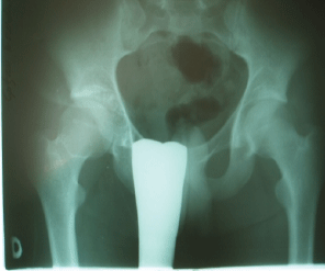
Figure 1: X-ray findings of PSC in right hip

Bukva B1* Brdar R1 Vrgoc G2 Krivokapic B3 Ducic S1 Abramovic D1
1University Children`s Hospital, Belgrade, Serbia, Europe*Corresponding author: Bukva B, University Children`s Hospital, Belgrade, Serbia, Europe, Tel: 00381642095947; E-mail: bojanbukva@yahoo.com
Synovial chondromatosis is an uncommon disorder characterised by the formation of multiple cartilaginous nodules within the synovium and most commonly affects large joints, such as the hip and knee, but it also may be localisated an extraarticulary. It usually present an adult population and is rare in children. The main symptoms are pain, swelling, and limitation of movements in the affected joint. Diagnosis is generally confirmed by the histologycal analysis after clinical and radiological examination. The autors report the synovial chondromatosis presenting in the hip of the 17 year-old boy and discuss the etiology, diagnosis, and treatment of this rare lesion based on current literature.
Synovial chondromatosis; Painful hip; Child
Synovial chondromatosis is a rare benign arthropathy affecting diarthrodial joints. It represents an idiopathic benign metaplasia of synovial tissue to cartilage, as there are no cartilage cells in normal synovial membrane [1]. It is caracterised by the formation of islands of metaplastic cartilage within the synovium. The cartilage is formed in the synovial membrane of joints, tendon sheats and bursae. It may develop primarly in the joint (intra-articular) or from extra-articular synovial tissue. Also, it is described the combined chondromatosis, an intra-articular and extra-articular chondromatosis [2]. Primary idiopathic synovial chondromatosis most commonly occurs within the 14 to 59 year-old age group, with a male/female ratio of 2/1 [3]. The involment in primary synovial chondromatosis is mostly monoarticular, affecting the large joint as the knee or the hip; but other joints such as elbow, shoulder and ankle may be affected also. Secondary chondromatosis is a conseqence of the trauma, osteonecrosis, pigmented villonodular synovitis or degenerative joint diseaseand it could be deverse affectation [4].
A 17-year-old boy presented complaining of the right hip pain and painfull right lumbo-sacral region. According to patient history the pain started approximately 6 months earlier, during daytime and during the night, mild to moderate intensity (VAT 2-5) without need to take analgetics. He could not recall traumatic event. At the physical examination of the right hip revealed painfull limitation of flexion, abduction and internal rotation compared with motions on the contralateral hip. The strenght of the quadriceps femoris was slightly reduced, and sensibility was unaffected. Blood analizies (full blood count, erythrocyte sedimentation rate, C-reactive protein, lactate-dehydrogenase and alkaline-phosphatase) were normal.
Radiologically, rounded loose bodies were presented in the right hip, with variable size and variable degrees of calcification and ossification. Some of the lesions have a radiolucent center due to incomplete calcification (Figure 1).
Figure 1: X-ray findings of PSC in right hip
MR imaging described intrasynovial mass that surround the femoral neck, especially in dorsal aspect. These mass was lobulated, separately till 15 mm of diameter with intrasynovial fluid. According to T2 hyper and T1 intermedial signal the synovia was hyperthrophic, with the edge amplification of signal in front of the femoral head and neck, and intraarticullary also. The edge of the right hip was slightly eroded (Figure 2).
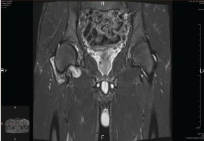
Figure 2: MRI findings of PSC of right hip
Intraoperatively, the mass excision was performed through the Smith Peterson incision. The lobulated mass was up to 20 cm of diameter, separately attached to synovia (Figure 3). The material was send to histopathological analysis.
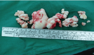
Figure 3: Intraoperative findings: rounded loose bodies and bodies attached to the synovial
Postoperatively the hip was not imobilized. Three days after surgery passive and active assisted range-of-motion exercises were started. Two months later, the patient had achieved total recovery of strenght with complete symetric restoration of motion.
The histologic examination of the bodies confirmed cartilaginous synovial metaplasia consistent with synovial chondromatosis. The bodies were composed of cartilage with calcification. No malignant transformation was present.
Primary synovial chondromatosis (PSC) is a benign, idiopathic, metaplastic disorder of the synovial membrane, with secondary formation of osteochondral nodules [3,5,6]. It could be different sizes, and if it presents a nodules over 1 cm of the diameter it is called giant solitary synovial chondromatosis [4]. Also, giant solitary synovial chondromatosis (size 1-20 cm) may be intra-articular or extra-articular.
According to Milgram, it has been postulated that PSC appears in 3 separate phases: phase 1 is active intrasynovial disease without the presence of free intraarticular bodies; phase 2 is a transitional stage, involving both an active intrasynovial proliferation and intraarticular loose bodies; and phase 3 is multiple loose bodies without obvious intrasynovial disease [6]. Because of the lack of obvious clinical symptoms and the difficulty of making the correct diagnosis on radiographs, phase 1 is rarely seen.
The etiology of PSC remains unclear as well. Werner et co-workers in a study on chondromatosis of the elbow, found the disease tends to affect the dominant extremity, which leads to the assumptation that biomechanical stress might be a factor in the formation of the SC [7]. Hereditary transmission might be another factor, as several authors increased familial clustering [7,8].
The exact pathogenesis of PSC is poorly understood, but it is generally accepted that the process involves a benign reactive metaplasia of the subintimal connective tissue of the synovial membrane. As primitive mesenchymal cells differentiate into mature chondrocytes, nodules of hyalinecartilage are formed, break off, and became loose bodies. These loose bodies, nourished by synovial fluid, grow inside the joint [6].
Clinically its presentation varies, depending on the site and whether it is extra or intra-articular. In all cases it results in either intra-articular or extra-articular mass. Intra-articular involment presents with joint pain, effusion, stiffness, crepitus, locking, giving way, and secondary degenerative changes with loose bodies. Most patients have a long clinical history before a correct diagnosis is made, and what is the most important, clinical features are not helpful in differentiating between this entity and synovial chondrosarcoma [9].
Imaging techniques for PSC include plain radiographs and MR imiging. On radiographs it appears as a large mass with or without feathery calcification, or a round calcified mass. The mass may indent or erode into the surrounding bone and gives an appearance of chondrosarcoma. In about 11 % of cases there is pressure erosion of the bone and in 88 % of cases discrete radio opacities are seen on plain radiographs [10]. MR imiging allows for a visualisation of uncalcified lesions or lesions in the initial phase, without the presence of loose bodies [11]. The appearance of synovial chondromatosis in MRI is variable and depends on the relative preponderance of synovial proliferation and loose body formation. The intrasynovial masses have lobulated borders and homogenous intermediate signal on T1-weighted images and high signal on T2- weighted images [12].
According to Kramer, there are three different MRI patterns, depending on the presence and distribution of various synovial chondromatosis components and the stage at which the process is being imaged [11]. Pattern A shows multi-nudeate cells, isointense or slightly hyperintense to muscle on T1WI and hyperintense on T2WI. Pattern B is similar to pattern A but with additional foci of signal voids due to presence of calcification. Pattern C shows foci of peripheral hypointensity and central fat signal intensity secondary to ossifications. Gadolinium-DTPA may be used for assessment of the synovial activity and to differentiate early synovial chondromatosis from a simple joint effusion [5,13,14].
The histology of synovial chondromatosis shows cellualr hyaline cartilage arranged in small clusters separated by fibrous tissue. Chondrocytes may show minor cellular atypia with irregular hyperchromatic nuclei and occasionally bi-nucleate or multi-nucleate cells. These changes may mimic malignancy and can lead to the erroneus diagnosis of chondrosarcoma with subsequent implications of tretatment. Bertoni et al. in 1990th described histological criteria to differentiate between benign and malignant transformation of synovial chondromatosis thus: malignant change is caracterised by marked cellular atypia; chondrocytes are arranged in sheets with loss of cluster appearance, presence of necrosis, mitoses and myxoid features and invasion in to adjacent tissues [15].
In treatment of PSC there are two options: an open surgical procedure with loose bodies removal and partial synoviectomy, which is described by several authors [7,16-18], and an arthroscopic treatment. An arthroscopic treatment has been reported for intraarticular PSC [16,19,20], but no long-term studies have compared open and arthroscopic treatment. Open surgical treatment is preferred in cases of extraarticular involvement with limited anatomic space [21].
Radiotherapy is beneficial for reccurent lesions and inhibition of FGF-9 has been suggested as a nonoperative method of treating primary synovial chondromatosis [22,23].
This probably saved the child from further surgical treatment and unnecessary chemotherapy.
In conclusion, PSC mostly affects large joints, the knee or hip, but atypical extra-articular or intra-articular involvment might occur. In case of missing calcifications only MRI allows for diagnosis of uncalcified lesions in the initial phase. Diagnosis as well as lesion character, benign or malign, have to be assured histologically. Regular follow-up examinations that include MR imiging are advised for up to 2 years. Secondary degenerative arthritis is by far the most common complication of longstanding disease [5,14,24,25]. The recurrence after excision has been reported also [6,14,26,27]. Malignant transformation to chondrosarcoma is a known serious complication however, it is rarely seen [5,14,24,25].
Orthopedic surgeons, radiologists and pathologists needs to be aware of this condition and expert histopathological opinion should be obtained, since this benign condition may be confused with malignancy.
Download Provisional PDF Here
Article Type: Case Report
Citation: Bukva B, Brdar R, Vrgoc G, Krivokapic B, Ducic S, et al. (2016) Primary Synovial Chondromatosis of the Hip in Adolescent-A Case Report. J Clin Case Stu 1(6): doi http://dx.doi. org/10.16966/2471-4925.119
Copyright: © 2016 Bukva B, et al. This is an open-access article distributed under the terms of the Creative Commons Attribution License, which permits unrestricted use, distribution, and reproduction in any medium, provided the original author and source are credited.
Publication history:
All Sci Forschen Journals are Open Access