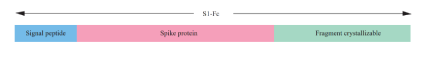
Figure 1: The schematic diagram of S1-Fc gene. It is composed of signal peptide, spike protein and Fc region.
Zhen-Wang Jie1,2,# Yu-Ting Lin1# Wei-Hong Sun1# Wan-Jie Yang1 Zi-Yan Zhang1 Kai Weng1 Yuan Chen1 Chen Li1 David Lin1,2*
1Hope Bio-Technology (Suzhou) Co. Ltd, Suzhou, Jiangsu, P.R. China*Corresponding author: David Lin, Hope Bio-Technology (Suzhou) Co. Ltd, Suzhou 215124, Jiangsu, P.R. China, Tel: +86-0512-81878332; E-mail: davidlinmd@aliyun.com
Coronavirus disease 2019 is pandemic across the world since the severe acute respiratory coronavirus-2 emerged in December 2019. The most efficient method to defend COVID 19 is vaccine. Here, we used HEK293 cell line to produce the recombinant protein and compared several vaccine candidates’ efficiency. The structures of the candidates were designed based on the S1 protein and RBD region. After three doses, the vaccines induced SARS-CoV-2 specific neutralizing antibodies in mice which presented high titers. The maximum neutralization activity could reach more than 90% without inflammation suggesting the great neutralizing ability against SARS-CoV-2.
Significance statement: The COVID-19 has threatened human beings since 2019. This paper reveals a new recombinant protein vaccine that could induce mice immunity against the coronavirus. It compared the efficacy of different potential vaccines expressed by human embryonic kidney 293 cell. It showed the difference of them and helps people learn more about the COVID-19 subunit vaccines.
COVID-19; Vaccine; HEK293; Recombinant protein
As the coronavirus disease 2019 (COVID 19) is pandemic across the world, over 103 million cases of COVID 19 have been reported and more than 2 million people have been claimed. It is urgent to develop the vaccine of COVID 19 to help human get rid of the threat of it. The pathogen severe acute respiratory syndrome coronavirus 2 (SARS-CoV-2) is a betacoronavirus responsible for the COVID 19 pandemic [1]. SARS-CoV-2 has 29,903nt encoding four structural proteins (nucleocapsid protein, membrane protein, spike protein and envelope protein) and sixteen non-structure proteins (nsp1-16) [2,3]. The entry of SARS-CoV-2 into cells is dependent on spike protein. It contains 2 subunits, S1 and S2. The S1 subunit consists of N-terminal domain (NTD) and receptor binding domain (RBD) which binds to the receptor angiotensin-converting enzyme 2 (ACE2) of host cells to determine infection entry [2-6]. It has been reported that most antibody generated by COVID 19 patients were binding the receptor binding domain (RBD) of S1 protein. S1 protein has been recognized as a successful target for vaccine development [4,7-12].
Several studies about the COVID 19 vaccine have been launched since 2020. There are 2 mRNA vaccines on the market and several protein vaccines [13-17]. These vaccines are developed on the basis of S protein and present a great protection. Though many effective vaccines are on the market, the mutation of virus and limited production still pose threat to the lives of billions of people [17]. In particular, the efficacy of current vaccine decreased in the defense of SARS-CoV-2 variant. The transmission speed of the mutant was fast.
Several studies have demonstrated that IgG-based scaffold can enhance the solubility and stability of fusion protein, generating robust and long-lasting immune responses [18-20]. The fragment crystallizable (Fc) region is required for killing of virus-infected cells by complement-dependent cytotoxicity (CDC) and by antibodydependent cellular cytotoxicity (ADCC) [21,22]. Some Fc-fusions target receptor-ligand interactions, working as agonists to directly stimulate receptor function to reduce or increase immune activity [23]. In this study, we compared the immune efficiency of S1-His, RBD and S1-Fc. To increase the efficacy of vaccine, we developed S1-Fc as vaccine through the combination of S protein and mutant IgG using transient expression system Expi293F cells. Expi293F cell line was favored by researchers for its high transfection efficiency and human-derived. Immunization of mice with three doses of S1-Fc with adjuvant induced dose level-dependent increases in pseudovirus neutralization titers and vitro test.
Human embryonic kidney HEK293F (Expi293F™ from Thermo Fisher) was cultured in Dulbecco’s modified Eagle’s medium (DMEM). Cell lines were tested for mycoplasma. Sterility contamination after receipt, before expansion and cryopreservation. Expi293F™ cells were grown in Expi293™ media with GlutaMAXTM (Gibco). The selected medium is the one with clear chemical composition, no serum, no protein and no animal origin, The cell transiently transfected using polyetherimide (PEI MaxTMPolysciences, Inc).
Human embryonic kidney HEK293T (from Thermo Fisher) was cultured in Dulbecco’s modified Eagle’s medium (DMEM) with GlutaMAX™ (Gibco) supplemented with 10% fetal bovine serum (FBS [Sigma-Aldrich]). When 293T cells grow to more than 80%, it could be passaged. DMEM medium containing 10% FBS, pancreatin, and PBS was heated in a 37°C water bath. The cells were rinsed once with 5ml of PBS. 3ml of pancreatin (0.5%) were added and gently shaken to make the pancreatin completely cover the cell surface of the culture flask for 1 minute. 5ml of DMEM medium containing 10% FBS was used to terminate the digestion. The cells were centrifuged and 6.5 × 10⁵ cells were obtained to culture. When the cells grow to 50%, it could be used to transfection.
The protein gene was merged as figure 1 and codon optimization was performed. Then the gene was inserted in the pcDNA3.4 plasmid. (Conducted by Genscript). The Expi293F Expression System which is a mammalian protein system was used as the host cell. The recombinant plasmid dissolved in 1.5ml Opti-MEM (DNA density: 3ug/ml) was mixed with PEI (transfection reagent). When the density of cells reached 3.0 × 10⁶ cells/ml, cell viability >95%, the mixture was added into the cell culture flask. After 6 days culture, the recombinant protein was obtained.
Figure 1: The schematic diagram of S1-Fc gene. It is composed of signal peptide, spike protein and Fc region.
Purification of the recombinant protein S1-Fcwas based on protein A affinity chromatography. The Monofinity A Resin from GenScript and AKTA AVANTTM were used to purify the fusion protein with flow rate 1ml/min. The procedure used was followed as the protocol described. Then the dialysis and ultrafiltration concentration were performed. Finally, the Superdex 200 installed on AKTA AVANT was used to the final purification. SDS-PAGE was used to measure the size of recombinant protein (SDS-PAGE kit Solarbio).
His Trap Excel affinity chromatography column (1ml pre-packed column, max flow rate: 4ml/min, max pressure: 0.5MPa) was used to capture and purify protein with His mark on AKTA. Equilibration buffer A: 20mM Tris, 150mM NaCl, pH 6.5, elution buffer B: 20mM Tris, 150mM NaCl, 500mM imidazole, pH 6.5 (equilibration, the buffer is degassed by ultrasound). The His column is flushed, balanced, and the flow rate is 1ml/min.
RBD was a commercialized protein purchased from Sinobiological (Cat: 40592-V08H118).
The protein was separated by HPLC-SEC method. The instrument was a Waters™ high-performance liquid chromatograph. Xbridge™ BEH 200 Å SEC (7.8*300mm, 3.5μm) SEC column was used, and the flow rate of the mobile phase (150mM phosphate containing 10% methanol) 0.86ml/min. After equilibrating the chromatographic column until the baseline is stable, load 10μl, detection wavelength: 214nm, and record the chromatogram and data with in 20 minutes. Use Empower software to process the test results, and calculate the purity of the protein according to the area normalization method.
The recombinant protein concentration was measured by “sandwich” capture assay enzyme-linked immunosorbent assay (ELISA). 96-well plate was coated with antibody (SinoBiological, Cat:40150-D003 ) using carbonate buffer containing 32mM sodium carbonate, 68mM sodium bicarbonate (pH 9.6) at 4°C overnight, then it was blocked by 5% bovine serum. The purification product and culture supernatant were added to wells in duplicate, followed by incubation for 2 h at 37°C. Plates were then incubated for 1 h with anti-spike IgG with Horseradish Peroxidase (HRP) conjugate (SinoBiological, Cat: 40150-D001-H). Lastly, developed with 3,3’,5,5’-tetramethylbenzidine (TMB), the optical density (OD) at 450 nm was measured (Molecular Devices, SpectraMax® iD3).
The method to measure titers of IgG was determined by indirect assay. The spike protein (SinoBiological, Cat: 40591-V08H) was coated on the 96-well plate and blocked by 5% bovine serum. Mouse sera, diluted 1:100 in phosphate buffer, were added to wells in duplicate, and 2-fold serial dilutions were performed, followed by incubation for 2 h at 37°C. Plates were then incubated for 1 h with goat anti-mouse IgG with HRP conjugate (SinoBiological, Cat: SSA007). TMB was added and OD was measured.
All the line charts and bar charts were drawn by using GraphPad. When the IgG line graphs were drawn, the IC50 was calculated by the function of log (inhibitor) vs. response - Variable slope (four parameters) in GraphPad.
The S protein binds to the cellular surface receptor ACE2 through the receptor binding domain (RBD), an essential step for membrane fusion [5,24]. The ACE2 binding test can assess the vaccine candidate ability of binding ACE2 to prevent the entry of virus. 6-well plate was coated with ACE2 (SinoBiological, Cat: LC14SE1109 ) using carbonate buffer containing 32mM sodium carbonate, 68mM sodium bicarbonate (pH 9.6) at 4°C overnight, then it was blocked by 5% bovine serum. The purification product was added to wells in different concentration, followed by incubation for 2 h at 37°C. When the blocking of serum was conducted, the serum of immunized mice were mixed with 62.5ug/ml S1-His and added to wells, followed by incubation for 2 h at 37°C. Plates were rinsed and then incubated for 1 h with anti-spike IgG with Horseradish Peroxidase (HRP) conjugate (SinoBiological, Cat: 40150-D001-H). Lastly, developed with 3,3’,5,5’-tetramethylbenzidine (TMB), the optical density (OD) at 450nm was measured (Molecular Devices, SpectraMax® iD3).
The 293T-ACE2 cells (SinoBiological, Cat: OEC001) were inoculated in a 96-well plate with 3-5 × 10^4/ml 200ul per well. After plating, the plate was placed in a 37°C, 5% CO2 incubator for 24 hours. The pseudovirus (SinoBiological, Cat: PSV001) was diluted by 2 times before use. The test sample was mixed with pseudovirus, incubating at room temperature for 1 hour. The supernatant of pre-laid 293T-ACE2 plate was aspirated and the pseudovirus mixture was added to the plate. The plate was incubated at 37°C for 48 hours. 50ul chromogenic agent (Fire-LumiTMkit, GenScript) were added per well. There were test samples, positive control and negative control. Inhibition rate (%)=1-(sample RLU average-negative RLU average)/(positive RLU average-negative RLU average).
Use the recombinant protein to perform animal immunization experiments. The flow chart of the immunization experiment is shown in figure 2. C57BL/6 male mice of at least 25g were used in the experiment. Recombinant protein was diluted in PBS mixing with aluminum adjuvant (v/v:3:1) and injected subcutaneous. Each mice was injected 100ul mixture. The weight and temperature of mice were monitored. The animal experiments were conducted by KCI BioTech. All animal procedures were approved by the KCI BioTech Institutional Animal Care and Use Committee, and performed according to the NIH Guide for the Care and Use of Laboratory Animals. All efforts were made to reduce the number of animals and to minimize animal suffering.

Figure 2: Pseudovirus neutralization titers of different serum. 30μl pseudovirus dilution mixed with 20ul 30 times diluted serum were used as test sample. The positive control was composed of 30ul pseudovirus dilution and 20ul PBS. The negative control was 50ul DMEM. Inhibition rate (%)=1-(sample RLU average-negative RLU average)/(positive RLU average-negative RLU average). The x axis shows the serum from different mice. Number # indicates the mouse number. AL indicates the serum from the mouse immunized with adjuvant aluminum hydroxide. S1-His+AL indicates the serum from the mouse immunized with S1-His with adjuvant. RBD+AL indicates the serum from the mouse immunized with RBD with adjuvant. S1-Fc+AL indicates the serum from the mouse immunized with S1-Fc with adjuvant.
Initially, the S1-His was developed as our potential vaccine which was inserted in pcDNA3.4 then transfected in HEK293T. In order to compare the effect of different host cell, the HEK293F and HEK293T was used to express the S1-His. The results showed that the recombinant protein production of HEK293F was higher than that of HEK293T (Figure 3A). After purification, the recombinant protein was tested by HPLC. The results were shown in figures 3B and C. The S1-His expressed by HEK293T was around 0.2mg/L and S1-His expressed by HEK293F was around 1.4mg/L. The molecular weight of S1-His ranged from 100kDa to 130kDa. After purification, the protein expressed by HEK293F was purer than HEK293T due to the supplement of serum in HEK293T culture. The advantage in purification makes us prefer to use HEK293F as a host cell.

Figure 3: (A) S1-His protein expression in different host cell (HEK293T and HEK293F). (B) S1-His protein expressed by HEK293T. Production of the S1 proteins and their characterizations by SDS-PAGE (left panels), AKTA-AVANT chromatography (middle panels), and HPLC (right panels). (C) S1-His protein expressed by HEK293F, with the characterizations by SDS-PAGE (left panels), AKTA-AVANT chromatography (middle panels), and HPLC (right panels). (D) S1-Fc protein expressed by HEK293F, with the characterizations by SDS-PAGE (left panels), AKTA-AVANT chromatography (middle panels), and HPLC (right panels). (E) S1-His and S1-Fcprotein expression in HEK293F.
The S1-Fc gene which was composed of a signal peptide, a spike protein of coronavirus, and a fragment crystallizable region was inserted in pcDNA3.4 plasmid. The molecular weight of S1-Fc was about 102.81 kDa. As we can learn from the figure 3D, the S1-Fc expressed by HEK293F ranged from 130kDa to 170kDa. It might indicate that the S1-Fc expressed by HEK293F have glycosylation. Since glycosylation was shown to influence several protein functions, including protein–protein interactions and protein activity, the cell line used for the production of recombinant proteins could have a significant effect on their function in vitro and in vivo. In previous research, it was shown that the protein expressed by HEK293 cell was rich in glycosylation [25]. The plasmid was transfected in HEK293F cell line and the recombinant protein was expressed at about 8mg/L (Figure 3E).
The expression of S1-Fc was higher than the expression of S1-His in HEK293F (Figure 3E). The results showed that the purification effect of protein A affinity chromatography column is better than that of His column purification. The purity of purified S1-Fc could reach 97%, and the purity of S1-His could reach 94.78% (Figures 3B and C).
The S protein binds to the cellular surface receptor ACE2 through the Receptor Binding Domain (RBD), an essential step for membrane fusion. Binding to the ACE2 can effectively prevent the entry of virus [5,25]. The ACE2 binding test can assess the binding ability of the vaccine candidate (Figure 4A). The results showed that the S1-His protein could bind to the ACE2. When the concentration reached 250ug/ml, the OD was around 2 and didn’t increase as the concentration increased. The results indicated that the recombinant protein could bind to the ACE2 and partly prevent the virus binding to ACE2. Thus, the entry of virus would decrease [25]. The serum after 3rd immunization of S1-His was used to incubate with S1-His, then the mixture was used to assess its binding ability (Figure 4B). The results showed that immunization with S1-His had ability to prevent S1 binding to the ACE2. The immunization of S1-His was effective to neutralize the binding of S1 and ACE2.

Figure 4: (A) Binding outcomes of the S1 protein with ACE2 receptors. The X-axis indicates the concentration of S1-His in μg/ml, while the Y-axis indicates the binding signal intensity in OD (A450) value. (B) The blocking effects of the S1 protein immunized mouse sera against the S1 protein binding to its ACE2 receptors. X-axis indicates different blocking agents, including PBS (without serum) as unblock control, PBS serum that was the mouse serum immunized with PBS, and two S1-His protein immunized mouse sera, 5ug/mouse and 25μug/ mouse, that were collected after the 3rd immunization with 5ug/ ml and 25ug/ml S1-His, respectively. The concentration of S1-His protein used in the blocking assay was 62.5ug/ml.
The immunization strategy was conducted as figure 5A depicted. The serums were collected and measured by ELISA. The aluminum hydroxide was used as the adjuvant in the immunization. The immunization experiments were divided into 4 groups: AL: the mice were immunized by aluminum hydroxide with PBS. RBD+AL: the mice were immunized by aluminum hydroxide and RBD protein mixture. S1-His+AL: the mice were immunized by aluminum hydroxide and S1-His protein mixture. S1-Fc+AL: the mice were immunized by aluminum hydroxide and S1-Fc protein mixture. All groups were immunized 3 times. The IgG titers were measured by ELISA shown in method. The serums were measured after each immunization and the results were shown in figure 5B. The x axis indicated the serum dilution times, and the y axis indicated the OD value.

Figure 5: The immunization of S1 protein. (A) immunization strategy of proteins. d-5: 5 days before the first immunization; d-0: time of first immunization;d13, d15, d22, d25, d32 indicate the number of days after the first immunization. The shapes above the timeline show the treatment of experiment. The red circle refers to the serum collection. The green arrow refers to the immunization of the recombinant protein. (B) The antibody titers of serum after1st immunized mice. (C) The antibody titers of serum after 2nd immunization. (D) The antibody titers of serum after third immunization.
After the first immunization, the IgG titers of 4 groups were low without any difference. The second boost made IgG titers of serum from S1-His+AL and S1-Fc+AL were higher than the adjuvant and RBD with adjuvant group. The IC50 of S1-His+AL was 1:1457 and S1-Fc+AL was 1: 1130. Thus, the efficiency of S1-His was a little higher than S1-Fc. After the third immunization, the titer of RBD+AL increased. The serum titers of three test groups were higher than AL. It indicated that three doses of RBD, S1-Fc and S1-His with adjuvant have immune effect. The result was consistent with the previous researches showing that RBD and spike protein have the potential to be the vaccine target [4,7,9,15,26-27]. Though the antibody of RBD was found in most COVID 19 patients’ serum, the single domain of RBD was not suitable as a vaccine due to its small size and poor immune response [7]. The IC50 values of 3rd immunization serum RBD+AL, S1-His+AL and S1-Fc+AL were 1:2064, 1:3518, 1:1967. The body condition of S1-Fc immunized mice and pathological slices of mice after immunization were shown in figure 6. The vaccination didn’t change the body condition significantly indicating the safety of the S1-Fc.

Figure 6: (A) the pathological section of S1-Fc immunized mice. The slices include lung, liver, brain, spleen, kidney and heart.(B) (C) (D) (E)were the body weight, food intake, body temperature and weight gain of mice which were immunized by different concentration of S1-Fc by days. Each group included 6 mice.
The pseudovirus neutralization was measured to assess whether the serum could defend the virus invasion. In the first trial of pseudovirus neutralization, the serums were measured after three immunizations. 30ul pseudovirus dilution mixed with 20ul 30 times diluted serum were used as test sample. The positive control was composed of 30ul pseudovirus dilution and 20ul PBS. The negative control was 50ul DMEM. Inhibition rate (%)=1-(sample RLU average-negative RLU average)/(positive RLU average-negative RLU average). The AL inhibition rate was -46% and -57% which indicated that the AL RLU average was higher than positive RLU average resulting the negative percentage (Figure 2).
There were 4 mice in S1-Fc+AL group. The serum from four S1- Fc+AL mice neutralized more pseudovirus than AL group. The four mice serum in S1-Fc+AL neutralized about 20%, -24%, 34% and 96% pseudovirus (Figure 2). There were four mice immunized with S1-His, and three of them neutralize more pseudovirus than AL.S1-His serums neutralized 16%, 60%, -7%, and -109% pseudovirus which indicated S1-His+AL less efficient than S1-Fc + AL. The serum from RBD + AL neutralized -3% and 52% pseudovirus. Therefore, the neutralization efficiency of S1-Fc exceled S1-His and RBD.
In order to find out whether the adjuvant affected the serum neutralization, we compared the serums (AL, S1-Fc and S1-Fc+AL). It was shown that adjuvant affected the immune effect (Figure 7). The S1- Fc+AL neutralized 30% pseudovirus, while S1-Fc and AL neutralized -24% pseudovirus respectively. The adjuvant plays an important role in the S1-Fc function.

Figure 7: The pseudovirus neutralization experiment of serum with and without adjuvant. S1-Fc means the serum is without adjuvant while S1-Fc+AL is with adjuvant.
Then, the second trial of mouse immunization was conducted to test the different dosage influence. Each group was immunized according to figure 5A respectively, and the immunization dosage were 1ug, 5ug and 10ugS1-Fc per dose. The test sample was composed of 30ul pseudovirus dilution and 20ul serum. The positive control was composed of 30ul pseudovirus dilution and 20ul serum from AL boost group. The negative control was 50ul DMEM. Inhibition rate (%)=1-(sample RLU average-negative RLU average)/(positive RLU average-negative RLU average). The results were shown in figure 8. After three immunizations, the 5ug group achieved 55%, 46%, 50%, 100%, 63% and 72% pseudovirus neutralization, the 1ug group achieved 45%, 47%, 28%, 72%, 69% and 51% pseudovirus neutralization, and 10ug group achieved 80%, 80%, 97%, 48%, 77%, and 50%. It was shown that as the dose increased, the neutralizing effect of the serum was better. After 3-dose immunization, the serum neutralizing effect got better.

Figure 8: The pseudovirus neutralization of 2nd trial of immunization. The test sample was composed of 30μl pseudovirus dilution and 20μl serumfrom S1-Fc immunized mice. The positive control was composed of 30μl pseudovirus dilution and 20μl serum from AL boost group. The negative control was 50μl DMEM. A: The percentage of pseudovirus that neutralized by serum from the 1st dose of immunization. B: The percentage of pseudovirusthat neutralized by serum from the 2nd dose of immunization. C: Percentage of pseudovirus that neutralized by serum from the 3rd dose of immunization.
Firstly, we compared the expression of S1-His in HEK293T and HEK293F. The HEK293F was chosen as the host cell to express the recombinant protein due to the culture of HEK293F without additional heterologous protein. After purification, S1-Fc was purer than S1-His expressed by HEK293F. The previous research showed that HEK293 cell produced more recombinant protein than CHO cell did [28]. HEK293F could be a potential cell for protein production. Now, our protein production is more than 8mg/L. With optimization in the procedure, the production will increase in the future. Since the functional SARS-CoV-2 S1 protein is predicted to have at least 16 N-glycosylation [11,29], the HEK293 was chosen as the host cell and the cell can provide more glycosylation.
To figure out the efficacy of S1-His and S1-Fc, the immunization and ACE2 binding assay were conducted. Generally, 3 dosesS1-Fc and adjuvant can effectively induce mice to produce neutralizing antibodies. Our research was consistent with the previous research that the spike protein fused with Fc region was found having a great immunogenicity [29]. In our research, we preliminarily compared the efficacy of S1-Fc, S1-His and RBD, then compared the efficiency of different doses of S1-Fc. The spike IgG fusion protein was more effective in SARS-CoV-2 neutralization than S1-His and RBD. The sequence of fusion protein was optimized and the HEK293 expression system indicated a great potential expressing the recombinant fusion protein. In the future, the virus challenge experiment will be conducted and the results will be reported.
David Lin is the presidents of Hope Bio-Technology (Suzhou) Co. Ltd. This research is sponsored by Hope Biomedical Technology (Suzhou)and Hope Bio-Technology (Suzhou) Co. Ltd. A patent covering S1-Fc vaccine has been applied. Remaining authors declare no competing interests.
All mouse studies were performed at Univ-bio LabEx™, and protocols were approved by the internal committee. Only animals with an unobjectionable health status were selected for testing procedures. All animal procedures were approved by the KCI BioTech Institutional Animal Care and Use Committee, and performed according to the NIH Guide for the Care and Use of Laboratory Animals. All efforts were made to reduce the number of animals and to minimize animal suffering. This study was carried out in accordance with the ARRIVE guidelines.
Download Provisional PDF Here
Article Type: RESEARCH ARTICLE
Citation: Jie ZW, Lin YT, Sun WH, Yang WJ, Zhang ZY, et al. (2022) A New Vaccine Targeting the S1 of the S Protein Induce Mice Immune Response against COVID-19. Int J Vaccine Immunizat 6(1): dx.doi.org/10.16966/2470-9948.128
Copyright: © 2022 Jie ZW, et al. This is an open-access article distributed under the terms of the Creative Commons Attribution License, which permits unrestricted use, distribution, and reproduction in any medium, provided the original author and source are credited.
Publication history:
All Sci Forschen Journals are Open Access