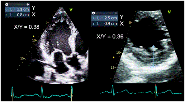
Figure 1: Determination of myocardial trabeculation using the Chin criterion (X: compacted thickness; Y: compacted plus non-compacted thickness) using apical (left) and transverse (right) sections.


José Maria Del Castillo1-3,* João Giffoni da Silveira Neto1-3 Renata Rodrigues Teixeira de Castro1-3 Caio Guedes de Sousa1-3 Issam Shehadeh1-3 Katarina Barros de Oliveira1-3 Djair Brindeiro Filho1-3
1ECOPE – Escola de Ecocardiografia de Pernambuco, Brazil*Corresponding author: José Maria Del Castillo, ECOPE – Escola de Ecocardiografia de Pernambuco, Rua Jorge de Lima 245 apto 303 Salute-Imbiribeira, Recife CEP 51.160-070, Brazil, Phone: +55 81 98163-4155, E-mail: [email protected]
Introduction: Myocardial hypertrabeculation, a hallmark of non-compaction cardiomyopathy, can also be found in healthy individuals, pregnant women, and athletes. By disrupting myofibrillar alignment, it may impair myocardial rotational mechanics. In athletes, however, overall performance appears unaffected.
Objective: We hypothesized that hypertrabeculation reduces ventricular torsion, compromising mechanical efficiency. To explore potential compensatory mechanisms preserving ventricular function, male athletes with and without hypertrabeculation were compared.
Methods: One hundred male athletes were enrolled: 50 with normally compacted myocardium and 50 meeting hypertrabeculation criteria (compacted/non-compaction ratio ≤ 0.5), engaged in predominantly aerobic or anaerobic training. Echocardiography assessed structural variables, systolic and diastolic function, myocardial strain, pulse wave velocity (PWV), mean arterial pressure, and myocardial work at rest. Statistical analysis used paired t-tests and Fisher’s exact test (p<0.05).
Results: Group A (no hypertrabeculation): 50 athletes (mean age 30 ± 8 years, Chin index 0.63 ± 0.09). Group B (hypertrabeculation): 50 athletes (mean age 31 ± 8 years, Chin index 0.41 ± 0.06). Demographics, cardiac dimensions, and ventricular function did not differ significantly. Eccentric remodeling occurred in 4% (Group A) vs. 6% (Group B); concentric remodeling in 10% vs. 8%. Borderline global longitudinal strain was observed in 20% of both groups. Ventricular twist <10 was present in 32% of athletes with hypertrabeculation vs. 2% without. PWV was reduced in the hypertrabeculation group, while mean arterial pressure remained similar. Myocardial work indices and efficiency showed no differences. No association was found between hypertrabeculation and ethnicity (p=0.69).
Conclusion: Athletes with ventricular hypertrabeculation exhibit preserved cardiac dimensions, volumes, and systolic/diastolic function, as well as comparable longitudinal, circumferential, and radial strain. However, apical rotation, twisting, and PWV are reduced. This reduction, combined with lower peripheral vascular resistance typical of athletes, may enhance ventricular ejection, serving as a compensatory mechanism for diminished apical rotation.
Athletes; Myocardial strain echocardiography; Apical rotation; Myocardial hypertrabeculation
The first echocardiographic description of increased myocardial trabeculation was published in 1984 by Engberding and Bender [1]. Subsequent studies associated hypertrabeculation with heart failure, introducing the concept of “myocardial non-compaction”[2]. Diagnostic criteria were initially proposed by Chin et al. in 1990 [3], later refined by Jenni, Stöllberger, and Paterick [4]. From the 2000s onward, however, hypertrabeculation has also been reported in individuals without cardiomyopathy, including healthy subjects [5], pregnant women, and athletes [6,7] highlighting the importance of careful interpretation of this finding [8].
During embryogenesis, compaction of the subendocardial myocardium begins around the tenth week of gestation, with the left ventricle (LV) nearly fully compacted by the fourth month [9]. The compacted layer consists of longitudinal fibers extending to the LV apex, then reflecting upward through the subepicardial surface (ascending apical band) in an oblique orientation. This arrangement underlies the LV’s twisting motion. In contrast, fibers in the central ventricular wall (basal band) are circular. The intersection of these fibers in a double helix with opposing directions defines the heart’s “helical” architecture [10,11], a structure confirmed by tractographybased magnetic resonance imaging [12].
Myocardial compaction is closely linked to LV performance, as torsion enhances mechanical efficiency while reducing energy expenditure. Yet, the presence of hypertrabeculation in athletes reported with a prevalence ranging from 9% [13] to 18% [14] challenges this paradigm. Despite altered myocardial architecture, no evidence of impaired ventricular function has been observed, suggesting the existence of compensatory mechanisms that sustain cardiac performance.
This study was designed to test the hypothesis that myocardial hypertrabeculation reduces ventricular torsion, thereby compromising mechanical efficiency. To explore this, we compared male athletes with and without hypertrabeculation, aiming to identify potential compensatory mechanisms that preserve ventricular function in the presence of altered myocardial architecture.
From a database of over 1,000 professional athletes across different sports, 100 male athletes were selected. The control group comprised 50 athletes with normally compacted myocardium (28 predominantly aerobic, 22 anaerobic), while the experimental group included 50 athletes meeting hypertrabeculation criteria (26 aerobic, 24 anaerobic).
Professional athletes were defined as individuals whose primary occupation and source of income is sport, with cardiorespiratory capacity (VO₂ max) >125% of the age-predicted value, determined by ergospirometry. All participants trained more than 25 hours per week, regardless of sport.
Hypertrabeculation was identified using the Chin et al. method, based on the ratio between the compacted layer thickness and total ventricular wall thickness (compacted+non-compacted). Ratios ≤ 0.5 were considered diagnostic (Figure 1).
Figure 1: Determination of myocardial trabeculation using the Chin criterion (X: compacted thickness; Y: compacted plus non-compacted thickness) using apical (left) and transverse (right) sections.
• Structural: LV and LA dimensions and volumes, LV mass, LV mass index, relative wall thickness.
• Systolic function: LV ejection fraction (EF), tricuspid annular plane systolic excursion (TAPSE).
• Diastolic function: Mitral Doppler and tissue Doppler parameters, E/e’ ratio, E-wave strain rate, LA reservoir strain, atrial stiffness index (E/e’/LA reservoir strain).
• Myocardial deformation: LV global longitudinal strain, basal and apical rotation, ventricular twist.
• Hemodynamics and function: Pulse wave velocity (PWV), mean arterial pressure (MAP=SBP+2×DBP)/3, and myocardial work indices (constructive, indexed, wasted, efficiency).
Echocardiographic examinations were performed with Vivid equipment and analyzed using Echopac 206 software (GE HealthCare Technologies Inc.). PWV was measured with the Arteris AOP system (Cardio Sistemas Coml. Ltda).
Data were classified as qualitative or quantitative. Quantitative variables were compared using paired t-tests (p<0.05). Associations between training type (aerobic vs. anaerobic), ethnicity, and hypertrabeculation status were analyzed using Fisher’s exact test (p<0.05).
Age <15 or >50 years; comorbidities (hypertension, diabetes mellitus, systemic disease, coronary artery disease, valvular or congenital heart disease); use of anabolic-androgenic steroids; VO₂ max <125% of predicted; or absence of regular training during evaluation.
All athletes included were professionals, training more than 25 hours per week. Of the total, 57 engaged predominantly in aerobic training, and all were asymptomatic. Regarding ethnicity, 56 were Caucasian and 44 of African descent.
• Group A (without hypertrabeculation): Chin index >0.5 (mean 0.63 ± 0.09). Comprised 50 male athletes, mean age 30 ± 8 years. Of these, 34 performed aerobic training (32 soccer players-18 Caucasian; 2 long-distance runners-1 Caucasian) and 16 anaerobic training (14 martial arts practitioners-8 Caucasian; 2 weightlifters-1 Caucasian).
• Group B (with hypertrabeculation): Chin index ≤ 0.5 (mean 0.41 ± 0.06). Included 50 male athletes, mean age 31 ± 8 years. Among them, 23 performed aerobic training (20 soccer players-10 Caucasian; 3 long-distance runners-all Caucasian) and 27 anaerobic training (21 martial arts practitioners-13 Caucasian; 5 weightlifters-2 Caucasian; 1 volleyball player-Afro-descendant).
Comparative data are summarized in table 1.
| Parameter | Non-hypertrabeculated | Hypertrabeculated | p Value |
| Age, years | 30.29 ± 8.63 | 31.29 ± 8.14 | 0.67 |
| BSA, m2 | 2.01 ± 0.23 | 2.05 ± 0.16 | 0.37 |
| BMI, kg/m2 | 25.83 ± 3.0 | 26.24 ± 3.24 | 0.47 |
| HR, bpm | 61.63 ± 9.42 | 62.71 ± 7.06 | 0.41 |
| Chin Index | 0.63 ± 0.09 | 0.41 ± 0.06 | <0.001 |
| LVDd, mm | 51.64 ± 4.82 | 53.86 ± 4.21 | 0.07 |
| IVSd, mm | 9.11 ± 1.20 | 9.57 ± 2.08 | 0.24 |
| LVPWd, mm | 9.04 ± 1.23 | 9.32 ± 1.28 | 0.35 |
| LVMass, g | 169.35 ± 34.60 | 194.28 ± 54.05 | 0.02 |
| LVMass[i], g/BSA | 85.15 ± 18.10 | 94.28 ± 24.82 | 0.09 |
| LVRWT | 0.36 ± 0.06 | 0.35 ± 0.06 | 0.75 |
| LVEF, % | 59.07 ± 5.77 | 59.68 ± 5.32 | 0.69 |
| LVEDV[i], mL/m2 | 77.93 ± 17.86 | 75.31 ± 12.11 | 0.51 |
| LVSV[i], mL/m2 | 45.79 ± 11.79 | 45.21 ± 9.20 | 0.84 |
| LAV[i], mL/m2 | 32.48 ± 8.62 | 28.67 ± 8.11 | 0.10 |
| TAPSE, cm | 2.36 ± 0.26 | 2.47 ± 0.35 | 0.19 |
| Mitral E wave, cm/s | 78.11 ± 15.17 | 77.86 ± 13.26 | 0.93 |
| Mitral A wave, cm/s | 50.04 ± 8.13 | 50.07 ± 12.94 | 0.99 |
| Mitral E/A | 1.59 ± 0.38 | 1.64 ± 0.47 | 0.66 |
| e’ wave avg, cm/s | 14.23 ± 2.93 | 13.36 ± 3.0 | 0.27 |
| E/e’ wave avg | 5.60 ± 1.06 | 6.01 ± 1.30 | 0.19 |
| LVGLS, % | -19.71 ± 1.98 | -18,45 ± 2.49 | 0.06 |
| LVGLSRs, s-1 | -1.15 ± 0.21 | -1.09 ± 0.14 | 0.19 |
| LVSRe, s-1 | 1.82 ± 0.51 | 1.83 ± 0.40 | 0.97 |
| LVMD, ms | 31.29 ± 12.96 | 39.66 ± 9.68 | 0.01 |
| LVGCS, % | -18.20 ± 2.97 | -17.03 ± 1.46 | 0.06 |
| LVGRS, % | 48.62 ± 17.38 | 44.12 ± 11.71 | 0.16 |
| RVLSlat, % | -24.39 ± 2.84 | -24.53 ± 3.25 | 0.94 |
| LALSr, % | 40.36 ± 10.18 | 36.79 ± 6.30 | 0.17 |
| LALSp, % | 12.71 ± 4.96 | 12.43 ± 4.08 | 0.81 |
| LAstiff | 0.15 ± 0.04 | 0.17 ± 0.05 | 0.11 |
| LVRotB, | -5.43 ± 2.68 | -4.67 ± 2.70 | 0.29 |
| LVRotA, | 9.71 ± 4.05 | 7.0 ± 3.42 | 0.008 |
| Twisting, | 15.14 ± 3.22 | 11.67 ± 3.41 | <0.001 |
| PWV, m/s | 6.88 ± 0.84 | 5.24 ± 0.46 | <0.001 |
| SBP, mmHg | 119.82 ± 8.33 | 123.0 ± 9.67 | 0.17 |
| DBP, mmHg | 77.50 ± 7.39 | 77.46 ± 9.49 | 0.99 |
| MAP, mmHg | 91.61 ± 7.38 | 92.64 ± 8.83 | 0.62 |
| GWI, mmHg% | 1698.89 ± 283.25 | 1726.43 ± 216.63 | 0.68 |
| GCW, mmHg% | 2062.46 ± 242.92 | 2000.75 ± 199.11 | 0.25 |
| GWW, mmHg% | 73.14 ± 43.29 | 80.36 ± 39.70 | 0.49 |
| GWE, % | 96.54 ± 1.10 | 96.21 ± 0.96 | 0.16 |
Table 1: Comparison between non-hypertrabeculated and trabeculated athletes.
BSA: Body Surface Area; BMI: Body Mass Index; HR: Heart Rate; LVDd: Diastolic Left Ventricular Diameter; IVSd: Diastolic Interventricular Septal Thickness; LVPWd: Diastolic Posterior Wall Thickness; LVMass: Left Ventricular Mass; LVMass[i]: Left Ventricular Mass Index; LVRWT: Left Ventricular Relative Wall Thickness; LVEF: Left Ventricular Ejection Fraction; LVEDV[i]: Left Ventricular End Diastolic Volume Index; LVSV[i]: Left Ventricular Stroke Volume Index; LAV[i]: Left Atrial Volume Index; TAPSE: Tricuspid Annular Plane Systolic Excursion; LVGLS: Left Ventricular Global Longitudinal Strain; LVGLSRs: Left Ventricular Global Longitudinal Systolic Strain Rate; LVSRe: Left Ventricular Early Diastolic Strain Rate; LVMD: Left Ventricular Mechanical Dispersion; LVGCS: Left Ventricular Global Circumferential Strain; LVGRS: Left Ventricular Global Radial Strain; RVLSlat: Right Ventricular Lateral Wall Longitudinal Strain; LALSr: Left Atrial Longitudinal Reservoir Strain; LALSp: Left Atrial Pump Strain; LAstiff: Left Atrial Stiffness Index; LVRotB: Left Ventricular Basal Rotation; LVRotA: Left Ventricular Apical Rotation; PWV: Pulse Wave Velocity; SBP: Systolic Blood Pressure; DBP: Diastolic Blood Pressure; MAP: Mean Arterial Pressure; GWI: Global Work Index; GCW: Global Constructive Work; GWW: Global Waste Work; GWE: Global Work Efficiency
No significant differences were observed in demographic parameters, cardiac dimensions, or ventricular function. The only difference was a higher unindexed ventricular mass in the hypertrabeculation group, which disappeared after indexing by body surface area.
Group A (control): 4% presented eccentric remodeling and 10% concentric remodeling. An increase in the indexed volume of the left ventricle (LV) was observed in 50% and of the left atrium (LA) in 28%. Global longitudinal strain of the LV was considered borderline (between -16 and -17%) in 20% of the athletes, with an ascending progression pattern from the base to the apex. In 2%, the ventricular twist was less than 10°. Longitudinal strain of the lateral wall of the RV was decreased in 26% of the athletes.
Group B (hypertrabeculation): 6% presented eccentric remodeling, 2% mild concentric hypertrophy, and 8% concentric remodeling. An increase in the indexed volume of the LV occurred in 54% and of the LA in 14%. Global longitudinal strain was borderline in 20%, also with ascending progression from the base to the apex. Ventricular twist was less than 10° in 32% of the athletes. Longitudinal strain of the right ventricular lateral wall was decreased in 16% of the athletes.
In the control group (without hypertrabeculation), 34 athletes (68%) engaged in aerobic training and 16 (32%) in anaerobic training. In contrast, among athletes with hypertrabeculation, 23 (46%) performed aerobic training and 27 (54%) anaerobic.
Analysis using Fisher’s exact test revealed a significantly higher proportion of aerobic training in Group A (p=0.03). However, within Group B, the incidence of hypertrabeculation did not differ between aerobic and anaerobic training modalities.
Pulse wave velocity (PWV) was significantly reduced in athletes with hypertrabeculation, indicating greater arterial compliance. Mean arterial pressure (MAP) did not differ between groups.
Indices of myocardial work including indexed, constructive, wasted work, and efficiency showed no significant differences between athletes with and without hypertrabeculation.
Of the 100 athletes, 56 were Caucasian and 44 of African descent. In the control group, 29 were Caucasian (58%) and 21 of African descent (42%). In the hypertrabeculation group, 27 were Caucasian (54%) and 23 of African descent (46%). Fisher’s exact test revealed no significant association between ethnicity and the presence of hypertrabeculation (p=0.69).
Recent studies have reported ventricular hypertrabeculation in athletes, pregnant women, and the general population, without necessarily indicating a pathological phenotype. Importantly, hypertrabeculation does not result from incomplete embryonic compaction. Jensen et al. [15] demonstrated that Atrial Natriuretic Factor (ANF/NPPA), highly expressed during embryonic trabeculation, is virtually absent in adult hypertrabeculation. This suggests that the condition arises from abnormal either pathological or adaptive growth of the already compacted myocardial layer, representing a redistribution of left ventricular (LV) subendocardial fibers.
In athletes, the central question is whether trabeculation confers mechanical advantages. Halaney et al. [16] showed that a compacted LV exhibits greater compliance, but the apex experiences higher stress and deformation, implying increased energy demand. Trabeculation may mitigate this by redistributing stress, protecting the apex from remodeling, and reducing myocardial workload.
In the present study, indices of myocardial work including total, constructive, wasted work, and efficiency did not differ between groups. However, individual tracings revealed greater apical activation in athletes without hypertrabeculation, suggesting increased oxygen consumption in this region [17] (Figure 2). Based on the hypothesis that elevated apical work reflects greater wall stress, hypertrabeculation may function as a load-redistribution mechanism, reducing apical activation and potentially preventing adverse remodeling over time.
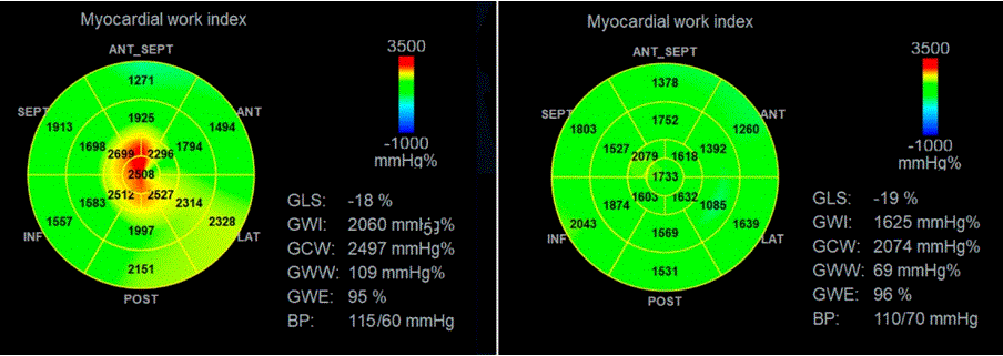
Figure 2: Comparative polar maps of the myocardial work index. In the athlete without hypertrabeculation (left), greater apical activation is evident.
In contrast, the athlete with hypertrabeculation (right) demonstrates a more homogeneous distribution of myocardial work.
Van Dalen et al. [18] demonstrated that LV rotation is essential for mechanical efficiency, yet significantly reduced in hypertrabeculation. In athletes with preserved systolic and diastolic function, this reduction must be offset by adaptive mechanisms that maintain cardiac output without loss of efficiency. While some studies report preserved apical rotation in adolescents [19], others describe reductions in both rotation and ventricular twist [20-22]. Gati et al. [23] identified hypertrabeculation in 18% of 1,146 athletes, with 8% meeting diagnostic criteria for non-compaction cardiomyopathy. It is noteworthy that, as a training adaptation, hypertrabeculation may reflect chronic increases in preload and afterload, without electrocardiographic abnormalities, symptoms, diastolic dysfunction, or reduced exercise capacity (peak VO₂ >120% of predicted). Conversely, athletes with suspected noncompaction cardiomyopathy exhibit electrocardiographic changes, systolic dysfunction, ventricular dilation, reduced ejection fraction, and myocardial fibrosis on MRI findings absent in the present cohort.
Participants were drawn from a database of over 1,000 professional athletes, restricted to men to avoid sex-related physiological confounders. Hypertrabeculation was identified using the Chin index, chosen for its simplicity and equivalence to the Paterick index.
No significant demographic differences were observed between groups. Increased indexed volumes of the left ventricle (LV) and left atrium (LA), as well as eccentric and concentric remodeling, were frequent findings in athletes [24]. Borderline values of right ventricular and LV longitudinal strain were also noted, consistent with previous reports of strain augmentation during exercise [25]. Diastolic function parameters remained normal in both groups, and the atrial stiffness index was within the lower limits of normal [26].
Myocardial Deformation. Athletes with hypertrabeculation showed a slight, non-significant reduction in LV longitudinal strain and LA reservoir strain, without changes in atrial stiffness. Rotational parameters revealed a non-significant decrease in basal rotation, but a significant reduction in apical rotation and ventricular twist, both below reference values [27].
These findings highlight the importance of investigating adaptive mechanisms that preserve overall cardiac performance despite reduced mechanical efficiency. Potential compensatory mechanisms include decreased afterload through exercise-induced regulation of systemic vascular resistance, adrenergic modulation, and enhanced myocardial inotropism via sympathetic stimulation [28].
It is important to emphasize that the present data were obtained at rest; analysis during exercise is essential for a more complete understanding. Drury et al. [29] demonstrated that untrained individuals show a marked increase in apical rotation and ventricular torsion during submaximal exercise, with basal rotation rising further at maximal effort. In endurance athletes, rotational parameters increase with training, but during intense competitive exercise (e.g., marathons) they decline, likely reflecting exhaustion. Santoro et al. [30] reported significant increases in basal and apical rotation and ventricular torsion after isotonic exercise, suggesting that the basal reduction observed at rest represents a contractile reserve designed to optimize systolic function during exertion. Notably, these findings were not directly linked to hypertrabeculation.
Regarding training modalities, all athletes practiced mixed regimens, with predominance of aerobic (soccer, running) or anaerobic (martial arts, weightlifting, volleyball) exercise. No differences in hypertrabeculation distribution were observed between training types.
Pulse wave velocity (PWV), a marker of arterial stiffness, does not directly measure peripheral vascular resistance (PVR), though it may be influenced by it. Thus, reduced PWV combined with low PVR a common condition in athletes may indicate decreased resistance to ventricular ejection. This adaptation could serve as a compensatory mechanism for reduced apical rotation, helping to preserve overall cardiac performance.
This study has several limitations that should be acknowledged:
• The relatively small sample size and the exclusive inclusion of male athletes may limit the generalizability of the findings.
• Despite the high proportion of Afro-descendant athletes in the sample, no predominance of hypertrabeculation was observed in this group, a finding that contrasts with reports in the literature.
• All assessments were performed at rest, which restricts the evaluation of myocardial rotational mechanics and potential changes in pulse wave velocity under exercise conditions.
Athletes with myocardial hypertrabeculation do not differ significantly from those without the condition in terms of cardiac dimensions, volumes, systolic and diastolic function, or longitudinal, circumferential, and radial strain. However, they exhibit a marked reduction in apical rotation and ventricular twist, accompanied by decreased pulse wave velocity, a marker of arterial stiffness. This reduction, combined with the lower peripheral vascular resistance commonly observed in athletes, may facilitate ventricular ejection and act as a compensatory mechanism for the loss of efficiency associated with diminished apical rotation.
Additional compensatory mechanisms for maintaining myocardial efficiency remain to be clarified. Potential contributors include modulation by beta- and alpha-adrenergic receptors and adaptations of the autonomic nervous system, which may enhance inotropism and support long-term ventricular performance.
Download PDF Here
Article Type: RESEARCH ARTICLE
Citation: Del Castillo JM, da Silveira Neto JG, de Castro RDT, de Sousa CG, Shehadeh I, et al. (2026) Myocardial Hypertrabeculation in Athletes. Ventricular Mechanics and Deformation Parameters. J Surg Open Access 10(1): dx.doi.org/10.16966/2470-0991.277
Copyright: © 2026 Del Castillo JM, et al. This is an open-access article distributed under the terms of the Creative Commons Attribution License, which permits unrestricted use, distribution, and reproduction in any medium, provided the original author and source are credited.
Publication history:
All Sci Forschen Journals are Open Access