
Figure 1: Flowchart for echocardiographic assessment upon exiting extracorporeal circulation.
Recommended sequence of echocardiographic evaluation before exiting extracorporeal circulation, Mena Ana, 2025.


Ana Mena1* Edgardo Venegas2 Milton Chango3
1Anesthesiologist, Hospital Carlos Andrada Marin, Quito - Ecuador*Corresponding author: Ana Mena, Anesthesiologist, Hospital Carlos Andrada Marin, Quito - Ecuador, E-mail: [email protected]
Weaning from cardiopulmonary bypass (CPB) represents one of the most critical and physiologically demanding phases of cardiac surgery. This transition constitutes the ultimate functional test of myocardial recovery, vascular tone, pulmonary function, and end-organ perfusion following extracorporeal circulation. Despite advances in surgical techniques, perfusion technology, and myocardial protection, separation from CPB remains a period associated with significant hemodynamic instability and adverse outcomes.
From an anesthetic perspective, successful weaning from CPB requires a comprehensive understanding of CPB-induced pathophysiological changes, meticulous preparation, advanced multimodal monitoring, and continuous multidisciplinary communication. The anesthesiologist plays a central role in integrating hemodynamic data, echocardiographic findings, metabolic status, and pharmacologic support to guide decision-making during this dynamic process [1-3].
This chapter focuses on the physiological basis, preparation, monitoring strategies, and stepwise approach to weaning from CPB, emphasizing practical algorithms, echocardiographic assessment, and troubleshooting strategies aligned with contemporary recommendations from the American Society of Anesthesiologists (ASA) and the European Association of Cardiothoracic Anaesthesiology and Intensive Care (EACTA).
The period preceding initiation of CPB is critical, as anesthetic decisions made during this phase directly influence myocardial protection, tissue perfusion, inflammatory activation, and the likelihood of successful separation from bypass. During this stage, the anesthesiologist functions as a perioperative physiologist, integrating clinical assessment, advanced monitoring, pharmacologic planning, and communication with surgical and perfusion teams [1,4].
Hemodynamic stability must be achieved prior to cannulation, with particular attention to heart rate control, maintenance of adequate mean arterial pressure (MAP), and optimization of preload. Both hypotension and tachycardia may compromise coronary perfusion and increase myocardial oxygen consumption, especially in patients with coronary artery disease, hypertrophic ventricles, or valvular stenosis [1-3].
Closed-loop communication with the surgical and perfusion teams is essential to confirm readiness for bypass initiation, anticoagulation adequacy, and contingency planning.
Transcatheter aortic valve replacement (TAVR) is an established therapeutic option for patients with severe aortic stenosis across a wide risk spectrum. Current evidence does not demonstrate systematic differences in long-term durability between transcatheter and surgical bioprosthetic valves, with comparable rates of structural valve deterioration reported up to 10 years. Nevertheless, interpretation of durability data is limited by heterogeneity in study design, definitions of structural valve degeneration, variable follow-up durations, and competing mortality risks [5].
Patient selection for TAVR should be individualized and based on a comprehensive Heart Team assessment. Advanced age (generally ≥ 70 years), the presence of significant comorbidities, and increased surgical risk favor a transcatheter approach. Comorbid conditions that increase operative risk include severe left ventricular dysfunction, pulmonary hypertension, chronic kidney disease, advanced chronic obstructive pulmonary disease, liver disease, prior cardiac surgery, and complex coronary artery disease not amenable to surgical revascularization. In addition, clinical frailty-independently associated with adverse surgical outcomes-represents a key determinant supporting TAVR over surgical aortic valve replacement (SAVR) [5].
Anatomical and procedural factors further influence the choice of intervention. TAVR is particularly favored in patients with conditions that render surgical access hazardous, such as porcelain aorta, severe chest wall deformities, hostile mediastinum, patent bypass grafts after prior coronary artery bypass surgery, or sequelae of mediastinal or thoracic radiotherapy. Radiation-induced cardiac disease is frequently associated with extensive mediastinal fibrosis, calcification of the aorta and cardiac valves, and impaired tissue healing, all of which significantly increase surgical morbidity and mortality [5].
The presence and complexity of concomitant coronary artery disease must be carefully considered. Non-complex coronary artery disease may be treated with percutaneous coronary intervention in conjunction with TAVR, whereas complex multivessel disease generally favors SAVR combined with coronary artery bypass grafting. Evidence from randomized data suggests that, in selected patients with severe aortic stenosis and concomitant coronary disease, a transcatheter strategy combined with PCI may be associated with lower short-term mortality and bleeding risk, although long-term conclusions remain limited [5].
Special anatomical subsets require cautious evaluation. Bicuspid aortic valve anatomy poses technical challenges for TAVR due to asymmetric calcification and elliptical annular geometry and has been largely excluded from pivotal randomized trials. Therefore, TAVR in bicuspid valves should be reserved for carefully selected patients treated at experienced centers. In contrast, surgical aortic valve replacement remains the standard of care for pure aortic regurgitation, with TAVR considered only in inoperable or prohibitive-risk patients [5].
Overall, the choice between TAVR and SAVR should integrate patient age, comorbid burden, frailty, anatomical suitability, and anticipated procedural risk, with particular relevance for perioperative planning and anesthetic management [5].
Key responsibilities and challenges: During the peri-induction and pre-CPB phase, the anesthesiologist orchestrates physiologic stability, comprehensive monitoring, pharmacologic management, and interdisciplinary communication. This period is characterized by rapid cardiovascular changes and heightened surgical stimulation, necessitating ongoing assessment and proactive management [3,4].
Hemodynamic stability and myocardial protection: Sustaining hemodynamic stability while balancing myocardial oxygen supply and demand is crucial. The anesthesiologist must control heart rate, blood pressure, preload, afterload, and ventricular contractility to prevent ischemia, ventricular dysfunction, and arrhythmias. Avoiding both hypotension (which can compromise coronary and cerebral perfusion) and hypertension or tachycardia (which increases myocardial oxygen consumption) is particularly important in patients with significant coronary artery disease, hypertrophic ventricles, or pressure-overload states [1-3].
Anticipation of surgical stimuli and hemodynamic: sternotomy, pericardiotomy, aortic manipulation, and cannulation may induce abrupt sympathetic responses and hemodynamic instability. The anesthesiologist must anticipate these events, adjusting anesthetic depth and vasoactive support to blunt adverse reactions and prevent ischemia or cardiovascular collapse [4,6,7].
Integration of monitoring and clinical data: Effective anesthetic management requires real-time synthesis of data from standard, invasive, and advanced monitoring modalities, including arterial waveform analysis, CVP trends, cerebral oximetry, depth-ofanesthesia monitoring, and echocardiography. It is vital to distinguish true physiological changes from artifacts, especially during periods of electrocautery, altered vascular tone, or non-pulsatile flow [8,9].
Communication and Team Coordination: Clear, closed-loop communication with surgical and perfusion teams is essential. The anesthesiologist communicates patient status, readiness for bypass, adequacy of anticoagulation, and potential challenges, as communication failures can lead to preventable adverse events [8,9].
Preparation for CPB and beyond: Preparation encompasses readying vasoactive drugs, inotropes, contingency plans for ventricular dysfunction or vasoplegia, and blood products. Maintaining proactive readiness in the dynamic operating environment is a continual challenge [9].
Standard monitoring: After induction, standard monitoring is established in line with cardiac anesthesia guidelines: continuous 5-lead ECG, pulse oximetry, and non-invasive blood pressure (NIBP). The 5-lead ECG aids in rhythm and ischemia surveillance, pulse oximetry tracks oxygen saturation and waveform quality, and NIBP provides backup after invasive monitoring is in place. Interpretation demands vigilance, as rapid hemodynamic shifts, electrocautery interference, and altered peripheral perfusion may limit reliability during key periods [9].
Arterial lines: Arterial catheter selection should prioritize highfidelity waveform analysis, rapid sampling, and reliability throughout CPB. The radial artery is preferred, utilizing a 20G catheter for most adults. In cases involving radial artery harvest for CABG, monitoring should be contralateral. Alternatives include brachial (higher thrombosis risk) and femoral arteries (reserved for select cases), with meticulous technique and monitoring required. Technical accuracy necessitates short, stiff, non-compliant tubing, proper transducer leveling, a pressurized flush system, and continuous waveform integrity assessment [9].
Central Venous Catheters (CVC): CVCs facilitate therapeutic access and allow CVP trend interpretation in context. The right internal jugular vein is preferred, with ultrasound guidance as standard. Catheter selection depends on infusion requirements, and large-bore introducers are used for rapid infusion and mechanical support in selected cases [9].
Pulmonary artery catheter (selected patients): PA catheters may be indicated for high-risk cases such as severe ventricular dysfunction, pulmonary hypertension, or complex valve surgery [9].
Peripheral venous access: Peripheral IV access remains essential for redundancy and rapid intervention, with at least two lines recommended (16–18G, upper extremity, securely fixed) [9].
Advanced monitoring: Advanced monitoring is selected according to risk, case complexity, and available resources. Since CPB alters perfusion, temperature, and autonomic regulation, reliance on advanced tools increases [9,10].
Near-Infrared Spectroscopy (NIRS): NIRS provides continuous monitoring of regional cerebral oxygenation, which is especially valuable in patients with carotid disease, advanced age, prior stroke, anemia, or extended CPB duration. Cerebral desaturation alerts may indicate hypotension, insufficient pump flow, hemodilution, hypoxia, CO2 changes, impaired autoregulation, or embolic events, prompting targeted intervention [11,12].
Transesophageal Echocardiography (TEE): TEE is introduced after induction and is indispensable for real-time evaluation of cardiac function, valvular anatomy, shunts, chamber size, and volume status. Pre-bypass, TEE confirms baseline function, guides cannula placement, and detects intracardiac air. Intraoperatively, it supports hemodynamic diagnosis and management, and during separation from CPB, it guides support decisions based on cardiac function and residual air [13-16].
Anesthetic depth and drugs: Maintaining appropriate anesthetic depth requires careful titration of hypnotics, opioids, and adjuncts to provide hypnosis, analgesia, amnesia, and immobility, while preserving cardiovascular stability. CPB alters drug pharmacokinetics due to hemodilution, hypothermia, changes in protein binding, reduced organ metabolism, and drug sequestration in the circuit, rendering fixed dosing unreliable [17,18].
Bispectral Index (BIS): BIS assists in assessing anesthetic depth during CPB, when traditional hemodynamic parameters are unreliable. BIS values are influenced by hypothermia, hemodilution, cerebral hypoperfusion, and electrocautery. Emphasis is placed on trend analysis to prevent awareness and avoid over-sedation during bypass and rewarming [11].
Hemodynamic management: The primary goal of hemodynamic management is myocardial protection and maintenance of end-organ perfusion during periods of intense stimulation and rapid physiologic transitions. Objectives include optimizing the myocardial oxygen supply-demand balance, sustaining coronary perfusion pressure, and avoiding extremes of heart rate and blood pressure to prevent ischemia, ventricular dysfunction, or neurological injury [10].
Anticoagulation: Effective anticoagulation is indispensable for safe CPB initiation and maintenance. Blood contact with non-endothelial circuit surfaces rapidly activates the coagulation cascade, necessitating systemic anticoagulation to prevent thrombosis and embolic events [10].
Heparin administration: Systemic anticoagulation is provided by intravenous unfractionated heparin (UFH) before cannulation. The standard initial dose is 300-400 IU/kg, allowing 3-5 minutes of circulation before coagulation assessment. Rapid bolus dosing is avoided to minimize hypotension and rare anaphylactoid reactions. If anticoagulation is insufficient, supplemental boluses (50-100 IU/kg) are administered, followed by repeat testing [10].
Verification of Activated Clotting Time (ACT): ACT is measured 3-5 minutes post-heparin administration, with a target of ≥ 480 seconds for CPB initiation. ACT is reassessed every 20-30 minutes during bypass. If ACT falls below target, additional UFH is administered and confirmed with repeat testing [10].
Management of heparin resistance: Heparin resistance-failure to reach target ACT with appropriate dosing-often results from antithrombin III deficiency. Management includes supplementation with ATIII concentrate or, if unavailable, fresh frozen plasma [10].
Clinical implications and patient safety: Inadequate anticoagulation may result in catastrophic complications such as circuit thrombosis, stroke, myocardial infarction, and organ failure. Management must be protocol-driven, anticipatory, and continuously reassessed, with the anesthesiologist central to dosing, monitoring, and troubleshooting to ensure patient safety and optimal outcomes [10].
Fluid balance, microcirculation, and endothelial integrity: Fluid management in cardiac surgery extends beyond macrocirculatory goals to encompass the preservation of microcirculation and endothelial function. CPB, surgical trauma, ischemia-reperfusion injury, and systemic inflammation can disrupt the endothelial glycocalyx, increasing capillary permeability, tissue edema, coagulation abnormalities, and organ dysfunction [10].
Excessive positive fluid balance before or during CPB causes hemodilution, edema, impaired oxygen diffusion, pulmonary dysfunction, and delayed recovery. Conversely, hypovolemia can compromise preload, cardiac output, and microvascular perfusion. The anesthesiologist should aim for a balanced, restrictive, and physiologydriven fluid strategy, favoring vasoactive support over excessive fluid administration [10].
Microcirculatory perfusion: Microcirculatory perfusion depends on cardiac output, arterial pressure, capillary recruitment, endothelial integrity, and blood rheology. Disruption of the glycocalyx increases capillary leak, diminishes oncotic gradients, and promotes tissue edema [17].
Rotational thromboelastometry (ROTEM): ROTEM offers realtime, viscoelastic assessment of clot formation, strength, and lysis— superior to conventional coagulation tests. In cardiac surgery, where CPB leads to hemodilution, platelet dysfunction, hypothermia, and factor consumption, ROTEM underpins goal-directed hemostatic management [1-3,18].
Role of ROTEM in CPB: ROTEM rapidly distinguishes between factor deficiency, platelet dysfunction, hypofibrinogenemia, hyperfibrinolysis, and residual heparin effect, enabling targeted transfusion and minimizing unnecessary blood product use and related complications [1-3].
• EXTEM: extrinsic pathway and platelet function
• INTEM: intrinsic pathway and residual heparin effect
• FIBTEM: fibrinogen contribution
• HEPTEM: differentiates heparin effect from other defects
ROTEM-guided algorithms decrease bleeding, transfusion needs, and re-exploration rates, forming the foundation of modern blood management protocols [1-3].
Final checks before initiating cardiopulmonary bypass: Before initiating CPB, a deliberate and structured verification process ensures patient safety and procedural readiness. This critical transition necessitates coordinated confirmation among anesthesia, surgical, and perfusion teams [1-3].
Coordination with the perfusionist: Communication with the perfusionist ensures the CPB circuit is primed, de-aired, and ready. Anticoagulation adequacy (ACT target met), pump flow targets, temperature strategy, and anticipated events are verbally confirmed using closed-loop communication [1-3].
Final surgical field assessment: The surgical field is inspected for readiness for cannulation, including appropriate exposure, absence of bleeding, correct cannulation site positioning, and agreement among team members to proceed [1-3].
Pre-cardiopulmonary bypass safety check: Transitioning to CPB is a pivotal moment requiring a focused safety pause. This checklist addresses physiologic readiness, technical preparedness, and team synchronization. Hemodynamic stability is reassessed, all monitoring is confirmed functional, vascular access and infusions are secured, and drug labeling is verified. Anticoagulation status is confirmed, with ACT at target and contingency plans for inadequate ACT or heparin resistance clarified [1-3].
Coordination with the perfusion team is reaffirmed, verifying the bypass circuit’s readiness and reviewing pump and temperature goals. TEE findings and the surgical field are re-examined. The process concludes with a verbal confirmation among the anesthesiologist, surgeon, and perfusionist that all conditions are optimal and no outstanding concerns exist. Only then is CPB initiated [1-3].
Preoperative anesthetic evaluation: The entire team must maintain awareness of the patient’s condition, comorbidities, and planned surgical procedure to ensure readiness for CPB initiation. Ideally, this evaluation should be performed collaboratively with the surgical team and specialized clinics. Inadequate preoperative assessment can result in patient harm [9].
• Airway management and mechanical ventilation with FiO2 0.6-1.0
• Standard and advanced monitoring:
• Arterial line
• Central venous access
• Transesophageal echocardiography (TEE) available
• Depth of anesthesia monitoring
• Cerebral oximetry
• Nasopharyngeal (core) and rectal (peripheral) temperature monitoring
• CVP / PAP as indicated
Unfractionated heparin anticoagulation
• Initial dose: 300-400 U/kg
• Target: ACT greater than 480 seconds
• Repeat ACT every 20-30 minutes
The perioperative effect of heparin on coagulation is monitored using ACT, which is influenced by patient and surgical factors such as temperature, hemodilution, and platelet count. Heparin dosing can affect hemostasis and postoperative bleeding risk, with variability in potency among suppliers contributing to differences in anticoagulant effect [1-3].
Pre-CPB checklist: Checklists should be used appropriately and adapted to the specific work environment, incorporating new technologies and periodic reviews according to institutional protocols. Effective checklist use relies on teamwork, communication, leadership support, and a culture of safety and incident reporting [1-3].
• Team communication: Anesthesiology, Surgery, Perfusion
• Verification of appropriate cannulas
• Target temperature
• Type of cardioplegia (crystalloid/blood, antegrade/retrograde)
• Target hematocrit during CPB (22–28%)
• Neurological protection strategy
• Blood products availability
Initiation of CPB (Connection): Cannulation
• Ascending aorta for arterial return
• Right atrium / vena cava for venous drainage
• Confirm position with TEE
Initiation: Gradually decrease pulmonary flow and increase pump flow
• Maintain MAP between 50-70 mmHg, adjusted for age and vascular condition
• Monitor arterial blood gases hourly and as needed
Cardiopulmonary bypass (CPB) plays a critical role in cardiac surgery by providing life support during induced cardiac arrest, thereby enabling complex surgical procedures. Despite its benefits, CPB can cause both local and systemic adverse effects, as its components may disrupt macro- and microcirculation, potentially resulting in hypoperfusion [1-3].
Modern CPB management utilizes goal-directed perfusion (GDP) strategies. GDP focuses on individualized hemodynamic management by targeting specific physiological parameters, with continuous monitoring to facilitate real-time adjustments. This method aims to optimize oxygenation and tissue perfusion during CPB, reducing the risk of inadequate perfusion and related physiological disturbances such as metabolic acidosis, elevated lactate levels, and organ ischemia [7-9].
GDP identifies, quantifies, and intervenes on key physiological variables, including oxygen delivery (DO2), oxygen consumption (VO2), and oxygen extraction ratio (ERO2), to minimize adverse effects and maintain homeostasis [7-9].
In addition to monitoring basic parameters like arterial blood pressure, pump flow, and blood gas analysis, advanced technologies are employed to enhance patient management during CPB. Measuring mixed venous oxygen saturation (SvO₂) and conducting continuous blood gas monitoring have become increasingly significant. Regional cerebral tissue oxygenation can be evaluated using near-infrared spectroscopy (NIRS), and monitoring oxygen delivery (DO₂) has been suggested by some investigators [7-9].
Current global parameters:
• SvO2: greater than 60-70%
• MAP: at least 65 mmHg
• PvO2: greater than 40 mmHg
• pH: 7.35-7.45
• pCO2: 35-45 mmHg
• Cardiac Index: 2.2-2.6 L/min/m2
• Lactate: less than 2.0 mmol/L
Integrated parameters:
• Oxygen Delivery Index (DO2i): at least 260-272 mL/min/m2
• Oxygen Consumption Index (VO2i): less than 60 mL/min/m2
• Carbon Dioxide Production Index (VCO2i): less than 60 mL/ min/m2
• DO2i / VCO2i: greater than 5
• VO2i / DO2i (ERO₂): less than 0.25
• VCO2i / VO2i (Respiratory Quotient): less than 1
During CPB, these variables should be indexed and monitored. Controlling these values guides interventions designed to maintain physiological homeostasis.
Definitions:
• CPB: Technique that temporarily replaces cardiopulmonary function.
• Non-pulsatile flow: 2.2-2.4 L/min/m2 under normothermic conditions.
• Hemodilution: Effect produced by circuit priming.
• MIT: Myocardial ischemia time during aortic cross-clamping.
• MUF: Modified ultrafiltration used to concentrate blood postCPB.
• ACT: Activated clotting time.
Maintenance of CPB:
Anesthesia and pharmacological management during CPB: CPB significantly alters the pharmacokinetics and pharmacodynamics of anesthetic drugs due to factors such as hemodilution, reduced plasma protein concentrations, altered clearance from hypothermia, and drug sequestration during pulmonary isolation. Cardiac anesthesiologists require specialized expertise to manage sedation, anesthetic depth, analgesia, and neuromuscular blockade. Additional considerations include corticosteroids, glycemic control, hemodynamic drugs, coagulation medications, and serum electrolytes. Proper management is essential for hemodynamic stability [7-9].
Hemodynamic parameters:
• Flow: 2.2-2.4 L/min/m2
• MAP: 50-70 mmHg
• SvO2: greater than 65%
• Ideal hematocrit: 22-28%
Thermoregulation: Normothermia or mild hypothermia: 28-34℃ depending on surgical requirements
Ventilation during CPB:
• Lungs at rest
• Minimal ventilatory settings or pause as needed
Metabolic management:
• Glucose: less than 180 mg/dL
• Ultrafiltration strategies applied as needed
Myocardial protection:
Cardioplegia: The main objectives of cardioplegia are to protect myocardial function from ischemic injury during CPB and to provide a still, bloodless surgical field. Standard induction of cardiac arrest involves administering a high potassium concentration (8-20 mEq/L) via crystalloid or blood solutions, causing sodium channel inactivation and depolarized arrest [7].
Cardioplegia solutions are categorized as pure crystalloid or bloodcrystalloid mixtures. Conventional blood cardioplegia is mixed in a 4:1 ratio, while Del Nido cardioplegia uses a 1:4 ratio with added lidocaine. Cardioplegia is administered every 20-30 minutes, with assessment of coronary filling and myocardial temperature [7].
Antegrade cardioplegia is delivered into the aortic root proximal to the cross-clamp at pressures of 60-100 mmHg or directly into the coronary ostia. Retrograde cardioplegia is administered via the coronary sinus catheter at flows of 200-400 mL/min and pressures of 30-50 mmHg. Higher pressures should be avoided to prevent venous thrombosis and injury [7].
Cardiac arrest induction is slower with retrograde cardioplegia (2-4 minutes) compared to antegrade (30-60 seconds). Experimental evidence suggests retrograde cardioplegia may result in incomplete right ventricular protection. In complex cases with prolonged crossclamp, severe coronary disease, poor collateralization, or aortic insufficiency, combined antegrade and retrograde methods may be used [7].
Pulmonary protection:
Respiratory failure is a frequent complication post-cardiac surgery, with a 9% incidence and a significant impact on mortality. CPB activates inflammatory and oxidative stress pathways, causing pulmonary ischemia-reperfusion injury. Additional risk factors include median sternotomy, internal mammary artery harvesting, myocardial cooling, and blood transfusion [7].
Strategies to mitigate pulmonary injury include CPB modifications and continuous positive airway pressure. Exposure of alveoli to 100% oxygen can lead to collapse and oxygen radical formation, increasing the risk of injury. Studies on intraoperative hyperoxia show mixed results, with some indicating longer postoperative ventilation times but no difference in ICU or hospital stay duration [7].
Non-invasive cerebral monitoring (NIRS):
Despite advancements, neurological complications remain a concern after cardiac surgery, leading to increased mortality and resource use. Near-infrared spectroscopy (NIRS) is an effective monitoring tool, with studies showing that cerebral oxygen desaturation correlates with postoperative neuropsychological dysfunction and longer hospital stays. NIRS-based management can lower major organ morbidity and mortality [1-3].
The cerebral autoregulation index (COx) has emerged as a valuable neuromonitoring parameter during cardiopulmonary bypass (CPB), enabling individualized blood pressure management based on cerebral autoregulatory status. COx-guided perfusion strategies allow identification of the optimal mean arterial pressure range that preserves cerebral autoregulation, thereby minimizing periods of hypo- or hyperperfusion during CPB [19].
Recent evidence indicates that COx-guided blood pressure management during CPB is associated with a reduced incidence and severity of postoperative delirium, particularly in patients undergoing complex procedures such as acute type A aortic dissection surgery. In addition to improved neurological outcomes, this individualized approach has demonstrated potential benefits in reducing postoperative complications and enhancing early recovery [19].
Although current findings are promising, available data are primarily derived from single-center or observational studies. Therefore, larger multicenter randomized trials are required to validate the clinical benefits of COx-guided perfusion and to define its role in routine CPB management [19].
Contemporary cardiopulmonary bypass (CPB) strategies have shifted from fixed high-flow perfusion toward individualized, goal-directed perfusion (GDP), with indexed oxygen delivery (DO2i) recognized as a key determinant of tissue oxygenation and postoperative organ function. Excessively high pump flows may contribute to blood trauma and inflammatory activation, whereas inadequate DO2 leads to impaired oxygen consumption (VO2), anaerobic metabolism, and lactic acidosis once the critical DO2 threshold is crossed [20-22].
Experimental and clinical studies have demonstrated a close relationship between DO2 and VO2 during CPB, defining a critical DO2 below which whole-body oxygen consumption becomes supply-dependent. This transition is associated with impaired tissue oxygenation and accumulation of lactate, underscoring the physiological relevance of maintaining DO2 above a critical threshold throughout CPB [21,22].
Accumulating clinical evidence supports the use of GDP protocols targeting DO₂i to reduce postoperative complications, particularly acute kidney injury (AKI). Ranucci, et al. identified a DO2i threshold of <272 mL/min/m2 during normothermic coronary surgery as an independent predictor of postoperative AKI. Subsequent studies reinforced the predictive value of DO2i by integrating carbon dioxide production (VCO2), showing that a DO2i <262 mL/min/m2 combined with a low DO2/VCO2 ratio during hypothermic CPB further increased the risk of end-organ dysfunction [21-23].
Beyond isolated nadir values, cumulative exposure to low DO2i quantified as time or area under the curve below the critical thresholdhas been shown to correlate with adverse outcomes, emphasizing the importance of continuous monitoring and timely corrective interventions. Together, these data support GDP as an evidencebased perfusion strategy aimed at preserving end-organ function by maintaining adequate oxygen delivery during CPB [21-23].
Hypotension during CPB:
• Increase flow
• Adjust vasopressors: norepinephrine, epinephrine
• Evaluate anemia/hemodilution
Air embolism:
• Stop pump
• Aspirate
• TEE assessment
Post-clamp ventricular fibrillation:
• Defibrillation: 10–20 Joules
Post-CPB coagulopathy:
• Transfusion guided by ROTEM/TEG
CPB-induced inflammatory response syndrome:
• Corticosteroids per protocol
• Ultrafiltration
• Strict temperature control
Documentation: All procedures performed during CPB must be documented in the following records:
• Perfusion record
• Anesthesia record
• Surgical record
• Informed consent forms
Monitoring and echocardiographic assessment during weaning:
Standard and advanced monitoring: Weaning from CPB requires continuous integration of standard and advanced monitoring modalities. Arterial pressure waveform analysis provides real-time assessment of systemic perfusion and vascular tone, while central venous pressure trends offer contextual information regarding preload [1-3].
Advanced monitoring techniques enhance physiological assessment during this period. Bispectral index (BIS) monitoring assists in titrating anesthetic depth when hemodynamic parameters are unreliable. Nearinfrared spectroscopy (NIRS) allows continuous evaluation of regional cerebral oxygenation, particularly in patients at increased neurological risk [1-3].
Goal-directed parameters such as mixed or central venous oxygen saturation, lactate trends, and indexed oxygen delivery further inform the adequacy of systemic perfusion during flow reduction [8,9].
• Arterial Blood Gases and Laboratory Parameters:
Correct electrolyte abnormalities (e.g., hypokalemia, hypocalcemia), optimize acid–base status, and ensure adequate hematocrit and hemoglobin levels prior to weaning.
• Temperature Management:
The patient should be rewarmed to near normothermia (core temperature >36 °C) using the CPB heat exchanger and external warming devices.
• Patient Positioning:
Place the patient in the Trendelenburg position before aortic declamping to facilitate venous return and reduce the risk of air embolism.
• Vasopressor, Antuarrythmics and Inotropic Support:Ensure that all vasoactive and inotropic agents are prepared, diluted, and readily available for immediate use.
• Cardiac Pacing:
Confirm appropriate pacemaker settings and functionality of epicardial pacing leads.
• Depth of Anesthesia:
Monitor and adjust the depth of anesthesia to ensure adequate hypnosis and prevent sympathetic activation during weaning.
• Monitor Function:
Correct monitor function with audible pulse oximetry.
Aortic Declamping:
The patient should remain in the Trendelenburg position during aortic declamping.
Ventilation and Valsalva Maneuver:
Perform a Valsalva maneuver before resuming regular mechanical ventilation to facilitate de-airing.
In cases of bicaval cannulation, ventilation should be resumed prior to declamping; with single venous cannulation, ventilation may be resumed immediately after declamping.
Magnesium Sulfate Administration:
Administer 2 g of magnesium sulfate to reduce the risk of reperfusion-related arrhythmias.
Respiratory Support:
Ensure effective mechanical ventilation and fully re-initiate ventilatory support following aortic declamping.
Caval Declamping:
Confirm adequate right ventricular filling and systolic function before proceeding with caval declamping.
Reperfusion Period:
Allow approximately 2 minutes of myocardial reperfusion, maintaining a mean arterial pressure (MAP) between 45 and 55 mmHg.
Air removal:
By ventilation and cardiac massage, guided by TEE
Cardiac and hemodynamic assessment:
• Assessment of Cardiac Function:
Evaluate heart rate, rhythm, and myocardial contractility, as well as preload and afterload conditions.
• Management of Arrhythmias:
Promptly treat ventricular or supraventricular tachyarrhythmias using electrical cardioversion or antiarrhythmic therapy as indicated. Preferred sinus rhythm; temporary epicardial pacing if needed Heart rate: 80–90 bpm.
• Hemodynamic Optimization:
Adjust preload, afterload, and contractility to ensure the heart can sustain adequate systemic perfusion without CPB support.
Transesophageal echocardiography (TEE) is indispensable during weaning from CPB. It enables real-time assessment of ventricular systolic and diastolic function, preload responsiveness, valvular competence, ventricular interdependence, and exclusion of intracardiac air [13-16].
TEE findings guide differentiation between causes of hemodynamic instability, including left or right ventricular dysfunction, hypovolemia, vasoplegia, dynamic outflow obstruction, or residual surgical lesions. Continuous echocardiographic reassessment during flow reduction is essential to guide timely intervention [13-16].
Weaning from CPB is a dynamic, stepwise, and iterative process rather than a single event. Following aortic declamping and an adequate period of myocardial reperfusion, mechanical ventilation is resumed, venous return optimized, and CPB flow gradually reduced as native cardiac output increases [1-3].
Hemodynamic targets during this phase typically include MAP between 60-80 mmHg (adjusted for patient factors), heart rate 70- 90 beats per minute, and maintenance of adequate filling pressures without ventricular distension. Vasoactive and inotropic agentssuch as norepinephrine, epinephrine, milrinone, or levosimendanare titrated based on ventricular performance and systemic vascular resistance [1-3,9].
TEE-guided assessment remains central throughout the weaning process, allowing immediate identification of ventricular dysfunction or residual air and guiding corrective maneuvers. If instability persists despite optimization, prompt consideration should be given to returning to CPB [13-16].
• Interdisciplinary Coordination:
Maintain continuous and clear communication among the anesthesiologist, surgeon, and perfusionist throughout the weaning process.
• Progressive Reduction of CPB Flow:
Gradually reduce CPB flow as native cardiac output increases. Adequate cardiac output and hemodynamic stability must be confirmed before further flow reduction.
• Evaluation of Tissue Perfusion:
Monitor indicators of adequate perfusion, including peripheral plethysmography, urine output, lactate trends, and arterial blood pressure [12].
• Continuous Echocardiographic Monitoring:
Reassess ventricular performance and filling using TEE during and after separation from CPB.
• Cardiac Pacing:
Test epicardial pacing leads and adjust pacing settings as needed to support heart rate and rhythm during weaning.
Before discontinuation of cardiopulmonary bypass, the aorta is declamped to restore coronary perfusion. This phase is critical for assessing cardiac and hemodynamic conditions in preparation for the gradual withdrawal of extracorporeal support. Following an adequate period of myocardial reperfusion, potential causes of hemodynamic instability-such as hypovolemia, myocardial depression, vasoplegia, or valvular dysfunction (Figure 1)-should be systematically evaluated. Once the underlying cause is identified, targeted therapeutic strategies can be implemented during and after separation from CPB (Tables 1,2).
Figure 1: Flowchart for echocardiographic assessment upon exiting extracorporeal circulation.
Recommended sequence of echocardiographic evaluation before exiting extracorporeal circulation, Mena Ana, 2025.
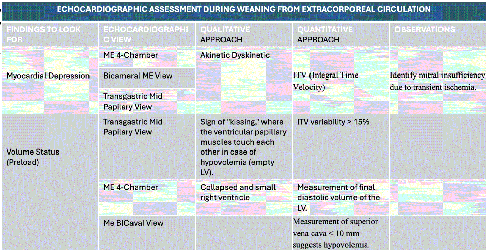
Table 1: Echocardiographic assessment in coronary revascularization during weaning from cardiopulmonary bypass.
Abbreviations: ME, Midesophageal; LV, left ventricle; ITV, Integral Time Velocity Echocardiographic Assessment in Coronary Revascularization during Weaning from Cardiopulmonary Bypass, Mena Ana, 2025
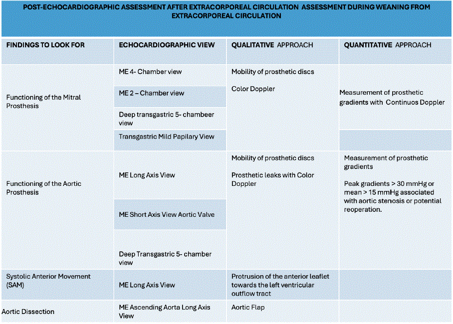
Table 2: Echocardiographic assessment in coronary revascularization post cardiopulmonary bypass.
Abbreviations: ME, Midesophageal Echocardiographic Assessment in Coronary Revascularization post Cardiopulmonary Bypass, Mena Ana, 2025
Transfusion decisions are based on:
• Hemoglobin and hematocrit levels
• Lactate levels
• Thromboelastometry (ROTEM/TEG)
• Active bleeding
• Consider fibrinogen (2-4 g) if less than 1.5 g/L
• Consider prothrombin complex concentrate (20-30 U/kg)
• Ionized calcium: 0.8-1.2 mmol/L or 8-12 mg/dL
• Use cell saver for intraoperative autotransfusion
• Maintain normothermia before and after CPB
• Reversal of Anticoagulation
Once stable separation from CPB has been achieved and surgical hemostasis is deemed satisfactory, anticoagulation should be reversed with protamine sulfate. Protamine administration must be performed in a controlled and titrated manner, with close monitoring for adverse reactions such as systemic hypotension, pulmonary hypertension, right ventricular dysfunction, or anaphylactoid responses. Activated clotting time (ACT) should be reassessed to confirm adequate reversal, in accordance with institutional protocols [6].
• Hemodynamic Surveillance and Stability
After complete discontinuation of extracorporeal circulation, continuous monitoring of arterial pressure, central venous pressure, pulmonary artery pressures (when available), mixed or central venous oxygen saturation, and urine output is essential. Maintenance of adequate mean arterial pressure and end-organ perfusion should follow goal-directed hemodynamic principles endorsed by ASA and EACTA [9].
• Contingency Strategy and Readiness to Reinitiate CPB
If hemodynamic instability occurs at any point during or after separation from CPB-manifested by low cardiac output, refractory hypotension, significant arrhythmias, ventricular failure, or echocardiographic evidence of inadequate cardiac performance—a prompt and coordinated decision should be made to return to CPB. The underlying etiology (e.g., hypovolemia, myocardial ischemia or stunning, vasoplegia, residual structural lesions, or electrolyte disturbances) must be identified and corrected prior to any subsequent attempt at weaning [24-26].
• In alignment with ASA and EACTA/EACTS principles, discontinuation of CPB should be viewed not as a single event but as a dynamic process requiring vigilance, structured reassessment, and shared decision-making to ensure patient safety and optimize perioperative outcomes [24-26] (Table 3).
| Pattern | Hemodynamic Findings | Likely Cause | Management |
| Low flow, high filling pressures | ↓ CI, ↑ CVP | LV dysfunction | Inotropes, afterload reduction |
| Low flow, low filling pressures | ↓ CI, ↓ CVP | Hypovolemia | Volume optimization |
| Normal/high flow, hypotension | MAP ↓, CI normal | Vasoplegia | Vasopressors |
| RV failure | ↑ CVP, septal shift | Pulmonary HTN, ischemia | Pulmonary vasodilators, inotropes |
Table 3: Hemodynamic patterns and targeted management during weaning.
Abbreviations: CI, Cardiac Index; CVP, Central Venous Pressure; LV, Left Ventricle; MAP, Media Arterial Pressure
In patients undergoing cardiac surgery with cardiopulmonary bypass, the venous-to-arterial carbon dioxide gradient (Pv-aCO2) and the Pv-aCO2/Ca-vO2 ratio have shown limited sensitivity for detecting changes in systemic blood flow and tissue oxygenation. Available evidence indicates that these parameters may remain relatively stable throughout surgery and are strongly influenced by dynamic physiological and pathophysiological conditions, including alterations in hemoglobin concentration, temperature, acid–base status, and microcirculatory flow.
The complex and non-linear relationship between partial pressure of carbon dioxide (PCO2) and carbon dioxide content (CCO2) further reduces the reliability of CO2-derived indices as markers of global perfusion. Additionally, poor agreement between simultaneous measurements obtained from central and mixed venous blood limits their interchangeability and clinical applicability
Given these limitations, CO2-derived variables should be interpreted with caution and should not be used in isolation to guide hemodynamic management or to assess tissue oxygenation during cardiac surgery. Multimodal monitoring strategies integrating hemodynamic, metabolic, and neuromonitoring parameters are preferable to optimize perfusion and postoperative outcomes.
Failure to separate from CPB requires rapid, structured evaluation using hemodynamic data and echocardiographic findings.
Low cardiac output with preserved systemic vascular resistance suggests myocardial dysfunction, whereas hypotension with normal or high flow is consistent with vasoplegia. Right ventricular failure should be suspected in the presence of elevated central venous pressure, septal shift, and reduced left ventricular filling [24-26].
If corrective measures fail, reinstitution of CPB allows myocardial recovery and reassessment before subsequent weaning attempts.
After successful separation from CPB, continuous hemodynamic surveillance is mandatory. TEE is repeated to confirm ventricular performance, exclude residual air, and assess surgical repair. Anticoagulation is reversed with protamine sulfate in a controlled, titrated manner, with close monitoring for adverse reactions.
Early complications following weaning include low cardiac output syndrome, vasoplegia, arrhythmias, coagulopathy, and pulmonary dysfunction. Rotational thromboelastometry (ROTEM) supports goaldirected hemostatic therapy and reduces unnecessary transfusion.
Maintenance of normothermia, adequate ventilation, and optimized oxygen delivery remains essential during this phase [24-26].
Patients receiving mechanical heart valves require lifelong anticoagulation with a vitamin K antagonist (VKA), guided by the international normalized ratio (INR), due to the high risk of valve thrombosis and systemic thromboembolism in the absence of anticoagulation. Post-operative anticoagulation should be initiated early, with bridging therapy using therapeutic unfractionated heparin (UFH) or low-molecular-weight heparin (LMWH) started within 24 hours after valve implantation, or as soon as surgical bleeding risk permits. Heparin therapy may be discontinued once the INR has been within the therapeutic range for two consecutive days. Available evidence suggests slightly lower bleeding rates with UFH compared with LMWH, although randomized data comparing bridging strategies remain limited [5].
INR targets should be individualized according to valve type, valve position, number of mechanical prostheses, patient-specific thromboembolic risk factors, and comorbidities. In patients who experience thromboembolic events despite adequate INR control and acceptable time in therapeutic range, intensification of VKA therapy or the addition of low-dose acetylsalicylic acid (75-100 mg/day) may be considered. Direct oral anticoagulants and dual antiplatelet therapy are contraindicated for thromboembolic prevention in patients with mechanical heart valves [5].
The complexity of long-term VKA therapy-characterized by a narrow therapeutic window, inter- and intra-individual variability, drug and food interactions, and the influence of ageing and comorbid conditions-necessitates structured patient education and close monitoring. Educational interventions and patient engagement have been shown to improve anticoagulation quality and adherence. INR self-monitoring or self-management may enhance therapeutic efficacy in selected, well-trained, and motivated patients, although safety benefits over standard care have not been consistently demonstrated [5].
The combination of VKA with antiplatelet therapy should be reserved for carefully selected patients, such as those with mechanical valves and concomitant acute coronary syndrome or symptomatic atherosclerotic disease with low bleeding risk. While the addition of antiplatelet agents may reduce major adverse cardiovascular events, it is consistently associated with a significantly increased risk of clinically relevant bleeding and should therefore be prescribed for the shortest effective duration, following an individualized risk– benefit assessment [5].
Lifelong clinical and echocardiographic follow-up is mandatory in all patients with prosthetic valves to detect prosthetic dysfunction, thrombosis, pannus formation, or progressive disease of other cardiac valves, with multimodality imaging playing a central role when complications are suspected [5].
Separation from cardiopulmonary bypass constitutes one of the most vulnerable periods of cardiac surgery. Successful weaning requires meticulous preparation, advanced monitoring, echocardiographic guidance, and coordinated multidisciplinary decision-making. From an anesthetic perspective, the process should be viewed as a structured, physiology-driven continuum rather than a discrete event.
Adherence to standardized protocols and checklists, combined with individualized hemodynamic and echocardiographic assessment, enhances patient safety and optimizes outcomes. Framed within ASA and EACTA recommendations, weaning from CPB is a shared responsibility that underscores the central role of the cardiac anesthesiologist in modern perioperative care.
Download PDF Here
Article Type: REVIEW ARTICLE
Citation: Mena A, Venegas E, Chango M (2026) Anesthetic Management and Weaning from Cardiopulmonary Bypass Principles, Monitoring, and Clinical Practice. J Surg Open Access 10(1): dx.doi.org/10.16966/2470-0991.275
Copyright: © 2026 Mena A, et al. This is an open-access article distributed under the terms of the Creative Commons Attribution License, which permits unrestricted use, distribution, and reproduction in any medium, provided the original author and source are credited.
Publication history:
All Sci Forschen Journals are Open Access