Abstract
Massive pneumothorax is an absolute emergency requiring immediate diagnosis and management. Clinical examination requires experience and a high level of suspicion to reach the diagnosis; however, it can be limited by the context in which it presents, the patient's characteristics and history, as well as differential diagnoses that can delay the diagnosis, or lead to incorrect diagnostic and therapeutic decisions that impact the final prognosis. Lung ultrasound has established itself as a noninvasive, safe, easily accessible, rapid, and reproducible diagnostic tool that has demonstrated high sensitivity and specificity in the diagnosis of pneumothorax when the “lung point” is identified. However, in complete pneumothorax, it is difficult to identify this sign due to its characteristic presentation, as it is obscured by the atelectatic lung due to the large amount of air accumulated between the two pleurae. We present the case of a patient with spontaneous complete left pneumothorax, which was initially asymptomatic. Ultrasound examination confirmed a suspected diagnosis due to mediastinal deviation, absence of pleural sliding artifact, absence of subpleural artifacts, absence of B lines, and the final diagnosis was established by direct visualization of the collapsed lung, compressed by air that generated the pneumothorax in zone 4 of the left hemithorax, which appears and disappears with respiratory movements, confirming the diagnosis by tomography. This ultrasound finding, called "atelectatic point", has not yet been described for the diagnosis of complete pneumothorax and is presented as a surrogate for the “lung point” in the diagnosis of this entity.
Keywords
Complete pneumothorax; Lung point; Atelectasis; Atelectatic point
Introduction
Pneumothorax is defined as the entry of air into the pleural cavity, and clinically, the symptoms do not necessarily correspond to its size. The etiologies can be varied, and the onset can be spontaneous [1]. According to the Spanish Society of Pulmonology and Thoracic Surgery (SEPAR), complete pneumothorax corresponds to the total collapse of the lung due to the amount of air accumulated between the pleurae. It constitutes an absolute emergency requiring immediate attention. The diagnosis is made with suspicion based on clinical data. It also requires imaging studies for confirmation, such as x-rays, CT scans, and currently ultrasound [1,2]. Along these lines, point-of-care ultrasonography has been used primarily by specialists in critical care areas, who, with adequate training, ask specific questions to resolve issues and facilitate decision-making, monitoring the progress of critically ill patients in real time [3,4].
In this way, it has been demonstrated that ultrasound is a safe, rapid, easily accessible, accurate, and economical noninvasive method that improves care outcomes in critical settings [4,5]. This is why lung ultrasound, based on the descriptions by Dr. Daniel A. Lichtenstein in his BLUE and FALLS protocols, has positioned this instrument as a diagnostic method of choice in critical areas to discern the etiology of respiratory failure between a pulmonary or cardiogenic origin [6-8]. The diagnostic criteria for pneumothorax have been clearly defined in the Evidence-Based Recommendations for Point-of-Care Lung Ultrasound [9] since 2012, and whose revision in 2022 gave rise to the New Guidelines and Consensus on Lung Ultrasound [10] giving certainty to the ultrasound signs determined by the absence of pleural sliding, B-line artifacts, the absence of a pulmonary pulse, and with high specificity in identifying the "lung point" [9,11]. In this aspect, by adding the ausence of these elements, the sensitivity reaches 88% and the specificity 100%, with a predictive value of 100% and a negative value of 99% [7].
In complete pneumothorax, complete pulmonary atelectasis coexists, hence why they have been considered synonymous or called total lung collapse. The spontaneous rupture of a bulla or the unidirectional entry of subpleural air, acting as a valve, has been suggested as an etiology. This progress to produce the mechanism of tension pneumothorax, until reaching the diagnostic clinic of the disease [12]. Atelectasis in ultrasound can be assessed by the absence of sliding of the pleural line; however, and unlike pneumothorax, the presence of the pulmonary pulse characterizes this entity and has been observed in patients with selective intubation [13].
Case Presentation
A 45-year-old male patient with no significant medical history was admitted to the emergency department with mechanical intestinal obstruction. He was clinically asymptomatic. However, during the patient transfer and reception protocol, alarming signs of 84% hypoxemia were identified when monitored by pulse oximetry. He had tachypnea of 22 breaths per minute, a heart rate of 91 beats per minute, and a systemic blood pressure of 96/54 mmHg. Physical examination showed no breath sounds in the left hemithorax, with no signs of respiratory dissociation. A bedside lung ultrasound was performed using a sector-array probe with Sonosite EDGE 2 equipment, in a cardiac and pulmonary protocol, with the following findings:
1. Cardiac evaluation revealed a right-deviated heart with small chambers and an inferior vena cava greater than 2 mm in diameter and less than 50% collapse.
2. Right hemithorax with pleural sliding artifact, with a thick and irregular pleural line and the presence of vertical B-line artifacts in the two anterior and two lateral zones.
3. Left hemithorax with absence of pleural sliding and horizontal reverberation artifacts or A-lines, with absence of vertical B-line artifacts, absence of pulmonary pulse, and the "stratosphere" sign, which is repeated in zones 1, 2, and 3. However, in zone 4, compressive pulmonary atelectasis is identified that appears and disappears with breathing, reminiscent of the "lung point" described above, pathognomonic for the diagnosis of pneumothorax.
The presence of ultrasound signs reflecting the loss of contact between the parietal and visceral pleura in zones 1, 2 and 3, the visualization of the collapsed lung with a "tissue"-like image, characterized by regular edges with a static air bronchogram of parallel distribution, with a pyramidal shape due to the effects of gravity and which maintains proximity to the posterior and lateral thoracic wall at the base, at the level of the posterior axillary line, translates into compressive atelectasis due to the complete pneumothorax in zone 4 of the left hemithorax. This last finding enables direct visualization by ultrasound of the collapsed lung due to a complete pneumothorax as an equivalent of the “lung point” and, which we have named as “atelectatic point”, since it represents the dynamic changes with breathing when there is a transition from the atelectatic lung to contact with the chest wall during expiration and the loss of said contact during inspiration, generating the typical image for pneumothorax.
This ultrasound finding, referred to as the "atelectatic point," has not been described so far for the diagnosis of complete pneumothorax. It is similar to the "lung point" for the diagnosis of this condition, as it is difficult to find due to the collapse of the parenchyma and obscuring the intersection of the pleura with the chest wall.
During the patient's workup, a simple abdominal CT scan was performed due to the initial condition he presented. The lung bases were intentionally analyzed in the image viewer, analyzing the pulmonary window and corroborating the ultrasound diagnosis. Finally, an emergency thoracotomy with pleural drainage was performed, producing the immediate release of pressurized air and with immediate hemodynamic and respiratory improvement of the patient, confirming again the lung expansion and resolution of the pneumothorax by means of ultrasound, finally documenting the sliding of the pleural line but also showing a thick and irregular pleural line, with the presence of vertical subpleural artifacts of the B-line type in both hemifields and which reflects interstitial syndrome, which raises suspicion of the existence of preexisting pathology.
Discussion
In the clinical case presented, the complete pneumothorax corresponds as defined by Kyoung MR, et al [12]. and will also lead to lung collapse causing total atelectasis. Let us remember that this atelectasis pathophysiologically occurs due to the absence of peripheral expansion of the lung and will be expressed in the x-ray study once all the trapped gas that occurs in the alveolar exchange has been absorbed, which begins when this exchange mechanism is stopped [13]. Due to this, in the images obtained ultrasound in the case, the lung can be seen as a collapsed mass in which a static or absent bronchogram is observed. The visceral pleura will not be in contact with the visceral pleura or will be too far medial that pleural sliding cannot be detected in the ultrasound image. It should not be strange, as in this case, in patients with total collapse of the lung, that the identification of this point called by the authors as “atelectatic point” could be a support point equivalent to the “lung point”, which could not be visualized, once identifying in the anterior zones the signs that reflect the loss of contact between the visceral and parietal pleura, and identifying in the region of the posterior axillary line the atelectatic lung, which is located in the anterolateral and inferior quadrants of the thorax [14], with the dynamic respiratory changes that make the image of a collapsed lung appear upon contact with the thoracic wall during expiration (Image A) and disappear with inspiration generating the typical image that is seen in pneumothorax (Image B). This finding is undoubtedly repeat over and over again with ventilatory dynamics, allowing us to assess its reproducibility in the case of complete pneumothorax, where an atelectatic lung will always coexist. This is a sign that we believe may have diagnostic value for complete pneumothorax. The images obtained were correlated with the simple CT scan performed on the patient as part of his comprehensive workup protocol (Image C).
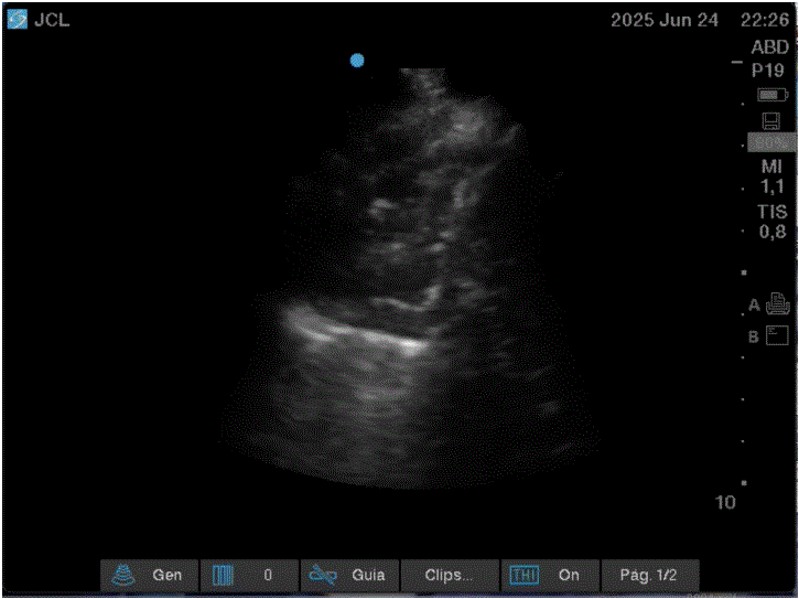
Image A: Patient in expiration in which the left lung is seen collapsed due to global compressive atelectasis, observing an isoechoic image, similar to tissue hepatization, with well-defined borders and heterogeneous content and with the presence of a static air bronchogram.
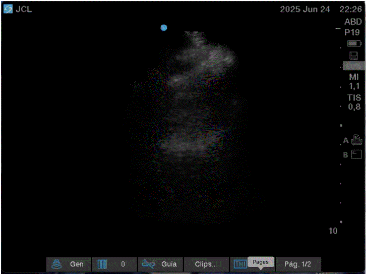
Image B: Patient in inspiration in which we observe the loss of pulmonary parenchyma, without sliding of the pleural line and without artifacts, as well as the absence of the lung pulse, characteristic of pneumothorax in the ultrasound.
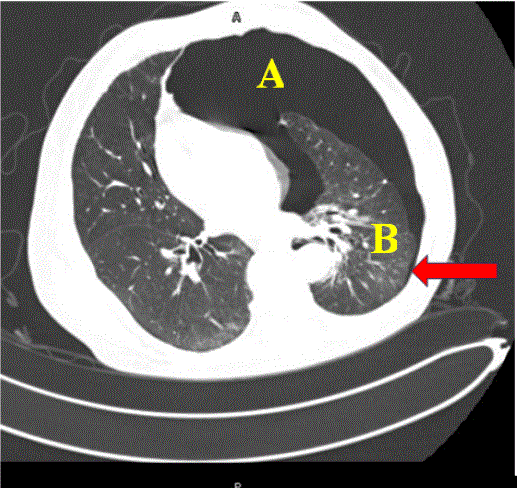
Image C: Simple tomography of the patient's chest where the pneumothorax area can be seen (A), the atelectatic area (B) and the area of the posterior axillary line where the dynamic changes with inspiration - expiration can be observed (arrow) that document the "atelectatic point" described in the text.
Finally, once the respiratory problem due to the pulmonary pathology was resolved with the placement of the endopleural tube, the patient was admitted to the hospitalization area, continuing his protocol and treatment by the general surgery service, for the disease that requested medical attention and corroborating the intestinal obstruction due to the history of previous surgeries and adhesion formation. During his hospitalization, he was evaluated by pulmonology performing a CT scan corroborating the resolution of the complete pneumothorax (Image D). At the same time, the suspicion of preexisting pathology was corroborated by observing data suggesting paraseptal pulmonary emphysema, with bullous disease of small elements due to multiple subpleural and paraseptal bullae. Finally, the tube was successfully removed and the occlusion disease was progressively resolved with conservative treatment and without requiring surgical intervention. The patient was discharged after 4 days of hospitalization and referred to the outpatient clinic for follow-up by pulmonology and surgery.
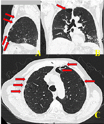
Image D: Simple chest CT study in longitudinal (A), coronal (B) and axial (C) sections where the proper placement of the endopleural tube that resolved the respiratory problem due to pneumothorax is observed and where multiple well-defined hypodense images are observed within the pulmonary parenchyma that corresponds to the bullous disease of small elements (arrows).
Conclusions
In complete pneumothorax, due to the large amount of air trapped between the pleurae, causing an atelectatic lung, it is difficult to identify the "lung point." However, the dynamic interaction between the atelectatic lung observed in the posterior axillary line and respiratory movements can be observed, giving rise to the image of a collapsed lung upon expiration and disappearing upon inspiration, showing the classic image of pneumothorax, a sign that has been named the "atelectatic point". It is also important to add that a multifocal ultrasound was performed, which included the evaluation of the cardiac cavity, giving rise to the deviation of the mediastinum to the contralateral side, still without giving the effect of tension pneumothorax, however, affecting the normal cardiac activity due to the increase in pressures of the thoracic cavities, which forces us to perform a complete and multifocal evaluation in this type of patients to have a clear overview of the hemodynamic and respiratory implications that this type of disease leads to and thus be able to identify complications early and offer timely care, reducing the risk of complications and reducing hospital stay times, lowering the costs of care due to the complications that may appear in these critical patients.
Data Availability Statement
The original contribution presented in the study are included in this article. Material further inquiries can be directed to the corresponding author.
Ethics Statement
This study is 100% descriptive and observational, so no experimental act was carried out and the protocols already studied and with level of evidence for the care of the patient involved were followed, fully adhering to the principles of competence, responsibility, honesty, integrity, impartiality, confidentiality and respect that are marked in the Codes of Ethics of the Mexican standards for patient care and the Declaration of Helsinki promulgated by the World Medical Association in 1964 for research on human beings.
Funding
The authors declare that they do not receive financial support for the research and/or publication of this article.
Conflict of Interest
The authors declare that the research was conducted in the absence of any commercial or financial relationships that could be construed as a potential conflict of interest.
Article Information
Article Type: CASE REPORT
Citation: Haro MJ, Tapia SM, de Jesús Montelongo F, Munguía JAC, Marines LAR, et al. (2025) Diagnosis of Complete Pneumothorax by Ultrasound: Description of the Atelectasia Point as the Equivalent of the Pulmonary Point. Case Report. J Surg Open Access 10(1): dx.doi. org/10.16966/2470-0991.273
Copyright: © 2025 Haro MJ, et al. This is an open-access article distributed under the terms of the Creative Commons Attribution License, which permits unrestricted use, distribution, and reproduction in any medium, provided the original author and source are credited.
Publication history:
Received date: 13 Oct, 2025
Accepted date: 24 Oct, 2025
Published date: 30 Oct, 2025
