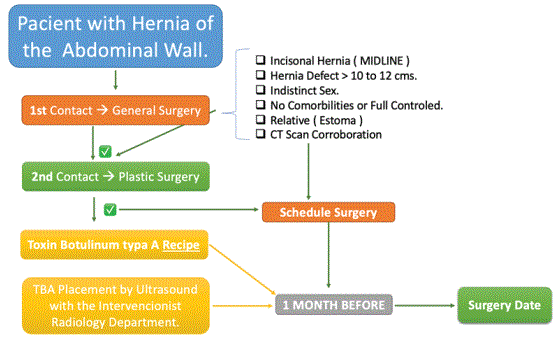
Algorithm 1: This is the management of how do we protocolize the patients, with all services.

Araujo-Lopez Adan1* Salazar-Del Pino Claudia Paulina2 Peña-González Ricardo Raciel3 Cardona-Ochoa Paulina4 GonzalezRodriguez Monica1 Rios-Lara R Luciano4 Torres-Piña Raymundo4 Ugalde-Vitelly Juan Antonio5
1Plastic Surgery Resident, Plastic and Reconstructive Surgery Service of the General Hospital of Mexico, “Dr. Eduardo Liceaga”, Mexico City, Mexico*Corresponding author: Araujo-Lopez Adan, Fourth year and Chief Resident, Plastic and Reconstructive Surgery Service of the General Hospital of Mexico, “Dr. Eduardo Liceaga”, Mexico City, Mexico, Tel: (+ 52) 55 2789 2000; E-mail: [email protected]
Introduction: The current incidence of incisional hernia is up to 11%. Botulinum toxin type A shows atrophy in the extrafusal and intrafusal muscle fibers. With the addition of Muscle Paralysis and improving strength, it is possible to perform an effective posterior component separation.
Objective: Describe an easy algorithm to treat complex abdominal wall reconstruction and improve the quality of adjacent tissues.
Algorithm 1: This is the management of how do we protocolize the patients, with all services.
Material and Methods: Fifty-nine patients were analyzed over 1 year in the General Surgery and Plastic and Reconstructive Surgery Service, patients with midline abdominal wall hernias, using the effective reconstruction algorithm, placing by ultrasound-guided botulinum toxin in the muscle transverse, 1 month later performing TAR, with Abdominoplasty for skin flap management, and comparing with the control group that did not apply botulinum toxin.
Discussion: Botox is a neurotoxin derived from the bacterium Clostridium botulinum (botulinum toxin type A) that has been observed at the sensory level causing atrophy in the extrafusal and intrafusal muscle fibers. Repair of the abdominal wall after incisional hernias has been a great challenge with recurrence rates of 11%. Performing the release of the transverse muscle has a recurrence of 6%, as well as the compensation of skin flaps are ideal for proper management.
Results: Within the observed analyzes, the results were divided into trans-surgical (99% of the patients closed the midline), immediate post-surgical (Pain, Seroma and Infection <1%) and late (with <3% recurrence) in the experimental group.
Conclusions: Within the study, we will be able to analyze that lowering the recurrence rate <3% in patients properly protocolized, is translated as an adequate and totally reproducible method in our environment.
Hernia; Botulinum toxin; Transverse; Abdominoplasty
An incisional hernia of the abdominal wall is any defect with or without an increase in volume in an area of a postoperative scar that is perceptible or palpable by clinical or imaging examination [1].
The current incidence of incisional hernia is up to 11% [2]. Repairing an incisional hernia has a probability of recurrence of 33%, the third and fourth attempts are associated with even greater recurrence [2], up to 64% after tension plasty and 32% after mesh plasty [2].
An incisional hernia can cause obstruction and strangulation of the intestine, which may require emergency surgery, and pain, discomfort, and cosmetic discomfort negatively impact the quality of life of patients [1]. The abdominal wall is the core of musculoskeletal posture, a protective barrier for the viscera, and the basis for mechanical respiration [1,2]. Maintaining constant intra-abdominal pressure allows support for breathing, locomotion of the trunk, urination and defecation, among other physiological functions [3], so that a postincisional hernia directly affects all levels of the organism [3].
Complex reconstruction of the abdominal wall requires reapproximation of the fascia to achieve primary closure of the defect without tension. Abdominal wall dysfunction is due to altered anatomy because of lateralization of the abdominal wall musculature and resulting loss of midline attachment to the linea alba of the aforementioned muscles [4].
Due to the high recurrence rate associated with the closure of hernial defects of the abdominal wall, novel techniques such as the use of pneumoperitoneum have been tried. This paper explores the use of preoperatively applied botulinum toxin to improve abdominal wall reconstruction and achieve tension-free midline wall coping [3,4].
A Universe of 59 patients operated by the set of two services, Plastic and General Surgery in the General Hospital of Mexico, during 1 year (from March 2022 to March 2023), in a clinical essay, prospective study.
The patients were divided by 2 groups, the experimental and the control, with 26 and 33 patients.
In the study we analyzed patients with a very similar characteristics; age between 18 to 65 years, any sex, no comorbidities, BMI <30 m/ k2, with midline hernia >12 cm2 visualized by a CT Scan. Relative contraindications where patients with intestinal stomas, (if they were young, no comorbidities, and BMI <30 m2).
The objective of this clinical essay is to make a perfect treatment in the knowledge of big hernia repair. It is not a hernia repair, every patient has to be perfect evaluated, and with the dissection during the surgery will be better for the closure of the middle line, and all the skin flaccidity will be out of it with the abdominoplasty.
All the study was made by the same surgeon, and groups of surgeon, including the General Surgery Team, and the Plastic Surgery Team, the surgery was made at the operation room, with and average of surgery time of 4 hours, with the beginning of the surgery by the general surgery team, if they have something to do at the bowels, and then we prepare the abdominal wall to the transversus abdominal release, and the abdominoplasty by the end.
We analyzed 59 patients, divides 2 groups, the experimental group with 26 patients, in the variables there where 7 feminine, and 18 male, with an average of age about 40 years, all patients in this group where evaluated, and admitted with a 2 filters of Services ([1), once they have the date of surgery,-1 Mont Before Surgery-the Toxin Botulinum Type A (Dysport 500 UI-Dilution-(Figure 1) has had injected by direct view (Lineal Transductor Ultrasound of 8 MHz of Skeletal Muscle Viewwith Radiology Intervention) at the transverse muscle by the 3 points (Figure 2) at the semilunar of the abdominal wall.
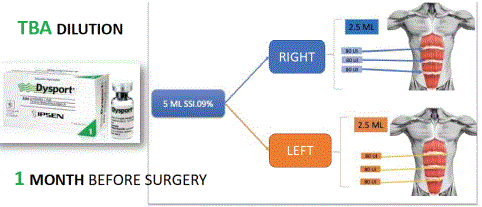
Figure 1: This is the Dilution and the sites we use to it was placed, with Ultrasound, in three different parts of the semilunaris linea and metameres located, with a total of 500 UI of Dysport.
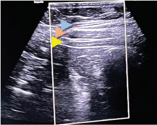
Figure 2: Image Ultrasound of the colocation of the TBA at the semilunar Linea and medial metameral doing the injection in the transversus muscle. Blue Arrow (External Oblique), Orange Arrow (Internal Oblique), Yellow Arrow (Transversus Muscle).
The Toxin Botulinum type A (TBA) It was placed by ultrasound, we take pictures before colocation (1 month) doing contraction (Figure 3), we measure the length of the hernia and compare with his Computerized Tomography Scan (CT), then we take another picturedoing contraction (1 month) (Figure 4) after the TBA, to measure what the length has decrease in millimeters.
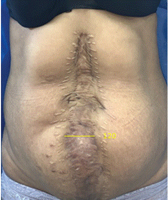
Figure 3: This Female has a Lineal Big Hernia, with contraction it has a defect of 120 mm in middle line.
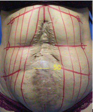
Figure 4: The same female with lineal big hernia a month after the surgery with TBA all over the Hernia, it decreases 50 mm.
The Control Group, with 33 patients, worth nothing that this is the normal treatment of patients in the General Surgery Team, but they do not place the TBA, and the TAR is not always the good option, not always get the midline close and no abdominoplasty made for the reconstruction.
With 11 patients of Feminine, and 21 of Male, and average of 42 patients. In this group, due to difficulty with reapproximating, only 15.1% of patient’s midline abdominal incisions were closed.
In both groups, regardless of the ability to reapproximate, mesh (Doble Component-Polytetrafluoroethylene (PTFE) and Polypropylene (Figure 5), and we use Polypropylene only “Macropores”) was used as a preventative measure to avoid recurrent abdominal hernia formation.
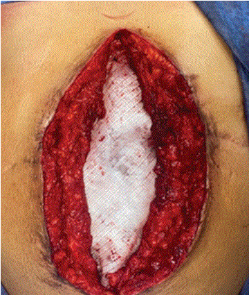
Figure 5: The sandwich technique in all patients, and the mesh collocated in all over the dissection of 30 × 30 cms.
The surgery was made by general surgeon at the beginning, they made all bowel restructure, and after that we (Plastic Surgery) preparer abdominal wall, and have the main purpose of it, that is to close the midline with the sandwich technique, using the mesh, and the abdominoplasty to the flaccidity of the skin.
The analysis of this study was with the hypothesis, we might not be sure if the dissection of the transversus abdominus will close the midline, in all patients, even the experimental group
In both groups, we analyze complications and improve of the surgery, in the surgery, after the surgery and we divided by immediately and lately.
For the main results in this study we describe it by the groups, in the Control Group; with Total of 33 patients; The Sex was 21 patients Male (63.6%), and Femenine 11 patients (33.3%). The average of the age was 42 years old.
The improvements in the Control Group during the surgery, were measured by the dissection was made during the surgery, and the muscle strength had during the dissection (The scale consists with 1=good, 2=mild, 3=weak, 4=flaccid), 18 patients (54.4% had weak), and 11 patients (33.3% had flaccid), none patients had good quality over the muscle strength (Graphic 1).
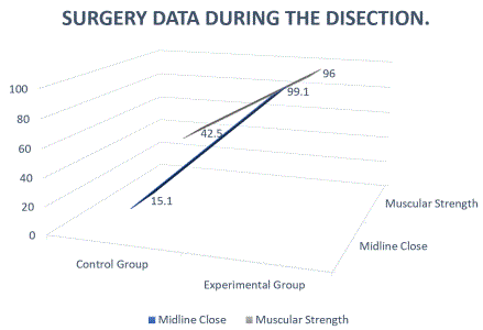
Graphic 1: Comparison between groups; In the Midline Close 15.1% had in the CG, and 99.1% had in the Experimental Group, The muscular strength was 42.% (weak) but in EG 96%.
The complications of the Control Group, we divide by immediately; about the pain, 27 patients had (81.8% had pain after the surgery), and the 15.15% didn´t had. The seroma in the patients were about 12 patients had (36.3% had), and the 60.6% did not had. And about the surgical wound infection 9 patients had (27.7% with infection) and the 23 did not had (68.9%) (Graphic 2).
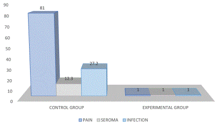
Graphic 2: Comparison between groups about early complications; Pain was in EVA >9 81% in the CG, and 1% in the EG, Seroma was in 12.3% of the CG and 1% in the EG, Surgical Site Infection was 27.7% of the CG and 1% in the EG.
The late complication of the Control Group, we measure at the 6 month, and the most important is the recurrence that happened in 14 patients (42.5%), and gone well with no recurrence of the hernia in 19 patients (57% ). And the visual aesthetics of the abdomen, in all patients (96.9% the 32 patients gone bad about), and only with we made the TAR and the abdominoplasty 3.8% of the sample like the visual aspect (Graphic 3).
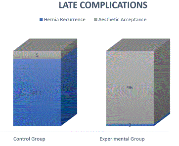
Graphic 3: Comparison between groups about lately complications; Hernia Recurrence was 42.4% of the patients of the CG, and 3% in the EG with the follow up at 6 months to 1 year, The Aesthetic acceptance of the abdominal wall was 5% in the CG and 96% in the EG.
The main results in the Experimental Group; the total are 26 patients, The Sex was 18 patients Male (69.3%), and Femenine 7 patients (26.9%). The average of the age was 40 years old.
The improvements in the Experimental Group during the surgery, were measured by the dissection was made during the surgery, and the muscle strength had during the dissection (The scale consists with 1=good, 2=mild, 3=weak, 4=flaccid), 19 patients (73.0% had good and strong), 6 patients (23.3% had mild), and 1 patient with weak (3.8%) most of the patients in the study get the muscle strength (Graphic 1).
The complications of the Experimental Group, we divides by immediately; about the pain, 25 patients did not had (96.5% did not pain after the surgery), and the 3.8% had. The seroma happened in just 1 patient about 3.8% of all the sample. And we did not have surgical wound infections (Graphic 2).
The late complication of the Experimental Group, we measure at the 6 months, and the most important is the recurrence that did not happened the patients in the sample. And the visual aesthetics of the abdomen, in all patients (100% the 26 patients gone good acceptance) (Graphic 3), (Figure 6).
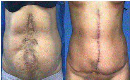
Figure 6: The before and after (6 months) of the most interesting case with 100% of acceptance in the patients.
Cases of the study that we can share in the clinical essay (Figures 6-8).
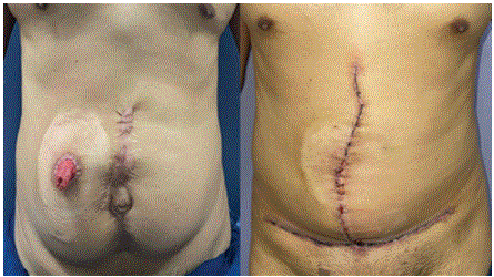
Figure 7: The before and after of a Male Case with bowel restitution.
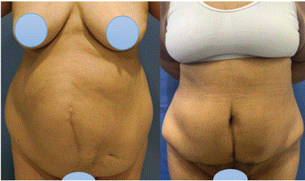
Figure 8: The before and after of anold Female case that get life changed and comorbidities too.
Botox is a neurotoxin derived from the Clostridium botulinum bacterium, divided into serotypes A-G whose main function is the inhibition of the release of acetylcholine through exocytosis, however, each of the different serotypes acts through different proteins having a variation in as to effect, duration and potency; subtypes A and B being the most used clinically [1].
Botulinum toxin type A has an affinity for cholinergic glycoproteins, temporarily inhibiting the transmission of impulses with a localized motor and autonomous effect, causing muscle flaccidity through temporary paralysis, reaching its peak at 4 weeks and lasting no longer than 6 months [2-4]. On the other hand, the mechanisms of action of botulinum toxin type A have been observed at the sensory level, causing atrophy in the extrafusal and intrafusal muscle fibers, and at the ganglionic level, being used in cases of hyperhidrosis and hypersalivation [4-6].
The repair of the abdominal wall after incisional hernias has been a great medical challenge over the years due to the high recurrence rate, between 30-70% as described by Ibarra Hurtado, and the risk that alternatives entail. surgical procedures which include component separation techniques, myofascial release, local tissue flaps, free flaps and the use of synthetic materials, which are associated with increased morbidity, infection, wound dehiscence and seroma formation [5-8]. The difficulty in carrying out the abdominal repair lies mainly in the muscular retraction to the side and the loss of the midline, for which the successful repair consists in facing the fascia without generating tension to restore the abdominal anatomy, function and enhance aesthetic appearance [7]. It is important to mention that fascial closure depends on many different factors, such as muscle and tissue quality, hernia size, loss of dominance, and surgical technique [5,7]. For this reason, in recent years a less invasive, safer for the patient and costeffective alternative for abdominal repair has been sought [8].
Wound infections are the main etiological factor for the development of recurrent incisional hernias. In one of the largest case series with 165 patients, 61% of the patients had a recurrent ventral hernia [6,7]. Other common etiologic factors for complex abdominal wall defects include trauma, infectious processes, and other abdominal catastrophes. In some instances, such defects represent life-threatening conditions with loss of control, persistent infections, exposed abdominal viscera, intestinal fistulas, and abdominal wall retraction [8]. Many patients are seriously ill with other significant medical problems such as sepsis, compromised nutritional status, immunosuppression, and cardiopulmonary problems. Such patients need timely and aggressive management to avoid further complications and deterioration that can affect the outcome of any reconstructive procedure [9].
In other patients, there is no loss of dominance but simply a loss of dominance with chronic and recurrent incisional hernias [9].
The use of type A botulinum toxin for abdominal wall repair was described for the first time in 2009 by Ibarra Hurtado in a prospective study conducted between 2007 and 2009 in 10 patients with abdominal hernias located in the midline and the guided application of botox. By electromyography mainly in the oblique muscles, being reassessed at 4 weeks, resulting in a significant reduction in the size of the transverse defect and surgery with less tension, facilitating closure towards the midline, showing no cases of recurrence at 9 months [8].
Subsequently, it has been shown that the administration of botulinum toxin 30 days prior to surgery achieves a significant elongation of the lateral abdominal wall of 3.2 cm per side, 6.3 cm of total elongation and a significant decrease in the width of the transverse hernia [7], which helps primary fascial closure of defects in 83% of cases without the need to require any other technique [4-8].
The literature maintains that the use of the toxin prior to surgery in the previously mentioned time interval has the ability to decrease intraabdominal pressure [5-7], increase the compliance of the abdominal wall and reduce the size of incisional hernias through chemical paralysis of the lateral muscles of the abdominal wall [4,5]; facilitating with this the medialization of the fascia and the possibility of primary closure without the need to separate the components, restoring the anatomy and the physiology of the abdominal wall, reversing muscle lateralization and fibrosis in a safe manner for patients, avoiding postsurgical complications and reducing the recurrence rate by nearly 100% [5,7].
On the other hand, due to the mechanisms of action in intrafusal muscle fibers, it has been suggested that the use of botulinum toxin could help reduce pain and use analgesics in post-surgical recovery. No respiratory complication secondary to the use of botulinum toxin in the anterolateral muscles of the abdominal wall has been demonstrated due to its role in accessory respiration [8,9].
Both CT fluoroscopy and ultrasonographic-guided injections appear to be adequate for targeted intramuscular BTX injection, with advantages and disadvantages of both techniques already illustrated; the lack of ionizing radiation with US as the major advantage, although with poor sonographic window in patients with challenging body habitus, potentially limits this technique in this patient population. The use of ultrasound guidance limits the risk of systemic distribution if injected intravascularly. The advantage of combining EMG guidance to ultrasonography adds the possibility of determining if a muscle is denervated or fibrotic and might be beneficial in patients with multiple recurrent hernias and especially in patients with a history of surgical component separation techniques [10].
The Transversus Abdominal Release allows for significant posterior rectus fascia advancement, wide lateral dissection, preservation of the neurovascular supply, avoids subcutaneous tissue undermining, and provides a large space for mesh sublay [11].
Myofascial advancement during component release was described as the most physiological reconstruction of large abdominal wall defects [11].
The TAR technique described herein is an extension of the retro muscular Rives-Stoppa repair to the posterior com- ponent separation technique. There are 3 main advantages to this approach. First, transversus abdominis muscle release results in significant medial mobilization of the posterior rectus sheath. Second, it allows for extensive lateral dissection in a potentially unlimited space between the transversus muscle and the underlying transversalis fascia/ peritoneum [11].
The main objective of this study, was done; creating a reliable, positive, learned technique, that can be teached by the head resident, to others, but the most important thought is about the perfect protocolization of the patients, and well know, that not all patients are good candidate for this technique, and the TBA is not a magical thing that close the abdominal wall without a big dissection, that the mesh is all.
With this technique, we demonstrate, that using the dilution (Figure 1), is the perfect management for it, that the safe placement of the TBA, has to be by ultrasound, it is not a rule that a Interventionist Radiology has to do that, actually we can made it by it owns, with ultrasound, to have a perfect placement and to be well distributed all over the muscle, making this lasting for the whole month and having the peak of action at the month.
The close of the midline is one of the big problems, in the big hernia repair, if the dissection is well done, you will have 5 o 7 cms of each side and can solve it. It the surgeon does not do and appropriate dissection the midline will not close.
Doing and appropriate management of the skin laxity is the most common problem in the hernia repair, doing a Lily Flower Abdominoplasty in 96% of all the patients, solve the close and compensation the abdomen flaccidity, and all the scars that they have as the previously surgeries.
One big thing in this article that we made the surgery, if the patients has done all the general surgery problems, and it’s going to be the last surgery, to make and properly management of all the abdominal wall (muscle, fat facias, and skin).
Download Provisional PDF Here
Article Type: RESEARCH ARTICLE
Citation: Araujo-Lopez A, Salazar-Del PCP, Peña-González RR, Cardona-Ochoa P, Gonzalez-Rodriguez M, et al. (2023) Botulunim Toxin Type A Followed by TAR (Transversus Abdominal Release) with Abdominoplasty as the Best Combination for Big Hernia Repair. J Surg Open Access 9(2): dx.doi.org/10.16966/2470-0991.270
Copyright: © 2023 Araujo-Lopez A, et al. This is an open-access article distributed under the terms of the Creative Commons Attribution License, which permits unrestricted use, distribution, and reproduction in any medium, provided the original author and source are credited.
Publication history:
All Sci Forschen Journals are Open Access