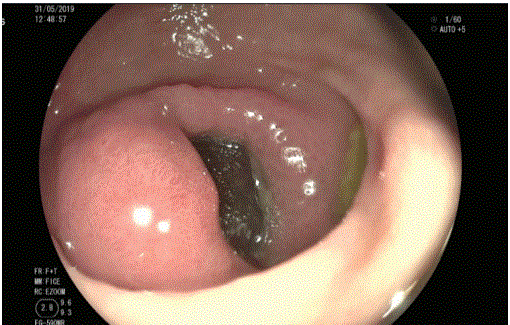
Figure 1: The endoscopic procedure shows an oval-shaped, 13 mm, stone impacted against the duodenum first portion.

Caltenco-Solís Raúl Beder1* Torrejón-Hernández Carlos ADRIAN2 Bizueto-Rosas Héctor2 Pérez-González Hugo Alonso3 Magaña-Salcedo Jaime Roberto1 Echeverry-Fernández Camilo Andrés1 Hidalgo-Delgado Jesús Nicolás1 Radilla-Flores MARIANA DEL CARMEN1 Gamboa-Ramírez Fernando1
1Senior resident, Department of General Surgery, Darío Fernández Hospital ISSSTE (Institute of Security and Social Services of State Workers), Mexico City, Mexico*Corresponding author: Caltenco-Solís Raúl Beder, Senior resident, Department of General Surgery, Darío Fernández Hospital ISSSTE (Institute of Security and Social Services of State Workers), PO 03900, Mexico City, Mexico, E-mail: caltenco@icloud.com
Introduction: Among the rare complications of cholecystitis is gallstone ileus and the least common is Bouveret syndrome, more frequent in older women. The diagnosis is fortuitous in most cases, so in patients with intestinal occlusion and medical history of cholecystitis, it should be considered as a possibility. The ideal treatment remains controversial.
Case: We present a 93-year-old patient, with intestinal occlusion symptoms and confirmed with Bouveret syndrome after endoscopic diagnosis, with unsuccessful initial treatment and subsequently resolved with open surgery through a jejunal enterotomy with stone extraction and good postoperative evolution.
Discussion: Clinically, these patients present with nonspecific symptoms such as vomiting, nausea, abdominal pain, and evidence of upper Gastrointestinal (GI) bleeding. Abdominal Computed Tomography (CT) scan is the reference diagnostic study. Endoscopy can be in some cases both diagnostic and therapeutic; the vast majority of cases are resolved with open surgery in one or two stages depending on the patient’s condition. Laparoscopic surgery can be beneficial but requires a mini-laparotomy to remove large stones.
Conclusion: The diagnosis of Bouveret syndrome requires a high index of suspicion in those patients with a history of cholelithiasis and symptoms of intestinal occlusion. There is the option of definitive diagnosis and treatment through endoscopic procedures, but these require the availability of diverse instruments and operator experience. For those cases with stones larger than 2.5 cm, a first intention open surgery approach is recommended.
Bouveret syndrome; Biliary ileus; Endoscopy; Open surgery
It is given the name of Bouveret’s Syndrome when a stone from the gallbladder or the bile duct is embedded in the duodenum causing intestinal occlusion; it is an extremely rare disease, approximately 300 cases have been reported in the international literature. This condition represents 1 to 3% of cases of gallstone ileus. The reported incidence of gallstone ileus is 0.3 to 0.5%; It was first described by Leon Bouveret in 1896 [1,2].
It is known as biliary ileum when the stone is generally embedded in a loop of the small intestine; a rare complication of cholelithiasis; uncommon cause of intestinal occlusion or acute abdomen (0.5-4%), secondary to the mechanical and obstructive effect of the stone in the intestinal lumen; the calculations can be multiple.
The name “ileus” can be imprecise since the occlusion can occur at different levels of the digestive tract [3,4]. Biliary ileum was first described in 1964 by Rasmus Bartholin as an autopsy finding [5].
Reissner and Cohen (1994) reported the frequency of obstruction sites: terminal ileum 60%, proximal ileum 24%, distal jejunum 9%, colon and rectum 2-4%, and duodenum 1-3%. The clinical picture is very nonspecific and depends on the site of obstruction. They report that only a third of patients present with colic-like pain, bloating, nausea, vomiting, anorexia, weight loss, jaundice, diarrhea, constipation, and even severity data such as cholangitis, perforation, peritonitis, and gastrointestinal bleeding [6]. It is known as a Mordor triad when there are three elements: a history of cholelithiasis, acute cholecystitis, and intestinal occlusion [7].
Rigler’s triad (1941) is made up of pneumobilia, intestinal occlusion data, and the identification of the stone in the right lower quadrant or ectopic position, as long as the stone is radiopaque (<10%) [5].
When the stone changes position on a second radiograph, it is known as Rigler’s Tetrad. The diagnosis of biliary ileum is considered with the presence of two signs of this triad.
When contrast studies are performed and there is the passage of contrast material into the biliary tract, it is known as Petren’s sign [6,7].
If the diameter of the stone released to the intestinal lumen by the enterobiliary fistula is equal to or greater than 2.5 cm, it can be detained at different points of the digestive system such as the stomach, duodenum, small intestine or colon; and it is considered as Barnard syndrome when it obstructs the ileocecal valve [5-8].
Biliary ileum is more frequent in patients older than 65 years, of the female sex compared to the male sex in a ratio of 3:1, even in some reports they report that it is 7:1. The mortality of the biliary ileum is approximately 12 to 30%, depending on the age, comorbidities and the site of impaction of the stone [5,9,10].
Controversy exists for both diagnostic methods and definitive treatment but, contrasted computed tomography is generally considered the method of choice, and surgical treatment is the ideal method for resolution, in one or two stages, depending on the patient’s condition, either open or laparoscopic depending on the experience of the surgeon, or it can be performed by endoscopic resolution [7,11- 14]. Treatment is initially aimed at solving the intestinal occlusion.
We present a 93-year-old female patient, allergic to penicillin; a medical history of systemic arterial hypertension, and recently diagnosed with Chronic Obstructive Pulmonary Disease (COPD); a surgical history of appendectomy and inguinal plasty. Her condition begins three days before admission with generalized abdominal pain, intolerance to the oral intake, nausea, and vomiting on 15 occasions with gastrobiliary characteristics, with a coffee ground aspect. During physical examination with normal peristalsis, pain on palpation in the epigastrium and right hypochondrium, with no evidence of peritoneal irritation. Initially with laboratory tests and normal abdominal radiography. Considering a history of upper gastrointestinal tract bleeding she is initiated with treatment including proton pump inhibitor and endoscopy of the esophagus and gastroduodenal was requested. Endoscopy reported: pylorus with edema of the mucous layer, stone impacted in the first portion of the duodenum (Figure 1); so the removal of the stone is attempted, achieving its capture using the 13 mm oval polypectomy loop device (Figure 2). Erythematous, friable, friable bulbar mucosa is observed; and the oval-shaped stone advanced without achieving its capture or crushing. A review is performed by advancing the endoscope to the second portion of the duodenum without being able to visualize the stone. The endoscopic procedure concludes with Bouveret’s syndrome, bulb, and duodenitis plus type I hiatal hernia.
Figure 1: The endoscopic procedure shows an oval-shaped, 13 mm, stone impacted against the duodenum first portion.
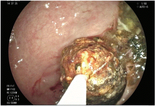
Figure 2: Polypectomy loop device attempting to catch stone during the endoscopic procedure.
She is admitted to our center again twelve days after presenting colic-type abdominal pain located at the epigastrium, radiating throughout the abdomen; nausea and vomiting of gastrobiliary content five times; during the physical examination with generalized pain on palpation and decreased peristalsis, normal laboratories. An abdominal CT scan is performed and the Rigler’s triad is identified: pneumobilia, intestinal occlusion, and a stone of 3 cm in the lower right quadrant (Figures 3-5).
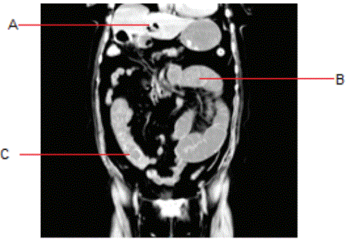
Figure 3: An abdominal CT scan showing the Rigler’s triad. A: Neumobilia; B: Small intestine dilated due to occlusion; C: Lower right quadrant with a gallstone.

Figure 4: An abdominal CT scan, coronal view, with Neumobilia (A) and Intestintal occlusion (B).
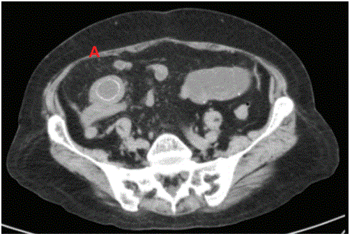
Figure 5: An abdominal CT scan, axial view, with right lower quadrant “donut” image, of a 3 cm stone (A).
After those findings, she undergoes open abdominal surgery, identifying a stone at 110 cm from the suspensory muscle of the duodenum (Figure 6). A longitudinal incision was made at 3 cm on the antimesenteric border in the jejunum to extract a 3 × 4 cm stone (Figure 6). An intestinal closure is performed in two planes with the Heinecke-Mikulicz technique (Figure 7); the entire path of the small intestine and colon is explored again, without another stone or pathology, so a Penrose type drainage is placed in the pelvic cavity and the abdominal cavity is closed as usual practice.
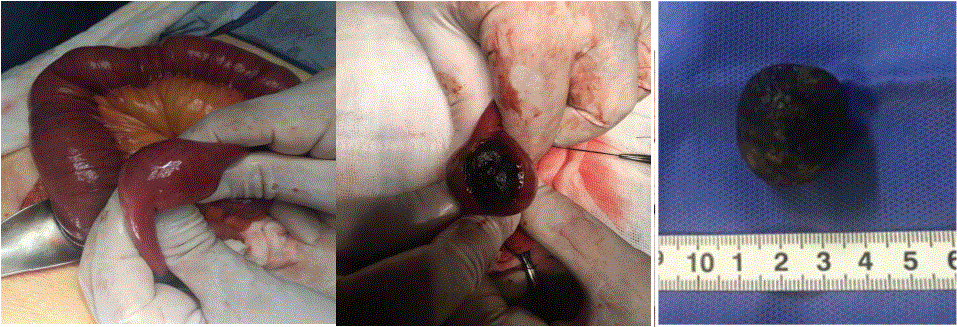
Figure 6: A stone found at 110 cm from duodenum (left), then showed from longitudinal enterotomy (center), after removal, being of 3 × 4 cm (right).
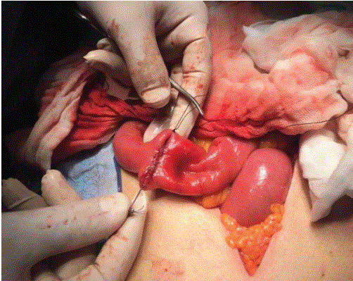
Figure 7: Intestinal closure by the Heinecke-Mikulicz technique.
It was decided to manage the patient with fasting for five days, to initiate progressive fluid intake on the sixth day, evolving without complications, so she is discharged from our center.
There are no specific data on laboratory tests for Bouveret syndrome; there may be leukocytosis, elevated liver enzymes, or electrolyte changes [12].
The clinical behavior of the biliary ileum varies depending on the level of intestinal occlusion; when the obstruction is at the duodenal level, vomiting, nausea, and abdominal pain occur in 70% of cases; less frequently data on gastrointestinal bleeding such as hematemesis and melena are observed [13,14].
The diagnosis requires a high index of suspicion, therefore, in most cases, it is a surgical or endoscopic finding [5]. Ideally, the diagnostic possibility is contemplated with the medical history of cholelithiasis and the intestinal occlusion presentation, and then performing an abdominal CT scan being the reference study.
We must bear in mind that the clinical data is nonspecific and that the initial diagnostic approach will be different in most cases; in patients with intestinal occlusion symptoms, abdominal radiographs can be requested in two projections, thus diagnosing only 21% of cases.
When this condition presents data of pain in the right upper quadrant or epigastrium, acute cholecystitis can be suspected, so the requested study would be an abdominal ultrasound that can detect structural changes in the gallbladder, pneumobilia, or a stone in the duodenum [2,3].
The simple abdominal CT scan has a sensitivity of 93% and specificity of 100%; it is feasible in a coronal view to detect the Rigler’s triad, as was initially described [5], when this study is accompanied by oral contrast medium, it can show the passage of the contrast medium through the fistula, to the gallbladder or the bile duct [5,7,9]. Finally, endoscopy can be diagnostic and therapeutic; it has been suggested as the initial or definitive procedure, as it offers a less invasive therapeutic option [15-17]. Stone impaction at the duodenal level is ideal for an endoscopic approach; once the impacted stone has been identified in the first portion of the duodenum, it should be attempted to displace it, followed by fracture of the stone, with the removal of the large fragments up to 1.5 cm, and avoid the attempt on those stones larger than this size so that they can be removed by esophagus; the minor fragments can be chosen to be left in the stomach for future radiological monitoring with close follow-up, to avoid possible complications.
The ideal is to have different equipment such as baskets, forceps, biliary balloons, laser lithotripsy, extracorporeal lithotripsy, or intracorporeal electrohydraulic lithotripsy, or the combination of these to offer different therapeutic procedures [3,17,18].
However, by means of endoscopy in Bouveret’s syndrome, at the moment, it does not correct the existing fistula, in addition, it is not possible to assess the existence of stones distally coupled with the high probability of failure since much experience is needed from the specialist to obtain good results. About 42% of patients in whom endoscopic stone removal is attempted, end up in surgical management.
The current treatment of choice for biliary ileus is enterolithotomy combined with exploratory laparotomy to assess the duodenum and small intestine and colon [1,8-11,18,19].
In surgical treatment, the intestinal occlusive process can be resolved first with enterotomy with the removal of the stone, and if the patient is stable and of low risk, cholecystectomy with fistula closure if needed. In unstable patients, delayed cholecystectomy is performed being the preferred behavior in most cases [20], since the combination of the two procedures in one surgery is reflected in increased mortality of up to 35%; however, when the fistula remains, it favors the recurrence of the biliary ileus or the appearance of cancer with a frequency of up to 15%. [8,9,12,14,15,18-20].
Currently, we should also consider laparoscopic surgery as an option for the treatment of this condition since in this option patient can have a faster recovery with this procedure. Although it has a high probability of conversion to open surgery due to being a rare clinical entity [1,12].
In our retrospective analysis of the case, given that it has been reported that stones larger than 2.5 cm cannot be crushed or extracted, the attempt to manipulate the stone conditioned its migration and subsequently impact and cause the intestinal occlusion symptoms. Despite our patient referring to evacuate one stone, there are reports on multiple stones and the fact that she required a second admission due to GI bleeding forced us to offer a definite approach by open surgery.
The diagnosis of Bouveret syndrome requires a high index of suspicion in those patients with a history of cholelithiasis and symptoms of intestinal occlusion. There is the option of definitive diagnosis and treatment through endoscopic procedures, but these require the availability of diverse instruments and operator experience. For those cases with stones larger than 2.5 cm, a first intention open surgery approach is recommended.
Authors declare no conflict of interest.
Download Provisional PDF Here
Article Type: CASE REPORT
Citation: Caltenco-Solís RB, Torrejón-Hernández CA, Bizueto-Rosas H, Pérez-González HA, Magaña-Salcedo JR, et al. (2020) Bouveret Syndrome Resolved by a Two-Staged Endoscopic and Surgical Approach. J Surg Open Access 6(5): dx.doi.org/10.16966/2470-0991.222
Copyright: © 2020 Caltenco-Solís RB, et al. This is an open-access article distributed under the terms of the Creative Commons Attribution License, which permits unrestricted use, distribution, and reproduction in any medium, provided the original author and source are credited.
Publication history:
All Sci Forschen Journals are Open Access