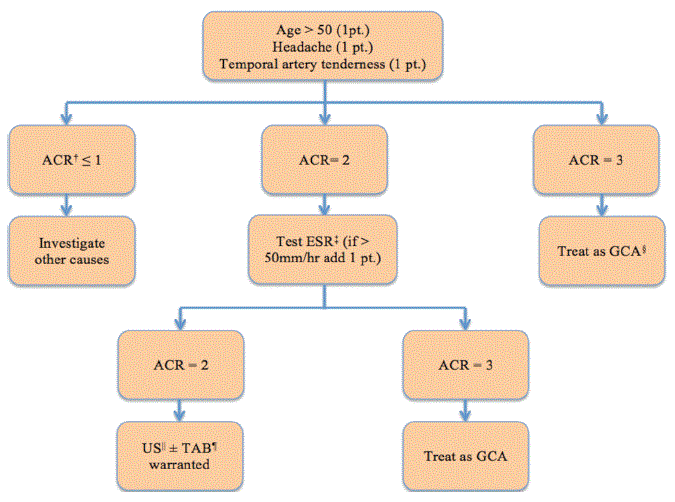
Figure 1: Revised American College of Rheumatology algorithm [15].

Ben McKernan* Tina Dilevska Genevieve Gibbons Thomas Bowles
Department of General Surgery, Albany Regional Hospital, WA Country Health Service, Australia*Corresponding author: Ben McKernan, Department of General Surgery, Albany Regional Hospital, WA Country Health Service, Warden Ave, Spencer Park WA 6330, Australia, Tel: +61 0898922222; E-mail: Benjamin.McKernan@health.wa.gov.au
Background: Giant Cell Arteritis (GCA) is a common chronic idiopathic vasculitis of medium to large vessels that can cause permanent visual loss and stroke. Temporal Artery Biopsy (TAB) is the gold standard diagnostic test with specificity up to 100%, but a poor sensitivity, which can range between 39-77%. TAB evaluates only a small section of a vessel in a systemic disease. Recent literature suggests that diagnostic Ultrasound (US) provides a cost-effective and safer alternative in the diagnosis of GCA (sensitivity 54-92%, specificity 83-96%). This study aims to review the impact of TAB in our regional center and emergency theatre utilization.
Method: A retrospective audit of all patients who underwent TAB at Albany Hospital between January 2009 and June 2019. Medical records were reviewed for histopathology and demographic data. All TAB were performed in theatre under local anaesthetic and 45% were performed by the trainee registrar.
Results: 62 TAB were performed on 61 patients. 70% of patients were female with a mean age of 69.8 years. One patient’s histopathology results were unattainable. 11 patients (18%) had positive histopathology that confirmed GCA. One of these patients had findings of treated arteritis. 49 out of 60 patients (81.7%) had negative histopathology findings.
Conclusion: The positive biopsy rate is slightly lower than most contemporary cohorts. There may be a role for training in vascular US in our regional center, which will not only save in theatre costs but also expedite patient diagnosis and treatment of GCA.
Temporal arteries; Giant cell arteritis; Ultrasonography; Cost-benefit analysis
Giant Cell Arteritis (GCA) is a chronic, idiopathic, granulomatous vasculitis of medium to large vessels [1]. It almost exclusively affects people over the age of 50 years, with a mean age of 72 years. The overall lifetime risk is 1% for females and 0.5% for males [1,2]. The aetiology of GCA is unknown, it has been proposed to be genetic, autoimmune or associated with an acute trigger (such as infection with varicella zoster) and it is characterised by local arterial inflammation, with thickening of the intima and media of the affected vessels [3- 5]. Patients with GCA have been shown to have higher rates of hospitalization compared to the general population [6].This is partly due to the increased risk of serious vascular and visual complications [6]. Early treatment with high dose corticosteroids is currently recommended in order to reduce the risk of visual loss [7]. However, this treatment regimen has been associated with adverse events in over 80% of patients, including cataract osteoporotic fractures, infections, hypertension and diabetes mellitus [8,9]. Therefore, a prompt diagnosis of GCA is important because if not recognized and treated early, ischaemic complications may result in permanent vision loss in 15%-25% of cases [10]. Conversely, misdiagnosis puts patients at unnecessary risk of sequaelae associated with high-dose corticosteroids.
In 1990 the American College of Rheumatology (ACR) developed criteria to distinguish GCA from other forms of vasculitis. The presence of three or more of the following features establishes a positive case: age 50 years or older, new-onset localized headache or head pain, temporal artery tenderness to palpation or decreased pulsation, ESR of at least 50 mm/h or a positive Temporal Artery Biopsy (TAB) result. These criteria have been shown to have a sensitivity of 93% and a specificity of 91% [11].
Despite the fact that the performance of the ACR criteria is predicated on a patient already having a diagnosis of vasculitis, it has frequently been used as a de facto initial diagnostic index and rationale for proceeding to TAB. Furthermore, whilst a positive TAB is not necessarily required to confirm GCA within this algorithm, it has a reputation for being the gold standard in diagnosis due to a specificity of up to 100%. A wide range of sensitivities for TAB in the diagnosis of GCA have been demonstrated in the literature [12]. Articles dating back to the 1980s advised sensitivities as low as 15% [13-15]. A 2019 meta-analysis of pooled TAB-positive GCA cases across 32 studies demonstrated a sensitivity of 77% [16]. A recent diagnostic accuracy and cost-effectiveness study quoted the lower-end of sensitivity as 39%, which would give a false negative result in as many as 61% of cases [17]. There are many reasons for this, including adequacy of the biopsy and the fact that it only evaluates a small section of a vessel in a systemic disease [17,18]. The sensitivity of TAB declines the longer treatment with corticosteroids has been given prior to biopsy [19]. Furthermore, TAB is an expensive and invasive procedure; it has a complication rate of 0.5% and carries serious risks such as facial nerve injury or stroke [20].
Innovation and enhancements in diagnostic techniques since the creation of the 1990 ACR criteria have led to better distinction between types of vasculitis, affecting the utility of established diagnostic means in identifying GCA [21].
Recent literature since the publication of the ACR criteria suggests that diagnostic ultrasound (US) could provide a more cost-effective and safer alternative to TAB in diagnosis of GCA, with a sensitivity that ranges between 54-92% and a specificity between 83-96% [12,17,18,20,22,23]. A current meta-analysis, which informed the 2018 European League against Rheumatism (EULAR) recommendations for imaging in large vessel vasculitis, demonstrated a sensitivity of 77% [23].As the prevalence of GCA in the general population is 0.25% [24] the false negative rate with US may be as low as 8%. The most specific finding on US is the ‘halo sign’, a dark halo around the vessel wall caused by inflammation. Additional consideration of stenosis and occlusions in the imaged vessels further increases the sensitivity of the test [25].
There is now enough evidence to indicate that TAB does not change the management of GCA even within the set ACR criteria [15,26]. US shows very promising utility in reducing the rate of unnecessary surgical procedures [27]. Various modified ACR algorithms integrating the use of US have been proposed to help improve the yield rates of TAB [15,28,29]. US is inexpensive, non-invasive, quicker and more widely available than TAB [27]. The recent UK cost-effectiveness study by Luqmani and colleagues compared TAB with US as diagnostic strategies. The calculated cost savings arising at the point of testing through use of ultrasound instead of biopsy (both alongside clinical judgement) was £456. This represented the difference between £514 for a biopsy and £58 for ultrasound per case [17]. According to the Australian Medicare Benefits Schedule (MBS), the fee for TAB in Australia is $343.75 [30]. Furthermore, the average direct cost of a ‘productive hour’ of theatre time may be estimated at $2004 [31]. The MBS fee for US of extra cranial vessels is $84.75 [30]. Thus the potential cost savings arising at the point of testing through use of ultrasound instead of biopsy in the Australian healthcare setting may be as high as $2263 per case.
The aim of this study was to establish the number of potential false negatives in the cohort of patients who underwent TAB in our center over a 10-year time frame and to provide a projected estimate of cost savings if US was used instead of TAB in this cohort.
In patients who have undergone temporal artery biopsy for diagnostic confirmation of GCA at our regional center:
Potential cost-savings were calculated from data provided by the Medicare Benefits Schedule and the Auditor-General’s Report on Victorian Public Hospital Operating Theatre Efficiency [30,31].
The rate of potential false negatives from histology data obtained by TAB should be less than 23-46%, which would be the projected false negative rate if US were utilized.
The lower-end of sensitivities were calculated from findings provided by a recent large multicenter study, which also contributed to much of the health economic analysis discussion [17].The upper-end of sensitivities were calculated from two recent meta-analyses [16,23]. The subsequent false negative rate was then derived by combining these figures with data from an epidemiological study of biopsyproven GCA patients [24].
Adult patients who had undergone TAB at our regional hospital between January 2009 and June 2019.
A list of patients over 18 years of age who had undergone temporal artery biopsy between January 2009 and June 2019 was requested from the Theatre Management System Manager.
All cases of temporal artery biopsy between January 2009 and June 2019 were provided from the Theatre Management System Manager. Medical records were then consulted for histopathology findings and demographic data.
This audit was approved by Standard 1 Governance on the 23rd of July 2019.
A total of 62 TAB were performed on 61 patients at this regional hospital between January 2009 and July 2019. One patient had a repeat TAB as the first was inconclusive. One patient’s histopathology results were unattainable; this case was excluded from the audit.
The average age of patients undergoing TAB was 69.8 years. 70% of patients were female.
11 patients (18%) had positive histopathology that confirmed GCA. One of these patients had findings of treated arteritis.
49 out of 60 patients (81.7%) had negative histopathology findings.
According to current literature, TAB has a reported sensitivity of between 39-77%. With a prevalence of 0.25% it therefore provides a false negative result in 23-61% of cases [16,17,24]. Thus between 11 and 30 cases represent potential false negatives.
If US had been utilized (false negative rate of 23-46%) [17,23,24] the number of potential false negatives would be between 11 and 22.
According to the MBS, the fee for TAB is $343.75 [30].Furthermore, the average direct cost of a ‘productive hour’ of theatre time may be estimated at $2004 [31].The MBS fee for US of extracranial vessels is $84.75 [30]. Between January 2009 and June 2019 a total of sixty-two TAB were performed. This represents a potential cost of $145,560.50. Use of ultrasound instead of biopsy in these cases represents potential savings of $140,306.
Between 11and 30 cases represent potential false negatives. For comparison, if US had been utilized (false negative rate of 23-46%) the number of potential false negatives would be between 11 and 22.
One of the most feared complications of untreated GCA is permanent vision loss as a result of ischaemia. This has been shown to occur in 15%-25% of cases [10]. Anterior ischemic optic neuropathy is the primary mechanism of vision loss [32]. The injury to sight is severe, and the prognosis following this is poor [33]. In our population of TAB potential false negatives, this represents between two and eight patients who would have been affected by this complication.
Inter-observer and intra-observer reliability is a commonly raised issue when considering the diagnostic performance and reliability of ultrasound across many conditions; GCA is no exception. Inter-observer and intra-observer agreement has been shown to be excellent in studies that have exclusively evaluated US in the diagnosis of GCA, particularly if a standardized training program is used for sonographers. In these studies, κ values were either >0.8 (implying virtually complete agreement) or disagreement occurred in only one out of every sixty cases [34-38]. Even the intra-class reliability of sonographers compared with pathologists reading TAB specimens has been shown to be equally consistent by Luqmani and colleagues: intra-class correlation coefficients were 0.61 and 0.62, respectively [17].
Two of the main benefits of US over TAB are time and cost. Not only can it take two weeks or more to receive the results of a biopsy, but a recent study has also shown the cost per case to be nearly ninetimes as much [17].
In our regional center over the ten-year audit timeframe, use of ultrasound instead of biopsy represents potential savings of $140,306. In Australia, the estimated population incidence of GCA for people over 50 is 3.2 per 100,000 person-years [39]. Thus in the Australian population of people aged over 50 (about 7.9 million) [40] over the same ten-year timeframe, it may be estimated that there were at least 2528 cases of GCA. If each TAB requires $2004 of theatre time and an MBS fee of $343.75, this represents a cost of over $5.9 million. Use of ultrasound instead of biopsy in all these cases represents potential savings to the Australian healthcare system of over $5.7 million.
New rheumatologic classification criteria for GCA are likely to include US imaging in addition to TAB [21]. The EULAR have recommended US at the first diagnostic test in their 2018 guidelines, given adequate expertise and equipment in the diagnosis of GCA [41]. Previous studies have cited the use of algorithms and combining the use of US into the pre-existing ACR algorithm alongside TAB to help improve its utility. Cristaudoand colleagues have proposed such a revision, shown in figure 1 [15].
Figure 1: Revised American College of Rheumatology algorithm [15].
Not only does US outperform TAB as a screening test for GCA, it is a far more cost-effective alternative. In centers with qualified sonographers, clinical examination and US will clearly exclude GCA in most patients. It provides the highest resolution of all imaging techniques and is especially suited to the imaging of small vessels such as temporal arteries. The most recently proposed revision of the ACR algorithm by Cristaudo and colleagues (Figure 1) is practical and easy to follow. It would be reasonable to incorporate the use of this algorithm into current practice.
The data collected was of relatively low resolution. Rather than the clinical diagnosis of GCA guiding whether or not a patient had GCA, the TAB result was used instead. The data does not make a case in its own right about the accuracy of TAB. The real sensitivity might have been more accurately estimated after some months of follow-up post-TAB. Furthermore, additional details about the patient cohort and results were not included. A more complete picture of the cohort might be attained by including data such as the number of transmural vs. adventitial vasculitis and the number of giant cells seen, for example.
There was no local US data to show that in our rural institution, US is reliable. The projected savings from US are an approximation only.
The authors are not recipients of a research scholarship. There are no potential or real conflicts of interest. This paper has been verbally presented by one of the authors at the Provincial Surgeons of Australia 2019 Annual Scientific Conference, on the 3rd of November 2019 in Ballarat, Victoria [42]. Minor changes have been made since this presentation.
Download Provisional PDF Here
Article Type: RESEARCH ARTICLE
Citation: McKernan B, Dilevska T, Gibbons G, Bowles T (2020) An Audit of Utility and Cost of Temporal Artery Biopsy in a Regional Centre. J Surg Open Access 6(4): dx.doi.org/10.16966/2470-0991.216
Copyright: © 2020 McKernan B, et al. This is an open-access article distributed under the terms of the Creative Commons Attribution License, which permits unrestricted use, distribution, and reproduction in any medium, provided the original author and source are credited.
Publication history:
All Sci Forschen Journals are Open Access