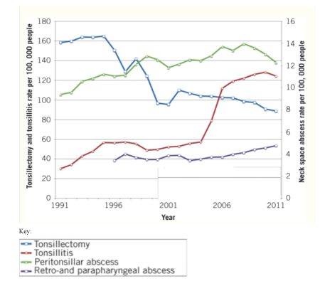
Graph 1: Overall trends in England for tonsillectomy (left vertical axis) against admissions for neck space abscesses (right vertical axis).

Anmolsingh R1 Ali A2 Edmiston R1 Rocke J1 Ranganathan B1 Kumar BN1*
1Department of ENT, Wigan and Leigh NHS Foundation Trust, UK*Corresponding author: Kumar BN, Department of ENT, Wigan and Leigh NHS Foundation Trust, Wigan, UK, E-mail: nirmalkumar@doctors.org.uk
Tonsillectomy and Adenoidectomy are common operative procedures in field of Ear, Nose and Throat (ENT) Surgery. Over 17000 tonsillectomy procedures were undertaken in England in the year 2014-2015 and the number for children is likely to be much higher [1]. Tonsillectomy may be performed for recurrent tonsillitis, histological diagnosis or for the management of snoring. Adenoidectomy is rarely performed in isolation but instead employed in combination with other interventions. Adenoidectomy and tonsillectomy (Adenotonsillectomy) is indicated for the paediatric population to manage obstructive sleep apnoea. Adenoidectomy may also be considered in the management of recurrent acute otitis media or persistent glue ear in conjunction with grommet insertion. Tonsillectomy and adenoidectomy are considered “basic” ENT procedures and are often undertaken by junior training grades as a result. These operations allow the development of surgical skills such as knot tying, instrument handling and electrocautery which are integral to the development of the ENT surgeon. This article will act as a resource not only for those undertaking adenotonsillar surgery, perhaps for the first time, but also other clinicians to allow full understanding of the indications for surgery in addition to potential complications and their management.
Tonsillar and adenoidal tissues are a part of the Waldeyer’s ring; a body of lymphoid tissue found in the pharynx. The palatine tonsils are dense compact bodies of lymphoid tissue that are located in the lateral wall of the oropharynx, bounded by the palatoglossus muscle anteriorly, the palatopharyngeus and superior constrictor muscles lie posteriorly and laterally. The adenoid is a median mass of Mucosa Associated Lymphoid Tissue (MALT), which is situated in the roof and posterior wall of the nasopharynx [2,3].
Tonsils and adenoids are immunocompetent tissues which form part of the immune system’s primary defence against ingested or inhaled foreign pathogens. They are involved in both humoral and cell mediated immunity. Removal of tonsils and adenoids may have some theoretical effect on immune function but there is little evidence to suggest any significant long term detriment following adenotonsillectomy [4,5].
Tonsillectomy is commonly performed as treatment for severe recurrent tonsillitis. Since the introduction of the Scottish Intercollegiate Guidelines Network (SIGN) 2010 [6], and the restriction in funding for surgical procedures thought to be of little clinical value [7], there has been a reduction in the number of tonsillectomies performed. However, during this time adenotonsillectomy for the management of obstructive sleep apnoea in the paediatric population has increased in prevalence.
SIGN indications for tonsillectomy suggest the following criteria for considering tonsillectomy [6]:
A repercussion of the decrease number of tonsillectomies performed has been an increasing number of admissions for tonsillitis and peri-tonsillar abscess. Millington AJ, et al. [8] in a period from 2003 to 2012, showed an increase of tonsillitis admissions locally by 118% in adults and 179% in children.
Graph 1 adapted Lau AS, et al. [9] demonstrating rates of admission increasing as rate of tonsillectomy decreases, while this does not demonstrate a direct association and there could be confounding factors involved it certainly is an interesting observation.
Graph 1: Overall trends in England for tonsillectomy (left vertical axis) against admissions for neck space abscesses (right vertical axis).
Obstructive Sleep Apnoea (OSA) is a disorder of breathing during sleep characterised by prolonged partial upper airway obstruction and/ or intermittent complete obstruction that disrupts normal ventilation during sleep and normal sleep pattern. Upper airway obstruction may result in distinct apnoeic events, hypopnoea, or increased work of breathing seen in upper airway resistance syndrome, all of which come under the umbrella of Sleep Related Breathing Disorders (SRBD) [10]. In children, hypertrophy of the tonsils and adenoid tissue is thought to be the commonest cause of obstructive sleep apnoea. The commonest indication for adenoidectomy and tonsillectomy in children is SRDB followed by recurrent infections [11].
Most children who develop peri-operative problems following adenotonsillectomy for have conditions that are apparent preoperatively. They can be easily identified as an ‘at risk’ group on clinical assessment in the outpatient clinic (e.g. underlying syndrome, severe co-existent condition, extremes of weight). The ‘normal child with severe OSA’ is also at risk of peri-operative problems but is much more difficult to identify. Overnight oximetry will identify a child with repeated falls in saturation below 80% [12].
‘At-risk’ children require treatment in a specialist centre and should be referred for further investigation and surgical management. Mirza et al. demonstrated the need to risk stratify paediatric OSA into mild, moderate and severe cases at a local District General Hospital (DGH). They concluded that >10% of patients were classified as severe at the DGH and required further intervention at a tertiary centre [10]. The suggested proforma used for this stratification was adapted from Robb PJ, et al. [13] (Figure 1).

Figure 1: Risk stratification criteria adapted from Robb PJ, et al. [13]
Children requiring surgery (usually tonsillectomy ± adenoidectomy) for symptoms suggestive of mild OSA can generally be managed safely in a DGH setting. If these children are managed as day case admissions, this should be on a morning operating list, where the anaesthesia and surgery are delivered or appropriately supervised by consultants with expertise in managing these operations. The children should be managed in a children’s day surgery ward or unit and operated upon during dedicated paediatric operating lists, where all staff have the requisite training and experience.
A proportion of children with Sleep related breathing disorders (SRBD) develop respiratory complications in the peri-operative period, severe enough to require an increased level of post-operative care (upto and including postoperative ventilation) not routinely provided in UK general hospitals. Children with severe OSA or significant co-morbidity must be referred for preoperative paediatric respiratory assessment since these children will usually require surgical management in a paediatric centre with direct and immediate access to PHDU and PICU.
Other indications for tonsillectomy may include histological analysis, chronic tonsillitis, for snoring, and recurrent peritonsillar abscess (quinsy). ‘Hot’ Tonsillectomy, during an active infective episode, is infrequently performed but is an acceptable practice to interval tonsillectomy. For example, in patients with an acute peritonsillar abscess which may not be amenable to aspiration, incision and drainage, or where there is re-accumulation following an initial successful attempt to incise and drain.
Adenoidectomy in the paediatric population is indicated for conditions including nasal obstruction, recurrent Otitis Media with Effusion (OME) and obstructive sleep apnoea. Depending on the indication, it may be combined with tonsillectomy and grommet insertion. NICE guidance states that Adenoidectomy should only be undertaken in patients with OME if persistent or frequent upper respiratory tract symptoms co-exist [14]. Rosenfield, et al. [15] recommend that adenoidectomy should not be performed in children with OME of less than 4 years of age unless they have a separate indication such as nasal obstruction or chronic adenoiditis.
Tonsillectomy is performed under general anaesthesia and can commonly be performed as a day case procedure both in adults and children. Cold steel dissection is the most commonly performed surgical technique, where extracapsular dissection is performed to remove the tonsil from the underlying muscles. Any subsequent bleeding is controlled by ties and/or bipolar diathermy. Surgery leaves exposed pharyngeal muscle within the tonsillar fossa which is allowed to heal by secondary intention over a period of 5-10 days.
Although the technique is the same, tonsillectomy in the adult population may be inherently more challenging, due to the degree of fibrosis and scarring between the tonsil and pharyngeal muscles, thought to be as a result of a greater number of recurrent infections resulting in a loss of the pseudocapsule.
Various advances in surgical technique have been developed with the aim of reducing intra-operative bleeding and subsequent postoperative morbidity. Many tonsillectomy techniques have been adopted and can include use of cold-steel dissection, guillotine excision, bipolar scissors, monopolar diathermy, harmonic scalpel and recently introduced coblation technique. Importantly, while diathermy is often used for both tonsillar dissection and haemostasis, its use may be reserved for precise haemostasis after a traditional cold steel dissection, in order to decrease thermal damage to surrounding tissues, which is thought to contribute to post-operative pain.
Diathermy techniques were first introduced around 40 years ago. In the last two decades, the use of these ‘hot’ techniques has increased dramatically in routine practice. The ability to dissect and control intra-operative bleeding with the same instrument is a likely important factor contributing towards their popularity despite suggestions that secondary haemorrhage rates were increased with its use. Diathermy allows blood loss during tonsillectomy to be minimised, which is a particularly important consideration in young children. The technique tends to be favoured by trainee surgeons because it is quicker to learn and requires less dexterity than cold steel dissection and ligation of vessels.
Coblation is a commonly used technology which combines lowtemperature radiofrequency energy with saline to create a plasma field. This plasma field is contained at the tip of the device and dissolves tissues at a molecular level, resulting in a precise dissection of tissues. The temperature generated in contact with tissues is generally 40-70 degrees Celsius as compared to electrocautery which generates temperatures of 400-600 degrees Celsius. Harmonic scalpel technology uses ultrasonic vibration at a frequency of 55,500 Hz, instead of electric current, to generate stress and friction in tissue producing heat and causing protein degradation. The concept for using coblation or harmonic scalpel technology over standard bipolar technology is that it minimizes thermal damage to the surrounding tissues which may result in less post-operative pain morbidity, encourage earlier return to normal function, such as eating and drinking, and enhance recovery. A Cochrane review demonstrated a slightly increased bleeding 24 hours after surgery but evidence was of a low quality [16]. A study by Walker P, et al. [17] demonstrated no difference in post operative bleeding rates between diathermy and the cold steel technique.
The traditional technique for ‘removal’ of adenoid tissue is via curettage, where a sharp metallic curette is inserted into the nasopharynx transorally and the adenoid tissue is blindly scraped from the posterior nasopharyngeal wall. Adenoidectomy can be more accurately described as a debulking procedure, where there is often some adenoid tissue left behind. Other alternatives do exist such as suction diathermy, coblation, microdebrider or laser. Haemostasis is generally achieved by packing of the nasopharynx for a few minutes before extubation.
There are numerous potential complications of adenotonsillar surgery which must be thoroughly discussed when gaining consent pre-operatively. The alternative of not performing surgery and continuing conservative treatment is also mentioned.
The National Tonsillectomy Audit (2003-2004) included 277 hospitals with a total of over 40,000 operations. This high-powered study showed an overall haemorrhage rate of 3.5% (Primary 0.5%, Secondary 3%, 0.1% had both) with an increased incidence using “hot” techniques [18] (Table 1).
| Technique | Postoperative Haemorrhage (%) |
Return to Operating Theatre (%) |
| Cold Steel Dissection with ties and packs | 1.3 | 1.0 |
| Cold steel dissection with diathermy haemostasis | 2.9 | 1.7 |
| Bipolar scissors | 3.9 | 2.4 |
| Monopolar Diathermy | 6.1 | 4.0 |
Table 1: Various Tonsillectomy Techniques and Hemorrhagic rate (adapted from National Prospective Tonsillectomy Audit 2005).
Primary haemorrhage: Primary haemorrhage is defined as a bleed within the first 24 hours following surgery. The usual cause of primary haemorrhage is usually secondary to opening up of a vessel or slipped ligature. Immediately postoperatively, excessive coughing upon awakening from general anaesthetic may increase the risk of bleeding due to increased intravascular pressures. Co-morbidities which may predispose a patient to increased intraoperative/ postoperative bleeding include bleeding diatheses (e.g. sickle cell disease, Von Willebrands disease) and difficult anatomy (e.g. Down’s syndrome, subglottic stenosis, Pierre Robbins Syndrome) which may make surgical access and control of bleeding more difficult. In such cases, preoperative optimisation is necessary to decrease the risk of unfavourable complications.
In adenoidectomy bleeding can be prevented by using a cautious diathermy and packing of the post nasal space with or without adrenaline intraoperatively. The usual causes are of primary bleeding are overzealous curetting of the adenoidal tissue and inadequate removal of adenoidal tissue. If bleeding cannot be controlled the post nasal space may need to be packed for 24 hours while the patient remains intubated on an intensive care environment.
A potentially life-threatening complication of adenotonsillar surgery is referred to as the “coroner’s clot”. A clot in the nasopharynx can dislodge and cause airway obstruction in recovery. It is easily prevented by ensuring adequate suctioning of the post nasal space intra-operatively usually with a suction catheter via the nasal cavity. This is also important when clearing the blood from the upper aero-digestive tract prior to extubation after arrest of secondary haemorrhage.
Secondary haemorrhage: Secondary post-tonsillectomy haemorrhage is defined as a bleed after 24 hours and usually tends not to occur beyond 2 weeks following surgery. The majority of these bleeds are self-limiting and usually do not require operative intervention. Patients presenting with secondary post-tonsillectomy haemorrhage are often admitted for observation, as a small selflimiting bleed may herald a larger more significant bleed. Patients with recurrent tonsillitis, ADHD or an older age are at increased risk [19]. A small minority of patients (1.9% suggested by the national prospective tonsillectomy audit 2005) may require return to theatre for haemostasis [18]. Infection is thought to be the cause of secondary haemorrhage, although patients rarely have any evidence of gross infection, systemic sepsis and inflammatory markers are usually not elevated beyond what would be expected in a patient who has had surgery. Antibiotics are commonly commenced prophylactically despite there being little clinical signs supporting their use, or evidence in the literature supporting this practice [20]. Other potential causes which have been suggested include sloughing of eschar, trauma secondary to ingested food or the use of Nonsteroidal Anti-Inflammatory Medications (NSAIDs) but Mudd PA, et al. [21] has recently disproved this. There remains debate and no consensus opinion has been reached nationally or internationally regarding the precipitants of secondary bleeding.
Post-operative bleeding is usually confirmed when blood is expectorated. The assessment of a child presenting with posttonsillectomy bleeding may be challenging, as they have a tendency to swallow blood, and may be difficult to assess due to both distress and non-compliance to examination. Other parameters such as an index of suspicion, excessive swallowing or haemodynamic instability may be helpful in making an objective assessment which may allude to underlying bleeding requiring intervention.
The immediate and initial management of the patient can vary depending on the severity of the bleeding and clinical condition of the patient. However, basic resuscitation principles form the foundation of management of the acutely unwell patient in the first instance, with the additional implementation of measures to control bleeding. The following algorithm (Figure 2) delineates the management of a posttonsillectomy bleeding. This may be applied to adults and the paediatric patient however in a child there are limits as to what invasive measures can be performed to control bleeding. If there is active bleeding that does not settle, or any haemodynamic compromise or reduced reserve, return to theatre should be considered earlier.
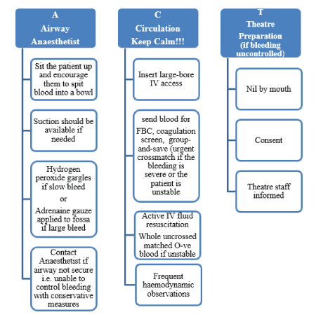
Figure 2: Emergency management of post Tonsillectomy bleed.
If the situation necessitates surgical intervention the following algorithm can be applied to achieve intraoperative haemostasis in the anaesthetised patient. (Figure 3)
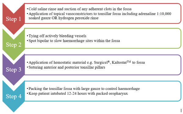
Figure 3: Stepwise approach to the intra-operative management of post tonsillectomy bleeding.
Before extubation, gastric contents are emptied using a Ryle’s tube and suction aspiration. There is an emetic effect associated with swallowed blood, and a patient who may vomit in the post-operative period may cause clinical uncertainty as it may suggest a further posttonsillectomy bleed.
Secondary haemorrhage rates for adenoidectomy range from 0.38 to 0.7% [22-23]. These bleeds can often be more challenging to manage than post-tonsillectomy bleeds and similarly possess life-threatening potential.
Pain does tend to be the most common post-operative source of morbidity following tonsillectomy. This has an impact on a patient’s oral intake and return to normal function. There have been numerous randomised controlled trials investigating post-operative analgesics, but there remains a debate regarding the optimal analgesia regime. Patients are generally counselled that post-operative pain may continue to increase until 7 days following surgery, and may persist up to 14 days. Clinicians in the UK adhere to the World Health Organization (WHO) analgesic ladder as a reference for prescribing analgesia. There has been a move away from the use of weak opiates, such as codeines, in the paediatric population which was demonstrated by a National Tonsillectomy Audit [24]. This was due to the Medicines and Healthcare Products Regulatory Agency (MHRA) altering codeine’s licence code following reports of deaths in children who had received this analgesic following tonsillectomy. Historically NSAIDs were avoided but no demonstrable relationship with their use and increase risk of post-operative haemorrhage has been shown in the literature [24].
Numerous adjuncts have been trialled to reduce pain post operatively in tonsillectomy including benzocaine lozenges and infiltration of the tonsillar fossa with local anaesthetic but studies invariably demonstrate no significant improvement [25,26].
Due to the trans-oral route taken in adenotonsillary surgery the structures of the oral cavity and pharynx are at risk of iatrogenic injury. Great care during instrumentation, particularly diathermy, and the use of a gum shield to protect the patient’s teeth are advised. The surgeon must also be aware of the possibility of Temporomandibular Joint (TMJ) dislocation after removal of the gag which makes it prudent to assess this before concluding the procedure. Manual reduction of this dislocation can be performed by pressing the mandible downwards, posteriorly and then finally upwards [27].
An extremely rare but theoretically complication is the occurrence of airway fires from the bipolar machine. It remains the surgeon’s responsibility to ensure there are protective measures and familiarity with bipolar settings. Management centres around removing the source of oxygen, extinguishing the fire (saline), assessing injuries and securing the airway. Often these patients will require management post operatively on Intensive Care primarily to assess associated lung injury [28].
Atlanto-axial subluxation is a rare complication but can occur when hyper-extending the neck or during forceful curetting. There is a higher risk with Down’s syndrome patients but pre-operative radiological investigation is not indicated in this sub-group. The condition is suspected if the patient presents with persistent neck pain and stiffness post operatively. C-Spine x-ray and Computed Tomography (CT) of the c-spine are used to confirm diagnosis. Early management includes cervical stabilisation and immobilisation as well as analgesia.
Early detection is the key to managing this complication since the long-term effect includes permanent and painful neck deformity which may require spinal fusion.
This complication can occur because of an increase in the size of the resonance chamber and scarring of the soft palate. It is usually most noticeable to vocal performing artists and as such this sub-group of patients should be counselled appropriately [29]. That said numerous studies have demonstrated that voice is not impacted by adenoidectomy or tonsillectomy and in some cases improves its quality [30,31].
Velopharyngeal Insufficiency (VPI) is improper closure of the velopharyngeal sphincter during speech. This can lead to hypernasal speech and in rare cases nasal regurgitation of food. Patients who already have palatal defects or previous palatal repairs are at an increased risk of VPI. The incidence is in the region of 1:1200 and 1:3000 following adenotonsillectomy and reduces to 1:10000 following adenoidectomy in isolation. Usually VPI resolves within 5 months of surgery and no active management other than referral to an experienced speech therapist is required. Those who do not improve may require surgical intervention [32].
Adenotonsillar surgery is a common surgical procedure in the UK with wide ranging indications and potential benefits for patients. We have surmised the various guidelines and recommendations for both tonsillectomy and adenoidectomy to provide one accessible resource for those undertaking these interventions.
Despite its comparatively short procedural time it carries numerous associated complications. A proper understanding of these risks is required for informed consent pre-operatively and proper operative technique intraoperatively. The surgeon must also have a good understanding of the presentation and management of these complications should they arise in the post-operative period.
Download Provisional PDF Here
Article Type: SHORT COMMUNICATION
Citation: Anmolsingh R, Ali A, Edmiston R, Rocke J, Ranganathan B, et al. (2018) Tonsillectomy and Adenoidectomy: Indications, Complications and their Management. J Surg Open Access 4(4): dx.doi.org/10.16966/2470-0991.173
Copyright: © 2018 Anmolsingh R, et al. This is an open-access article distributed under the terms of the Creative Commons Attribution License, which permits unrestricted use, distribution, and reproduction in any medium, provided the original author and source are credited.
Publication history:
All Sci Forschen Journals are Open Access