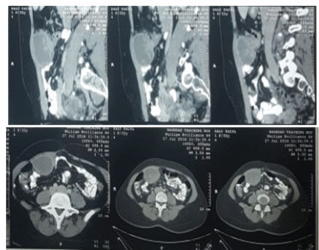
Figure 1: Abdominal CT scan shows mass of endometriosis.

Nawfal S Al-Khayat1 Ali E Joda2*
1Consultant general and pediatric surgeon, former head of pediatric surgery department at central child teaching hospital, Baghdad, Iraq*Corresponding author: Ali E Joda, Pediatric surgeon at central child teaching hospital, senior lecturer at Mustansiriayh medical college, Baghdad, Iraq, E-mail: ali.egab.joda@gmail.com
Background: The ectopic growth of functional endometrial tissue outside the uterine cavity is called endometriosis. It could be in pelvic or extra-pelvic locations. Abdominal wall endometriosis is rare and mostly reported following various surgical procedures mostly cesarean section or pelvic surgery. A few case reports have described abdominal wall endometriosis following laparoscopy at trocar port sites. We report a new case of abdominal wall endometriosis with review of the literatures.
Case report: This is a report of abdominal wall endometriosis at the laparoscopic port site in a 34-year-old woman who had previous diagnostic laparoscopy 4 years ago for gynecological problem when she presented with recurrent attacks of mild abdominal pain and discomfort for 4 months duration. Operative intervention was decided depending on ultrasound and abdominal CT scan findings, we performed a complete surgical resection of the lesion with a free macroscopic margin.
Conclusion: Due to a wide range of mimicking conditions and a relative rarity, a significant delay is often observed from the onset of symptoms to proper diagnosis. Although rare, if a painful mass in the surgical scar, such as the trocar site, is found in women of reproductive age with a history of pelvic or obstetric surgery, the physician should consider endometriosis.
Endometriosis; Abdominal wall; Trocar site; Laparoscopy
Endometriosis, classically defined as ectopic segmental growth of functioning endometrial tissue in extra-uterine location causing infertility, chronic pelvic pain, menstrual abnormalities and dyspareunia [1]. It can be classified as intra-pelvic or extra-pelvic. The most common locations of intra-pelvic endometriosis are the gynecologic organs like ovaries, fallopian tubes, uterine ligaments, rectovaginal septum and pelvic peritoneum. The extra-pelvic implantation of endometrial tissue frequently involves the alimentary tract with greater omentum, and surgical scars, while it is seldom found in remote sites such as the kidney, lung, skin, and nasal cavity [2]. The most common extra-pelvic localization of endometriosis is abdominal organs [3]. It is well known that endometriosis may migrate and implant in various anatomic locations including surgical scars [4]. So endometriosis of abdominal wall indicates the embedding of ectopic endometrial tissue in the layers of abdominal wall following various surgical procedures mostly cesarean section or pelvic surgery [5,6]. The incidence of scar endometriosis is estimated to be 0.03-1.5% of all women with past surgical history of cesarean delivery [4,6]. With the current era of increased use of laparoscopy, a few case reports have described development of abdominal wall endometriosis at port sites [7-10]. To our knowledge, only 19 case reports have been reported in the literature since the first reported case in 1990 [11]. Its clinical diagnosis is often confused with abscess, lipoma, hematoma, sebaceous cyst, suture granuloma, hernia, lymphoma, etc, [12]. Here in we report an additional case of extra-pelvic endometriosis on the trocar port site of previous diagnostic laparoscopy for infertility, with additional review of the literature about this often-misdiagnosed condition.
Case report
A 34-year-old woman presented with recurrent attacks of mild abdominal pain and discomfort for 4 months duration, she reported that her pain was relieved temporary by intake of non-steroidal anti-inflammatory drugs prescribed by physicians before she consulted me. There was no loss of weight, no vomiting, normal appetite, normal bowel motion and no significant urinary or gynecological problems. On examination, the abdomen was soft, not distended and there was a palpable mass about 7 cm in diameter felt at the right upper quadrant just above the umbilicus, it was mobile slightly transversely but fixed longitudinally with no other significant finding. Her past medical history was uneventful apart from one caesarian section done 6 years ago and one diagnostic laparoscopy 4 years ago for secondary infertility after her last pregnancy (which was negative and inconclusive because of inadequate visualization and abdominal adhesions). The routine biochemical tests, beta human chorionic gonadotropin (β-HCG) and carcinoma antigen (CA-125) were within normal ranges. Ultrasonography revealed a well-defined rounded mass located in the right upper abdomen just below the rectus abdominis muscle, it was non-homogenous, partly cystic with thin wall and internal echogenic content inside and show no vascularity in color Doppler ultrasound. Abdominal CT scan revealed localized tumefaction about 6 by 5 cm above the umbilicus and just to the right of the midline (Figure 1) so surgical treatment was recommended. Operative intervention was done through upper right Para median incision just near the mass, on exploring the peritoneal cavity, there was a dense mass attached to the posterior surface of anterior abdominal wall, attached to rectus muscle just under the scar of a previous laparoscopic port site, with adhesion to the omentum, small bowel and transverse colon. Sharp dissection and separation of adhesion done carefully without injury to small bowel or colon then complete excision of the mass with safety margin done and sent for histopathological examination (Figure 2), the abdomen closed in layers. Serum test for mycobacterial organism was negative. The postoperative period was uneventful and she was discharged the day after surgery. The result of histopathology was confirmative of endometriosis, it revealed hyperplastic fibrosis, with scattered foci of stromal tubular and cystic glandular endometrial tissues within connective tissue. So she referred to her gynecologist for further management and hormonal therapy. After one-year follow up, there was no recurrence of endometriosis.
Figure 1: Abdominal CT scan shows mass of endometriosis.
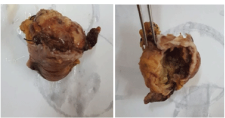
Figure 2: Completely resected endometriosis mass.
Discussion
The ectopic growth of endometrial tissue was first described by Rokitansky in 1861 and classified as internal or external according to the involvement of the myometrium, it can also be classified as pelvic or extra-pelvic according to its location [13- 15]. As previously stated, the extra-pelvic endometriosis refers to endometriotic implants in the alimentary tract, urinary tract, pulmonary structures, abdominal wall, skin, and even the central nervous system [2]. Localization of endometriosis in the abdominal wall is rare and often related to surgical scars. Following the current widespread use of laparoscopy by gynecologists, some cases of trocar site endometriosis have been reported in the literature, some of these patients already have pelvic endometriosis and a significant proportion of these patients discovered to have simultaneous presence of asymptomatic endometriosis during laparoscopy [13,16]. Cesarean section is the surgical procedure most frequently associated with abdominal wall endometriosis, with an incidence of approximately 0.03%-1% [15,17]. The condition also has been reported after episiotomy at the perineal site [18] or with other types of surgical scars, including the scars resulting from diagnostic laparoscopy, tubal ligation, hysterectomy, inguinal herniorrhaphy, laparotomy, and the needle tract of diagnostic amniocentesis in third trimester.
The question is how endometriosis implantation could occur four years after a diagnostic laparoscopy in a patient with no evidence of previous endometriosis as in our case? Or even in a patient without a uterus due to hysterectomy as reported by HoHyoung Lee et al.? [19]. The precise etiology of endometriosis remains controversial, and many theories have been proposed, including cellular immunity, coelomic metaplasia, implantation or retrograde menstruation, vascular and lymphatic metastasis, and dissemination [13,15,20] but Direct transplantation theory is probably the mechanism responsible for the development of a scar endometriosis following surgery [20]. The implantation theory is based on the usual presence of a retrograde menstrual reflux through the fallopian tubes in 80-90% of cases. Normally, refluxed endometrial tissue is cleared from the peritoneum by the immune system, and the dysregulation of this clearance mechanism has been implicated in the implantation and growth of endometrial cells [21]. The ectopic implant probably requires a combination of dysfunctions of immunologic defense mechanisms with a genetic component [22,23] Trocar port site endometriosis might develop as a result of peritoneal seeding of cells because of pneumoperitoneum or from direct contact of the excised lesion with the port tract. Several authors suggested that the practice of insufflating CO2 into the peritoneal cavity causes cell aerosolization and may encourage tumor cell shedding [9,13]. Murthy et al. and Hubens et al. suggested that a higher probability of tumor growth on the peritoneum is associated with gas laparoscopy as opposed to gasless laparoscopy procedures [24,25]. It has also been suggested that localized tissue ischemia renders that tissue conductive to implantation [26]. Another proposal is that cutaneous endometriosis may arise from endometrial tissue transported via lymphatic or vascular channels [27]. In the case of our patient, we suggest that microscopic endometriosis or asymptomatic undetected endometriosis, disseminated to the trocar site during the previous laparoscopic surgery. Thus, our case supports the aerosolization theory that pneumoperitoneum influences the implantation of free intraperitoneal endometriotic cells.
The first case of trocar site endometriosis was reported by Denton et al. in 1990 [11]. Endometriotic lesions exclusively confined at the trocar site are rare. During searches in the internet databases we collect 19 cases of trocar site endometriosis. Table 1 summarizes the data from the cases that have been described in the literature since the first report by Denton et al.
| Reference | year | age | complaints | Endometriosis | Previous | Interval | Location |
| history | operation | (m) | |||||
| Denton et al. [11] | 1990 | 37 | Pain, swelling | Yes | Lap sterilization | 12 | Near umbilical Trocar |
| Healy et al. [32] | 1995 | 23 | Cyclic painful Mass | No | Diagnostic Lap | 9 | Near umbilical Trocar |
| Wakefield et al. [31] | 1996 | 32 | Cyclic painful Mass | yes | Diagnostic Lap | 7 | Umbilical trocar |
| Martınez et al. [26] | 1998 | 35 | Groin masses | Yes | Diagnostic Lap | 11 | Suprapubic Trocar |
| Majeski et al. [29] | 2004 | 44 | Cyclic painful mass | No | Lap myomectomy | 48 | Umbilical trocar |
| Farace et al. [8] | 2005 | 37 | Nodular mass | yes | Lap cholecystectomy | 8 | Right trocar |
| Sirito et al. [28] | 2005 | 26 | Swelling | Yes | Lap cyst excision | 24 | Suprapubic Trocar |
| Barbaros et al. [9] | 2005 | 40 | Cyclic painful Mass | Yes | Lap cyst excision | 24 | Umbilical trocar |
| Strelec et al. [7] | 2009 | 24 | Cyclic painful Mass | Yes | Lap cyst excision | 24 | Suprapubic Trocar |
| Busard et al. [16] | 2010 | 37 | Cyclic pain | Yes | Diagnostic Lap | unknown | Right trocar |
| Akbulut et al. [13] | 2010 | 30 | Cyclic pain, Mass | No | Lap appendectomy | 8 | Right trocar |
| Akbulut et al. [13] | 2010 | 37 | Pain, mass | Yes | Lap cyst excision | 4 | Right trocar |
| Mederios et al. [10] | 2011 | 34 | Pain, swelling | Yes | Diagnostic Lap | 18 | Left trocar |
| Mederios et al. [10] | 2011 | 26 | Pain, swelling | Yes | Diagnostic Lap | 6 | Right trocar |
| Mederios et al. [10] | 2011 | 21 | Pain, swelling | Yes | Diagnostic Lap | 12 | Right trocar |
| Arif Emre et al. [34] | 2012 | 20 | Pain, swelling | Yes | Lap cyst Excision | 18 | Left trocar |
| Damir Eljuga et al. [41] | 2012 | 36 | Abdominal & inguinal pain | No | Diagnostic lap | 48 | Left trocar |
| Ho-Hyoung et al. [19] | 2012 | 42 | Mass, cyclic pain | No | Lap myomectomy | 8 | Suprapubic Trocar |
| Cozzolino et al. [35] | 2015 | 38 | Palpable nodule pain | Yes | Lap cyst excision | 12 | Right trocar |
| Our case | 2017 | 34 | Pain, swelling | No | Diagnostic lap | 48 | Right trocar |
Table 1: Cases of port site endometriosis as described in the reported literature.
General surgeons are usually encounter such patients with surgical scar endometriosis for suspicion of abdominal wall hematoma, suture granuloma and incisional or inguinal hernia. The diagnosis of scar endometriosis is highly suggestive in patients with the classic presentation of a palpable mass, cyclic pain, and a previous incision, especially of cesarean delivery or a gynecologic procedure that opened the uterine cavity or patient with history of treated pelvic endometriosis. Generally, symptoms of this rare condition are often nonspecific, but chronic pain is described as worsening during menses. However, in some series, almost 50% of patients reported noncyclic pain, not related to menstrual cycle [7, 28-36].
Endometriosis arising from an abdominal wall scar can be detected using ultrasonography USG, computed tomography CT, ultrasound guided fine-needle aspiration FNA, and magnetic resonance imaging MRI [13]. The sonographic appearance of endometriosis mass is polymorphic and can be described as cystic, polycystic, mixed, or solid with irregular borders, heterogeneous texture characterized by scattered internal hyper-echogenic foci, peripheral hyperechogenic ring and contain some scanty internal vascularity on Doppler examination according to the sonographic features described in the literature [37]. The CT and MRI features of abdominal wall endometriosis are often nonspecific and not pathognomonic for endometriosis because both showing just a solid enhancing mass and its appearance depends on the phase of the menstrual cycle, the proportion of stromal and glandular elements, the amount of bleeding, and the degree of inflammatory and fibrotic response [38]. In our case, we depend on ultrasound as it is simple, inexpensive, safe method and is considered sufficient to indicate the need for surgery as reported by the review of literature [35]. The major role of CT and MRI may be to determine the extension of the mass preoperatively. Although useful, CT, USG, and MRI cannot provide a definitive preoperative diagnosis while ultrasoundguided FNA is a fast and accurate diagnostic method for vague abdominal wall masses, where malignancy can be excluded and definitive diagnosis can to be determined [9]. The serum level of tumor marker Ca-125 is normal or slightly increased in such cases [37]; and our finding of negative serum level of Ca-125 is in agreement with the literature.
Really there is wide differential diagnosis of surgical scar endometriosis and is often confused with other pathologic conditions, such as a suture granuloma, abscess, inguinal or incisional hernia, soft-tissue sarcoma, desmoid tumor, lipoma, metastatic tumor, and sebaceous cyst. Therefore, the histopathologic confirmation of endometriosis should be settled [13]. The microscopic diagnosis should have at least two of the following three histologic features: 1) Endometrial type glands. 2) Endometrial type stroma. 3) Evidence of chronic hemorrhage (hemosiderine laden macrophage). The glandular epithelium commonly has metablastic changes (tubal, mucinous, squamous) and when there is atypical enometriosis, the epithelial lining of the glands may show enlargement with abundant eosinophilic cytoblasm, cellular stratification and hyperchomatic nuclei. Sometimes we may notice “burnt out” changes which are central necrosis with surrounding fibrosis and pseudoxanthoma cells but lacking confirmatory features of typical endometriosis. “Liesegang ring” appearance is acellular ring-like structures sometimes seen in areas of chronic inflammation of endometriosis. The endometrial glandular cells can be differentiated from other structures by using immunohistochemical markers: like cytokeratin 7, cytokeratin 18, estrogen and progesterone receptors. The cells of the glandular epithelium showed intense positivity for CK7 and CK18, progesterone but moderate positivity for estrogen [39].
Due to the relative rarity of this condition and the wide spectrum of mimicking conditions, there is significant delay from the onset of disease to correct diagnosis. In our case, this delay lasted for at least 4 months. Authors found that the delay in diagnosis could be associated with changes in ultrasound appearance of endometrioma, making the diagnostic procedure even more challenging. In our case, a symptomatic lesion was observed to develop four years after her diagnostic laparoscopy, yet there was no endometriosis lesion noted at that time.
The treatment of choice for endometriosis of the trocar site is a wide local excision of the lesion with at least 5-10 mm of healthy tissue as surgical margin, even for recurrent disease and Great attention must be paid not to break the mass during excision to prevent the re-implantation of microscopic endometrial cells [35,40]. Medical therapy is also used in the treatment of scar endometriosis and includes non-steroidal anti-inflammatory agents, oral contraceptives, gonadotropin-releasing hormone analogues, aromatase inhibitors, and radiofrequency ablation therapy [13]. Hormonal treatment without surgery offers only temporary relief of symptoms with recurrence after cessation of treatment [17].
The review of the literature [35] revealed that the development of trocar site endometriosis can be prevented by Routinely used techniques such as introducing and removing instruments and excised lesions within the sheath to avoid contact with the abdominal wall, using an Endobag during removing specimens from abdominal cavity or removal of endometriotic lesions, and exsufflating or washing the abdomen while the trocars are in place to prevent endometriotic implants, especially in patients with a history of endometriosis or in whom pelvic endometriosis is revealed during laparoscopy. Postoperative follow-up with a gynecologist is recommended, as concomitant pelvic endometriosis may be encountered in patients with abdominal wall endometriosis in a surgical scar [41].
Our experience agrees with the literature that although rare, endometriosis of the trocar port site should be included in the list of the differential diagnosis of abdominal wall mass in a fertile woman of reproductive age with a history of pelvic or obstetric surgery even in the absence of previous endometriosis. A significant delay is often observed from the onset of symptoms to proper diagnosis due to a wide range of mimicking conditions and a relative rarity, like any other chronic disease, it can lead to significant morbidity, impairment of the quality of life and many diagnostic pitfalls. Hence, one should consider it in all cases with unexplained pain, especially after previous cesarean delivery or history of endometriosis surgery. Wide excision is the treatment of choice to avoid recurrence.
Download Provisional PDF Here
Article Type: CASE REPORT
Citation: Al-Khayat NS, Joda AE (2018) Abdominal Wall Endometrioma at Laparoscopic Port Site: Case Report with Literature Review. J Surg Open Access 4(1): dx.doi. org/10.16966/2470-0991.164
Copyright: © 2018 Al-Khayat NS, et al. This is an open-access article distributed under the terms of the Creative Commons Attribution License, which permits unrestricted use, distribution, and reproduction in any medium, provided the original author and source are credited.
Publication history:
All Sci Forschen Journals are Open Access