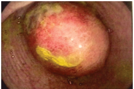
Figure 1: Colonoscopic image of exophytic endoluminal tumoration, partially obstructing the lumen of the organ, oval in shape with neovascularization, intense exanthema, and areas of extensive erosion covered with fibrin.

Renato Roriz da Silva1* Tiago Costa do Amaral2 Marcos Alberto de Mendonça Veiga2
1Department of Medicine, Division of surgery, Federal University of Rondônia, Rondonia, Brazil*Corresponding author: Roriz-Silva R, Department of Medicine, Division of surgery, Federal University of Rondônia, Hospital de Base of Porto Velho city, Rondonia, Brazil, Tel: 55 (69) 32165446; E-mail: roriz-silva@unir.br
Lipoma of the colon is a rare type of benign gastrointestinal tumor, occurring in around 0.035-4.4% of the population, according to statistics. In most cases, it is identified by colonoscopy as a yellowish sub epithelial nodule, and has no associated symptoms. This tumor of the colon occasionally produces symptoms, which may be associated with large lesions. The main symptoms are abdominal pain, changes in bowel habits, and lower digestive tract bleeding. Treatment can be performed via colonoscopy or surgical approach. The objective is to report a case of a large, symptomatic giant colorectal lipoma of unusual location (sigmoid colon), and of the treatment performed.
Abdominal pain; Colon; Colonic neoplasms; Differential diagnosis; Lipoma; Surgical procedures
Lipoma of the colon is a benign gastrointestinal tumor, first described by Bauer in 1757 [1]. It occurs in around 0.035-4.4% of the population, according to autopsy reports [2]. In 90% of cases, lipoma grows in the submucosal [3], making its correct diagnosis and proper therapeutic management difficult [4]. They are more common in women in their 50s and 60s, and are usually detected during autopsy, colonoscopy, or surgery [5]. In general, the gastrointestinal lipoma is less than two centimeters in diameter, sessile or pedunculated, and is located in the right colon [1,5,6]. Although usually asymptomatic, it can present symptoms when it is larger than 2 cm in diameter. The main symptoms are abdominal pain, changes in bowel habits and bleeding [1]. Episodes of intussusception or intestinal obstruction may also be observed in patients with giant lipoma in whom the initial manifestation of the tumor may be diagnosed as acute surgical abdomen. In this paper, we report the case of a patient with a symptomatic giant lipoma of the sigmoid colon that was surgically resected.
A female patient, 66 years of age, was admitted to the general surgery Division of the Hospital de Base by Dr. Ary Pinheiro, in Porto Velho, Brazil. She presented with abdominal pain in the lower left quadrant associated with hematochezia over the past three months and had a weight loss of 4 kg during that period. Neither the physical examination nor the initial laboratory tests revealed any changes. Colonoscopy identified an endoluminal mass located about 35 cm from the anal border with a wide base (3 cm), a smooth and slightly reddened surface - with areas of surface erosion - thinly covered with fibrin, partially obstructing the lumen of the organ (Figure 1). There was no evidence of change in the other colonic segments. A biopsy taken during the colonoscopy was inconclusive.
Figure 1: Colonoscopic image of exophytic endoluminal tumoration, partially obstructing the lumen of the organ, oval in shape with neovascularization, intense exanthema, and areas of extensive erosion covered with fibrin.
The total abdomen tomography (Abdominal CT) - with oral and venous contrast - identified a mass with attenuation of adipose tissue in the topography of the sigmoid colon, measuring around 5.4 cm at its greatest diameter, without invading the pericolonic tissues or enlarged lymph nodes (Figure 2). The treatment proposed was a surgical resection, as an endoscopic resection was made impossible by the wide base of the lesion and the lack of definition of the histopathological study of the biopsy sample taken during the colonoscopy. No other intracavity abnormalities were identified during surgery. A segmental colectomy (sigmoidectomy) with colorectal anastomoses was performed in two planes - the total plane with Polypropylene 2-0 and the sero-muscular plane with Polygalactin 3-0 (Figure 3). A sentinel drain was positioned at the bottom of the pouch of Douglas. Based on the biopsy of the surgical piece, it was concluded that it was a giant submucosal lipoma of the left colon measuring 6.0 × 4.8 × 4.2 cm, without the presence of atypical cells. The patient presented satisfactory postoperative progress and after removal of the abdominal drain was discharged on the fifth day following surgery. Her case has been followed has for 1 year and has remained free from symptomless.
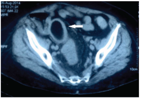
Figure 2: Axial abdominal tomography of the pelvic region with nodular image, completely filling the intestinal lumen, hypodense and surrounded by a hyperdense halo (arrow).
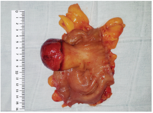
Figure 3: Surgical piece (colonic segment) with a parietal tumor of the sigmoid colon, measuring 6.0 × 4.8 × 4.2 cm, pedunculated with a wide base (3 cm).
Lipoma of the colon is a rare benign gastrointestinal tumor occurring in between 0.035 and 4.4% of the population according to autopsy reports first described by Bauer in 1757 [1,2,7,8]. Lipomas of the colon are benign tumors with extremely low potential for malignancy [9]. They are usually identified in the digestive system of female patients (66.7%) between 50 and 65 years of age [10], as in the case reported here. They are generally less than two centimeters in diameter, and of sessile or pedunculated form [1,5,6]. Colonic presentation occurs in 65-75% of digestive system cases, with the right colon being the most common, followed by the transverse, descending, and sigmoid colons, and the rectum, respectively [11]. In this present case, the size of more than 2 cm, and the location in the sigmoid colon, are atypical characteristics of lipomas, and is not a differential diagnosis that is generally considered in masses in the left colon. The lesion is usually unique, but multiple lesions may occur in 10% to 25% of the patients [12].
Lipomas arise from adipocytes of the submucosa in 90% of the cases, or from the subserosa [11]. These lesions are more common in females and are asymptomatic in the vast majority of cases (94%) [13]. They are usually detected during an autopsy, colonoscopy, or surgery [2,14,15]. However, a small percentage of cases may be symptomatic, particularly when larger than 2 centimeters in diameter [3-5,7-10]. The possibility of the occurrence of symptoms is directly proportional to the size of the tumor; for example tumors larger than 4 cm are symptomatic in 75% of patients [13]. The symptoms are non-specific, the most common one being abdominal pain, and may be associated with changes in intestinal transit (diarrhea or constipation). This occurs due to a partial reduction in the passage of the intestinal content [16]. Other acute clinical manifestations may be identified due to intussusception or bleeding [11]. Pain can be caused by intermittent intussusceptions [13]. Bleeding may occur in these cases because of mucosal and submucosal distension caused by tumor growth, with a consequent decrease in blood flow at mucosal points that can cause ulcerations [13]. However, these ulcerations rarely cause clinically apparent bleeding or chronic anemia [1,6,17]. Bleeding may be apparent when there is traumatic necrosis of the surface of the mucosa [16]. Episodes of intussusception or intestinal obstruction may be the first manifestations of complications from a lipoma of the colon and they can lead to an abdominal emergency [1]. There are many other complications that are rarely reported, such as self-amputation and prolapsed [10]. In the case presented, even though the tumor was larger than 4 cm, the patient had no symptoms of subocclusion or intussusception, but did present with moderate hematochezia and significant pain in the lower right quadrant, possibly due to ischemia of the mucosa and the mass effect because of size of the tumor.
A preoperative diagnosis is based mainly on the colonoscopy. Typical lipomas appear as smooth, spherical, light yellowish polyps of varied sizes, with or without a peduncle [18]. Endoscopic biopsies of colonic lipomas provide only limited diagnosis, and the results of the pathology are generally normal, or show an ulcerated mucosa [2]. Three endoscopic signs can assist in the diagnosis of a lipoma: the “pillow” sign, which occurs when the mass is pressed and results in a depression; the “tent” sign, which occurs when the mucosa over the lesion is pinched and pulled towards the lumen, resulting in a tent-like appearance; and the “naked fat” sign, which occurs when the fat is coarsely extruded following the biopsy [6,19,20]. Pressure exerted on the lesion may compress the superficial blood vessels and the distinct yellow color of the fat may be revealed [1]. Imaging exams facilitate diagnosis; however, diagnostic confirmation can only be obtained after excision of tissue [4]. An oval defect can be observed using bariumcontrast x-ray, and non-vascular, hyperechoic, and submucosal lesions observed using endoscopic ultrasound can assist the diagnosis, though they are not specific to lipomas of the colon [5]. Endoscopic ultrasound (EUS) typically shows a hyperechoic lesion originating in the submucosal layer. Lipomas with a heterogeneous or atypical hypoechoic appearance in EUS have been documented in literature [21]. The pathognomonic sign of a colonic lipoma is the “squeeze” sign, which consists of a radiolucent spherical filling defect with well-defined margins that can change in size and shape in response to peristalsis following the administration of a barium enema [16]. For large lipomas of the colon, abdominal CT is capable of detecting masses with thin margins in the density of the homogeneous fat, and magnetic resonance distinguishes lesions in the intensity of the adipose tissue [5]. Abdominal CT is considered the most widely used non-invasive diagnostic method for diagnoses which include complications such as intussusceptions [22]. It enables an ovoid mass, though limited, to be differentiated from the regular density of fat [3]. This test visualizes well-defined, ovoid homogeneous intramural lesions, with a fat density of between -40 and -120 UH [15]. But for small lipomas, the diagnostic value of TC is low [1].
Rarely a giant Lipoma is identified in the left colon but colonic adenocarcinoma is the main differential diagnosis in this situation. In this case, the patient presented symptoms and the colonoscopy did not rule out the possibility of malignant neoplasia. This hypothesis can be corroborated when the CT reveals an atypical appearance of lipoma with blurring of the tumoral or perilesional fat, secondary to fatty necrosis or infarction of the lipoma, which can simulate infiltration and tumor necrosis [23]. For diagnostic differentiation in this situation, proper histological sampling obtained from the interior of the mass is required - which is only possible after resection of the tumor [6]. Another important differential diagnosis of giant lipoma of colon is Gastrointestinal stromal Tumor of the Colon (GIST). The main location is the rectum but cases have been reported in the colon [24,25]. They frequently are transmural tumors with intraluminal mass and can present symptoms like giant lipoma: lower GI bleeding, abdominal pain, perforation and obstruction. Biopsy of suspected tumors has been controvertial and surgery is the main treatment to patients with primary GIST with no evidences of metastasis.
Treatment of lipomas of the colon involves observation in asymptomatic cases and endoscopic or surgical resection in symptomatic cases. In this case report we believe that it was impossible to do a conservative treatment because patient had evident symptoms and the lipoma in question could produce a colonic intussusception, obstrution or enterorrhagia. Endoscopic resection of giant lipomas involves a high risk of complications, especially perforation, as compared to resection of adenomatous polyps of the mucosa. This is due to the location of the lipoma in the submucosa and the need for extensive dissection with the use of excessive fulguration, due to the high water content present in the lipoma [26]. The endoscopic removal of a sub mucosal lipoma, elective up to 2 cm in diameter, is reported to be safe and appropriate; however, in larger lesions the risk of complications associated with uncontrolled bleeding and perforation make the procedure controversial [27]. In 2013, Kim et al. published a series of three cases of successful partial endoscopic resection of giant colorectal lipomas, but these did not have wide bases like in the case presented here [25].
The main treatment in symptomatic cases as well as in cases of intussusception or intestinal obstruction, or even when there is diagnostic doubt as to the benignity of the tumor, is surgery [23]. The principal surgical techniques used are local excision and segmental colectomy with primary anastomosis. In cases of diagnostic doubt or acute colocolic intussusception, segmental colectomy should be the proposed treatment [14], as was performed in this case. However, local excision should be considered whenever possible, in order to limited morbidity [23]. The prognosis is excellent and there are no reports of recurrence in literature and Laparoscopic resection were recently reported as an alternative to laparotomy for giant colorectal lipomas [3].
In this case, the surgical approach which was selected was a segmental colectomy and primary anastomosis, because the findings from transoperative palpation of the tumor were typical of lipoma (consistency, regularity, and precise borders), which helped in the decision to perform less radical surgery.
Conclusions
Lipoma of the colon is rarely symptomatic and its occurrence is uncommon in the left colon, especially when it is larger than 5 cm in diameter. It should be included in the differential diagnosis of colorectal diseases in the elderly population, even though this type of presentation is rare. In the presence of ulcerated lesions in large tumors, as in the case presented here, a surgical approach is clearly indicated, particularly when colonoscopy with a biopsy is unable to confirm the presence or absence of malignancy of the tumor.
Download Provisional PDF Here
Article Type: Case Report
Citation: Roriz-Silva R, Amaral TC, Veiga MAM (2017) Giant Colorectal Lipoma of Left Colon: Case Report and Literature Review. J Surg Open Access 3(3): doi http://dx.doi.org/10.16966/2470-0991.149
Copyright: © 2017 Roriz-Silva R, et al. This is an open-access article distributed under the terms of the Creative Commons Attribution License, which permits unrestricted use, distribution, and reproduction in any medium, provided the original author and source are credited.
Publication history:
All Sci Forschen Journals are Open Access