Figure 1: Multiple cystic lesions with displacement of abdominal structures.

Moisés A Perdomo Galván1 E Simón Nacif2 H Bizueto Rosas1,3* M Romero López1 Jorge A Villar-Tapia1 M Bárcenas Díaz1 J Rivera Bañuelos1 Y Gómez Solís1 A López Borel1 P Cardona Ochoa1 O Gutiérrez Olivares1 A Jiménez Canet1
1General Surgery Service, General Hospital “Dr. DaríoFernández Fierro”, ISSSTE, Mexico City, Mexico*Corresponding author: H Bizueto Rosas, General Surgery Service, General Hospital “Dr. DaríoFernández Fierro”, ISSSTE, Mexico City, Mexico, E-mail: dr_bizueto_h@yahoo.com
The following case over view summarizes diagnostic approach of a large omental cyst regarding a 52-year-old male with vague abdominal pain. While most literature reports incidental diagnosis of this pathology, there are clinical and radiologic data which can lead to its consideration as a differential diagnosis. Cyst throughout omental leaves are a rare condition usually found in patients who seek medical attention due to abdominal distention in addition to a painless abdominal tumor. The most frequent sign at the clinical exploration suspecting an omental cyst is described as an abdominal tumor not attached to deep structures. The approach to the diagnosis of an omental cyst incorporates a complete radiologic overview with ultrasonography and CT scan. Full resection of the tumor is established as the therapeutic of choice. CT is a key component in the election of either laparotomy or laparoscopic approach. In our patient, a loculated cystic tumor measuring 23 cm × 25 cm × 9.9 cm was reported in CT scan. Due to the radiologic findings, we opted an open approach. After 5-day surveillance in this unit, the patient was sent home. The tumor was identified as a loculated inclusion-type cyst by histopathology. A 6-month follow-up showed no recurrence of cystic lesions upon radiologic study.
Omental cyst; Cystic lesion
An omental cyst is defined as any cystic lesion located between the mesenteric leaves, being more commonly found at the ileum. It’s described as a rare entity, with unknown etiology and unspecific symptomatology; reporting an incidence of 1 in 140,000 hospitalized patients with intraabdominal pathology. The first report was published in 1852 by Gairdner. Diagnostic criteria based on radiologic studies such as ultrasonography, tomography and magnetic resonance. The laparoscopic approach is considered the “gold standard”, leaving open procedures to cases in which total resection is not possible through minimal invasive surgery.
52-year-old male was admitted to the emergency department with a 48-hour evolution of vague abdominal pain, irradiated to the back. Presenting in addition nausea without vomiting and low-grade fever, referring progressive growth of abdominal diameter over a 1-year-period before hospital admittance. During the general survey he denied chronic diseases and reported an umbilical hernia repair 1 month ago.
During the physical examination, an abdominal tumor was found, comprising 2/3 of the abdomen, with homogenous surface, not adhered to deep structures. No evidence of acute abdomen was found during initial evaluation, only mild-generalized tenderness. CBC (Complete blood count) revealing leukocyte elevation (15 × 103/mcl) and mild anemia (Hb 10.9 g/dl, Htc 33%). Normal AFP (Alpha-Fetoprotein Test), CEA (Carcinoembryonic antigen) and CA (Cancer antigen) 125.
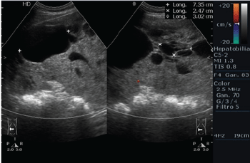
During his stay at the emergency room an ultrasonography study was made describing a large (over 28.5 cm), heterogeneous, cystic image located on upper abdomen displacing adjacent structures (Figures 1-5).
Figure 1: Multiple cystic lesions with displacement of abdominal structures.
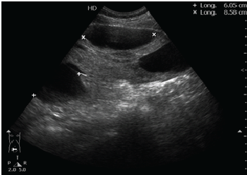
Figure 2: Cystic like image with hypoechoic content.
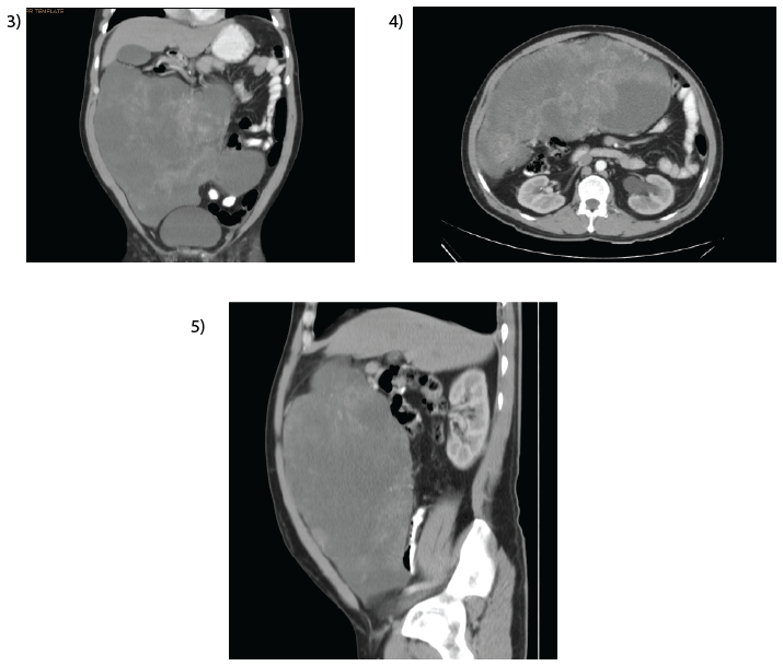
Figures 3, 4 & 5: As part of pre-operative protocol an abdominal CT scan was ordered. The study reported a hypodense, lobulated, welldefined lesion, measuring 23 × 9.9 × 25 cm, with apparent peritoneal dependence and low-moderated contrast enhancement was observed.
Due to radiological findings, laparotomy was performed. Under general anesthesia, we proceeded with a middle-line 20 cm incision for adequate exposure. Reporting a greater omentum-dependent tumor, multi loculated, adhered to the gastric surface over the greater curvature over a 2-cm area. Complete excision of the tumor was realized with harmonic scalpel 1 cm from its origin without complications to report. The gastric adherence was managed with cold scalpel resection presenting minimal erosion of the serosa which was reinforced by 2-0 silk Lembert suture (Figures 6,7).
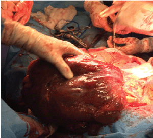
Figure 6: Cystic tumor arising from greater omentum.
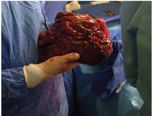
Figure 7: Cystic tumor with a size of 35 × 30 × 13 cm.
The definitive histologic exam reported a 4,200 g, tumor with a size of 35 × 30 × 13 cm. The tumor had a nodular surface, and was composed of cystic lesions with serous content.
Due to the findings during procedure, the patient followed a 72-hourfasting period, after which regular diet was tolerated without further complications. Patient was discharged the fifth day, after the procedure.
Six months post-operatively, the patient was asymptomatic and with no signs of recurrence.
Most cysts of the omentum are of lymphatic or mesothelial origin. All are rare. Representing 30% of intra abdominal cysts. The first excision of an omental cyst was performed by Tillaux [1]. Omental cysts represent an infrequent type of tumor which is strongly linked to cyst located at the mesenteric root and in the retroperitoneal region due to their shared lymphatic origin [1]. A small percentage of this patient present at emergency units with the diagnosis of acute abdomen with a wide range of manifestations such as bowel obstruction or a shock state due to bleeding or perforation [2].
On CT, both mesenteric and omental cysts can occur along the mesenteric root, retroperitoneal region and omental leaves without a clear region predominance reported. They appear as cystic, simple or multilocular, lesions with a variable wall thickness. Their macroscopic cystic characteristics give a wide range of differential diagnosis which includes many other cystic lesions (pancreatic pseudocyst, ovarian, urachal, peritoneal inclusion, teratoma, urogenital, and hydatid) also being considered hemangioma, endometriosis, and loculated ascites (usually tuberculous) [3]. Benign cystic mesotheliomas are to be considered. Being originated from serous lining structures (pleural, pericardial, peritoneal). They affect peritoneal (intra or extra) space, omental leaves and, abdominal viscera including the pelvic region. Defined ultimately at the histopathologic studies [4].
When surgical resection is needed, CT imaging is an important tool to select which patients will benefit from a laparoscopic or open approach. Other options for treatment comprise marsupialization, enucleation, and drainage [3].
Even if the patient has no symptoms, resection, either by laparoscopy or laparotomy, is usually indicated due to risk of complications such as rupture, bleeding and infection. Malignant transformation of omental cysts is an infrequent event with isolated reported cases of sarcoma and adenocarcinoma developed from this entity [5-8].
Omental cyst represents a rare entity of intra-abdominal tumor. Early onset symptomatology and physical findings should be considered for taking in consideration this pathology alongside the whole differential diagnosis spectra for each case. Also, the CT scan represents a key element in the evaluation of any patient with an abdominal tumor, not only for a diagnostic purpose, but also to determine the ideal surgical approach.
Download Provisional PDF Here
Article Type: Case Report
Citation: Galvan MAP, Nacif ES, Rosas HB, Lopez MR, Villar-Tapia JA, et al. (2016) Large Omental CystCase Report and Literature Review. J Surg Open Access 3(1): doi http://dx.doi.org/10.16966/2470- 0991.137
Copyright: © 2016 Galvan MAP, et al. This is an open-access article distributed under the terms of the Creative Commons Attribution License, which permits unrestricted use, distribution, and reproduction in any medium, provided the original author and source are credited.
Publication history:
All Sci Forschen Journals are Open Access