Figure 1: Clinical photograph


Seema Khanna Satendra Tiwary Puneet Ajay K Khanna*
Department of General surgery, Institute of Medical sciences, Banaras Hindu University, Varanasi 221 005, India*Corresponding author: Ajay K Khanna, Department of General surgery, Institute of Medical sciences, Banaras Hindu University, Varanasi 221 005, India, Tel: 09415201954; E-mail: akhannabhu@gmail.com
Hydatid cyst disease caused by Echinococcus granulosus may at times affect unusual sites and present a diagnostic challenge especially if found at rare locations. A 51 year male presented with a slowly progressive swelling in the upper medial aspect of the left thigh without pain and any discharge. There was no history of trauma and any dog keeping. Examination revealed a large 10 × 12 cm soft tissue swelling which was non-tender, non-pulsatile, without any features of acute inflammation figure 1. Overlying skin was normal. No bruit or venous hum was heard.

Figure 1: Clinical photograph
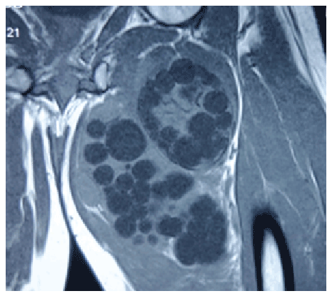
Routine blood investigations, ultrasound of the abdomen and X-ray of chest was normal. Ultrasound of the area showed multicystic swelling. FNAC of swelling revealed a cystic lesion and reported as parasitic or lymphatic cyst. On serological tests, IgG for hydatid was raised with a value of 74.20 IU. MRI of thigh revealed multiple, large, multicystic well defined masses in adductor muscle compartment in the left thigh. Largest cyst was 83 × 69 × 103 mm in size. Multiple, variable cysts were seen divided by septa. No obvious bone or joint involvement was noticed figure 2. MRI features were suggestive of hydatid mass.

Figure 2: MRI Picture
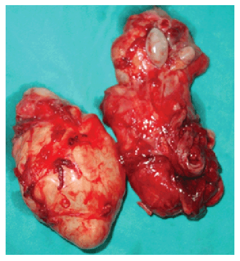
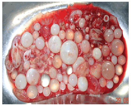
Patient was operated under general anesthesia. Hydatid cyst of approximately 25 × 15 × 15 cm was found to be located in the medial aspect of left thigh with multiple daughter cysts in the adductor muscle compartment figure 3 and 4. No neurovascular involvement was found. Care was taken to avoid spillage. 10% Betadine soaked mops were placed around the cyst and hypertonic saline (3% NaCl) instilled in it. Pericystectomy with drain placement done.

Figure 3: Excised Specimen

Figure 4: Multiple Daughter Cysts
Post-operative course was uneventful. Drain was removed on postoperative day four. Patient was discharged on antihelminthic Albendazole 400 mg twice a day for 1 month. Histopathology report revealed a germinal layer, lamellated ectocyst with fibrous outer layer. Marked foreign body giant cell reaction was observed, confirming the diagnosis of hydatid cyst.
Primary hydrated cysts in muscle are an uncommon manifestation, accounting for only 3% of all patients with hydatidosis [1]. Muscular hydatidosis usually are found to occur as isolated lesions without hepatic or pulmonary lesion [2]. The most common locations for development of parasitic cysts are the liver (65%-75%) and lung (25%-30%) [3], the involvement of other organs including brain, heart, kidney, bone, skeletal muscle, breast, thyroid comprise only about 10% and are considered as unusual presentation [4].
Growth of cyst in different organs of body is variable and is dependent on patient factors, parasite factors, host reaction and presence of any complications. Compressible organs like lung and brain facilitate the growth of cyst [5]. Constant contraction of muscles and production of lactic acid probably retards the growth of the cyst in the muscle. Hydatid cyst have been reported in neck, trunk and proximal part of limbs, may be because of relatively less muscle contraction in these areas [6,7]. Hydatidosis presenting as a painless, gradually progressive swelling should be included in the differential diagnosis of soft tissue swellings especially if the patient gives history of agricultural background or has been in contact with livestock.
MRI is the examination modality of choice due to it’s ability to demonstrate most of the features of hydatid cyst except calcifications. The classic MRI findings include a multivesicular cyst, a low intensity rim (“rim sign”) on T2 –weighted images or a detached membrane [8,9]. The most pathognomonic sign is the presence of daughter cysts within the larger cysts. The “rim sign” differentiates hydatosis from non- parasitic cysts in liver and lung. This represents the parasitic membrane (germinal layer) and a collagen rich membrane as a host response (pericyst). Comert et al. first described the “water lily sign” as a pathognomonic sign for intramuscular hydatid cyst [10], the collapsed parasitic membranes which are hypointense on all T1weighted images represent a non viable cyst and is described as a ‘Snake sign or Serpent sign” [11].
The treatment of muscle hydatid cyst is surgical excision i.e. pericystectomy while avoiding spillage of hydatid fluid and daughter cysts. This should be followed by antihelminthic medication to prevent recurrences. Mohamed et al. have analysed the efficacy of albendazole alone versus combined albendazole and praziquantel in human hydatidosis are of the view that praziquantel prevents encystment of protoscoleses following perioperative spillage [12]. Percutaneous aspiration, infusion of scolicidal agent and reaspiration (PAIR) under imaging is another option available for inoperable cases.
Adductor muscle hyadatidosis is a rare presentation of this disease and has to be included in the differential diagnosis of soft tissue swellings.
Download Provisional PDF Here
Aritcle Type: Guest Editorial
Citation: Khanna S, Tiwary S, Puneet, Khanna AK (2015) Adductor Muscle Echinococcosis: A Rare Presentation, J Surg Open Access 2(2): doi http://dx.doi. org/10.16966/2470-0991.e104
Copyright: © 2016 Khanna S, et al. This is an open-access article distributed under the terms of the Creative Commons Attribution License, which permits unrestricted use, distribution, and reproduction in any medium, provided the original author and source are credited.
Publication history:
All Sci Forschen Journals are Open Access