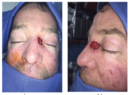
Figure 1: Case 1: Preoperative defect: a) Anteroposterior view; b) Lateral view

Laurel Karian Ramazi O Datiashvili*
Department of Surgery, Division of Plastic Surgery, Rutgers University/New Jersey Medical School, New Jersey, USA*Corresponding author: Ramazi O. Datiashvili, Department of Surgery, Division of Plastic Surgery, New Jersey Medical School, The State University of New Jersey, Rutgers, 140 Bergen Street, ACC E-1620, Newark, NJ 07103, USA, Tel: (973)972-5377; Fax: (973)972-8268; E-mail: datiasro@njms.rutgers.edu
Reconstruction of large soft tissue defects of the medial canthal area represents a challenge, because of the thin skin, concave contour, and proximity to important nearby structures. Tension-free closure is important in avoiding distortion of the eyelid and nasal ala. Skin graft closure is not aesthetically appealing, and there are limitations in the use of local tissues for reconstruction. We present our experience (two cases) of reconstruction of very large medial canthal defects with a combination of a V-Y cheek advancement flap and glabellar rotation flap, with satisfactory aesthetic and functional results.
Medial canthal defect; Glabellar flap; V-Y advancement flap
Reconstruction of skin defects of the proximal nasal sidewall and medial canthal area represents a challenge to the plastic surgeon due to limitations of skin laxity in the adjacent areas. This is especially true for large defects in this area. Reconstruction strategy requires consideration of several factors, such as maintaining the aesthetic subunits and contour of the nasal sidewall and the eye, avoiding distortion of nearby anatomic structures like the eyelid or nasal ala [1].
Several reconstruction techniques have been proposed, including healing by secondary intention for small defects, full-thickness skin grafts, and a variety of local flaps. Local flaps are generally preferred over skin grafts because they provide a superior color, texture and contour match [2,3]. However, closure must be free of tension to avoid complications such as ectropion or disfiguring elevation of the alar rim. In cases of very large soft tissue defects of the medial canthal area, one flap is often insufficient to provide tension-free closure without distortion of nearby structures.
Here we present two cases involving large soft tissue defects of the medial canthal and nasal sidewall area that were too large for reconstruction using a single flap. These defects were reconstructed using a combination of a glabellar flap and a V-Y cheek advancement flap
The glabellar flap is designed in a triangular shape over the area of maximal skin laxity the in the glabellar region. The midpoint of the triangle is midline, extending a few centimeters above the superior border of the eyebrow. A template is used to assess the amount of rotation of the flap, and the midpoint of the triangle may be readjusted more superiorly as needed for a flap rotation. Local anesthetic is injected into all areas of the flap except the base. The flap is elevated in the subcutaneous plane, with conservative flap elevation near the base, using blunt dissection to preserve blood supply. This flap is rotated into the superior aspect of the defect and tacked with a few sutures prior to design of the second flap.
The V-Y flap is then designed in the traditional fashion in the cheek, based on the area of coverage needed for the remaining defect. The medial border lies along the lateral nasal sidewall. When large V-Y flaps are needed, the medial incision may be extended along the nasolabial fold. Undermining is done to the extent needed for flap advancement, keeping of the underlying blood supply intact. The V-Y flap is then advanced to meet the inferior border of the glabellar flap and closed with permanent interrupted sutures.
A 52 year old male presented with a recurrent basal cell carcinoma of the left medial canthal area extending onto the nasal sidewall. The lesion was excised with a 3 mm margin and closed temporarily with Biobrane one week prior to reconstruction, in order to ensure negative margins on final pathology. The total defect measured 2 × 2.5 cm, with the medial extent only 1-2 mm from the palpebral fissure (Figures 1a and 1b). As the defect was quite large and involved two aesthetic units, a medial canthal region and a nasal side wall, and keeping in mind a quality of surrounding skin (telangiectasia), we opt, upon discussion with the patient, a local tissue transfer and arrangement as a first choice of treatment in favor of skin grafting. A glabellar flap measuring 3.5 × 2 cm was elevated as described above, but was insufficient to completely close the defect. In order to avoid unnecessary facial distortion a V-Y cheek advancement flap measuring 3.5 × 1.2 cm was then designed, mobilized and elevated to meet the edge of the glabellar flap (Figures 2a-2c). Postoperatively, there were no complications and the patient was very happy having an aesthetically pleasing result (Figures 3a and 3b).

Figure 1: Case 1: Preoperative defect: a) Anteroposterior view; b) Lateral view
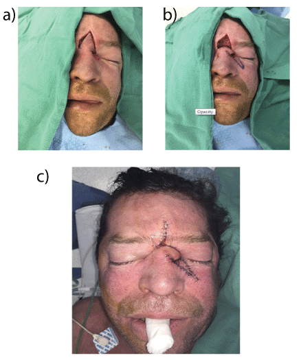
Figure 2: (a, b, c) Case 1: intraoperative photographs showing elevation and inset of glabellar and V-Y flaps.
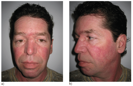
Figure 3: Case 1: postoperative views at 1.5 months: a) Anteroposteiror view; b) Lateral view
A 70 year old female presented with a large basal cell carcinoma of the right medial canthal area extending onto the cheek. After excision with a 3 mm margin, the total defect measured 5 × 2 cm (Figure 4). In this case the lateral border of the defect was again only a few millimeters from the palpebral fissure. A glabellar flap measuring 3 × 2 cm was elevated in a subcutaneous plane, but was insufficient to completely close the defect. A V-Y cheek advancement flap measuring 3.5 × 1.5 cm was elevated in a subcutaneous plane and mobilized to meet the edge of the glabellar flap. Postoperatively, there were no complications (Figures 5a and 5b). The patient has mild pin cushioning of the scar, and she is undergoing scar management.
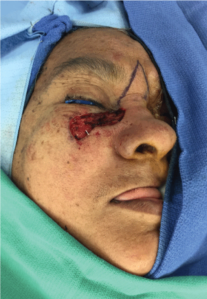
Figure 4: Case 2: Preoperative defect and flap design
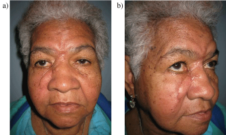
Figure 5: Case 2: Postoperative views at 1 month: a) Anteroposteiror view; b) Lateral view
Reconstruction of large soft tissue defects of the medial canthal area represents a challenge as tension-free closure is required to avoid distortion of the lower eyelid and nasal ala. Reconstruction options include healing by secondary intention for small defects, full-thickness skin grafts, and local skin flaps. Local skin flap is the reconstructive method of choice as they provide the best color and texture match, and help avoid contour irregularities by providing skin of a similar thickness [2,3].
Local skin flaps used for reconstructing the medial canthal area include bilobed flaps harvested from the nasal dorsum, forehead flaps, glabellar flaps, and cheek advancement flaps such as the V-Y flap [4-6]. While the bilobed flap offers an aesthetically pleasing result, it is not wide enough to reconstruct large defects of the medial canthal area, and the forehead flap is thick in comparison to the thin skin of the medial canthus. Glabellar flaps and V-Y advancement flaps are both commonly employed to reconstruct large defects in this area and have been used in successfully for very large defects [7-9].
The glabellar flap, based on the contralateral angular artery, takes advantage of laxity in the nasion and provides reliable coverage for superior nasal sidewall and medial canthal skin defects [4]. V-Y cheek advancement flaps take advantage of laxity in the cheek, and are best for more inferior nasal sidewall defects. Neither the glabellar flap nor the V-Y cheek advancement flap is sufficient alone to reconstruct a very large defect of the medial canthal region. Combining the two flaps is a useful alternative for reconstructing such defects, with satisfactory aesthetic and functional results.
Download Provisional PDF Here
Article Type: Case Series
Citation: Karian L, Datiashvili R (2016) Combination of Glabellar and V-Y Cheek Advancement Flaps for Large Skin Defects of the Medial Canthal Area. J Surg Open Access 2(6): doi http://dx.doi. org/10.16966/2470-0991.133
Copyright: © 2016 Karian L, et al. This is an open-access article distributed under the terms of the Creative Commons Attribution License, which permits unrestricted use, distribution, and reproduction in any medium, provided the original author and source are credited.
Publication history:
All Sci Forschen Journals are Open Access