Abstract
Upper gastrointestinal series (UGIS) with barium is a routine investigation, and the incidence of serious complications associated with this
procedure is very rare. We report 2 cases of perforation of the large intestine after UGIS with barium, and review relevant literature.
Case 1: A 72-year-old woman presented with nausea and vomiting that had started on the 4th day after UGIS with barium. She was brought
to our hospital by ambulance. The abdomen was rigid with muscle guarding. Computed tomography (CT) of the abdomen revealed retention of
barium in the sigmoid colon, with free air at its periphery. Upon diagnosis of pan peritonitis related to perforation of the digestive tract, emergency
partial sigmoidectomy and colostomy were performed on the same day. The patient recovered uneventfully and was discharged on postoperative
day 15. Colostomy closure was performed 6 months after the first surgery.
Case 2: A 66-year-old woman presented with abdominal pain on the day after UGIS with barium. She was brought to our hospital by ambulance.
The abdomen was rigid with muscle guarding. Abdominal CT revealed barium outflow into the abdominal cavity, and free air at its periphery. Upon
diagnosis of pan peritonitis related to perforation of the digestive tract, emergency partial sigmoidectomy and colostomy were performed on the
same day. Postoperatively, the patient received antimicrobial therapy for protracted fever, and was discharged on postoperative day 24.
Keywords
Barium; Upper gastrointestinal series; Perforation of the large intestine
List of Abbreviations
CT: Computed Tomography; UGIS: Upper Gastrointestinal Series; WBC: White Blood Cell ; CRP: C-Reactive
Protein
Background
Upper gastrointestinal series (UGIS) with barium represents a routine
investigation, and the incidences of serious complications associated with
this procedure are very rare. Since perforation of the large intestine caused
by barium ingestion can progress to severe peritonitis, prompt diagnosis
and treatment are needed. We report 2 cases of large intestinal perforation
after UGIS with barium, and provide a review of the relevant literature.
Case Presentation
Case 1
The patient was a 73-year-old woman.
Chief complaint: Abdominal pain, nausea, vomiting
Past history: Appendectomy for appendicitis at the age of 18 years.
Current history: The patient underwent UGIS with barium at a health
checkup. Four days later, she presented with abdominal pain, nausea, and
vomiting, and was emergently transported to our center.
Observations at admission: The following were noted: height, 155.0 cm;
weight, 54.5 kg; body temperature, 36.0°C; blood pressure, 152/87 mmHg;
and pulse rate, 60 beats/min. The patient appeared to have extreme pain,
and her abdomen was rigid.
Blood test findings at admission: The white blood cell (WBC) count
was13390/µL and the levels of C-reactive protein (CRP) were 25 ng/mL,
indicating increased inflammation.
Contrast-enhanced computed tomography (CT) findings: CT showed
a 3-cm barium stool in the sigmoid colon, barium leakage, and free air
surrounding the stool (Figure 1). Emergency surgery was performed
on the same day, under the diagnosis of barium peritonitis caused by
perforation of the sigmoid colon.
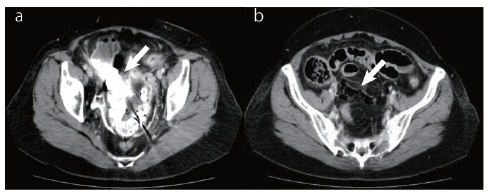
Figure 1: Abdominal computed tomography at admission for a
72-year-old woman with perforation of the large intestine after upper
gastrointestinal series with barium: a) A 3-cm barium stool (arrow)
is noted in the sigmoid colon, with barium leakage and free air
surrounding the stool; b) Free air is observed in the pelvis (arrow).
Surgical findings: Laparotomy showed ascetic fluid clouded by barium
and stools. Intra peritoneal examination revealed hard stool in the
sigmoid colon, with a perforation at the same site. No ischemic change or
tumor was noted in the surrounding area. After barium that adhered to
the colonic wall was carefully removed, partial sigmoidectomy, colostomy,
and intra peritoneal irrigation drainage were performed.
Histopathological findings: In the resected specimen, the barium stool
was retained within the sigmoid colon (Figure 2). Upon removal, a
perforation with a diameter of 15 mm was observed, with fibrosis and
inflammation of the surrounding area. Since no findings indicative of
diverticulum, malignant tumor, or ischemia were noted, a condition
caused by the barium stool was suspected.
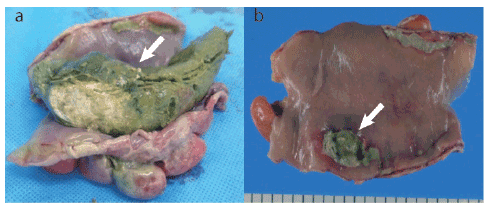
Figure 2: Resected specimen from a 72-year-old woman with perforation
of the large intestine after upper gastrointestinal series with barium: a)
Barium stool is retained in the sigmoid colon (arrow); b) Perforation of the
sigmoid colon, 15 mm in diameter, is noted after removal of the barium
stool, with fibrosis and inflammation of the surrounding area (arrow).
Postoperative course: The patient had an uneventful course; she resumed
food intake on postoperative day 6 and was discharged on postoperative
day 15. Colostomy closure was performed 6 months after the first surgery.
Case 2
The patient was a 66-year-old woman.
Chief complaint: Pain in the lower abdomen, vomiting.
Past history: The patient received medication for chronic renal failure,
hypertension, and gout.
Current history: The patient underwent UGIS with barium as part of
a health checkup. The next day, she presented with abdominal pain and
vomiting, and was emergently transported to our center.
Symptoms at admission: The following were noted: height, 147.0 cm;
weight, 58.0 kg; body temperature, 37.1°C; blood pressure, 122/75 mmHg;
and pulse rate, 60 beats/min. Abdominal examination revealed tenderness
in the medial lower abdomen, with rigidity and guarding.
Blood test findings at admission: The WBC count was 4920/µL, and CRP
levels were 0.02 ng/mL.
Plain abdominal CT scan findings: Barium stool retention, barium
leakage in the mesentery, and intramesenteric emphysema were observed
(Figure 3). The patient underwent emergency surgery under the
diagnosis of barium peritonitis caused by perforation of the sigmoid
colon.
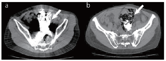
Figure 3: Abdominal computed tomography at admission for 66-year-old
woman with perforation of the large intestine after upper gastrointestinal
series with barium: a) Barium stool is retained in the sigmoid colon
(arrow); b) Barium leakage in the mesentery of the sigmoid colon and
intramesenteric emphysema are observed (arrow).
Surgical findings: Laparotomy revealed a large amount of cloudy ascetic
fluid. Intra peritoneal examination showed inflammatory thickening
of the sigmoid colon to the rectosigmoid mesentery, which indicated
perforation at the same site. Dissection of the mesentery at the same
site revealed a large amount of barium stool. There was a relatively large
perforation of 20 mm in diameter, and a vascular necrosis caused by
barium stool retention was suspected. After barium that adhered to the
site was carefully removed, the patient underwent partial sigmoidectomy,
colostomy, and peritoneal lavage and drainage.
Histopathological findings: A 20 × 18-mm perforation was noted in the
resected specimen of sigmoid colon (Figure 4). The area surrounding the
perforation showed inflammation in all layers; however, no diverticulum
or malignancy was noted.
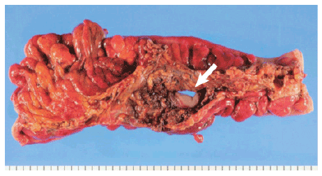
Figure 4: Resected specimen from a 66-year-old woman with perforation
of the large intestine after upper gastrointestinal series with barium: A
perforation of 20 × 18 mm is noted, with inflammation in all layers of the
surrounding area. No diverticulum or malignant finding is noted (arrow).
Postoperative course: Postoperatively, the patient was treated with
antimicrobial therapy for protracted fever. Her condition gradually
improved, and she was discharged on postoperative day 24.
Discussion
UGIS with barium is a procedure commonly performed for upper
gastrointestinal examination at health checkups in Japan. UGIS
with barium is not used for gastric cancer screening in Europe and
America. Administration of barium can cause adverse reactions such as
constipation, transient diarrhea, abdominal pain, intestinal obstruction,
gastrointestinal perforation, and peritonitis. Nonetheless, the incidence
of gastrointestinal perforation and concurrent peritonitis is very low.
Colonic perforation after UGIS is very rare, occurring in 3 out of 1.01
million people. Therefore, UGIS is considered relatively safe [1]. We
further examine relevant literature regarding such complications of UGIS
with barium.
Because UGIS with barium is conducted more often in Japan, we
primarily examined Japanese reports. We searched the database of the
Japan Medical Abstracts Society for original articles published between
1983 and2014. The following 3 keywords (in Japanese) were used in
the search: “barium,” “perforation of the large intestine,” and “upper
gastrointestinal series.” Thirty-four reports of concurrent perforation
of the gastrointestinal tract after UGIS with barium were found [2-31].
A summary of all cases of perforation of the gastrointestinal tract after
UGIS with barium in Japan is given in table 1. The median age of the
patients was 65 years (range, 40–90 years); 10 patients were younger than
60 years, and 26 patients were aged 60 years or older, indicating that the
complication occurs more commonly in these elderly individuals. Of the
36 patients, including the 2 patients we report on here, 4 were male, and
32 were female, indicating that the complication occurs predominantly in
women. Moreover, the most common site of perforation was the sigmoid
colon. Both our patients were elderly women, and the perforation site was
indeed the sigmoid colon.
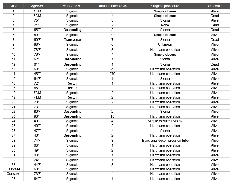
Table 1: Overview of reported cases of perforation of the large intestine after upper gastrointestinal series (UGIS) with barium, without organic disease
Age given in years. Duration after UGIS given in days
With respect to timing, perforation of the gastrointestinal tract
occurred within 4 days of UGIS in 30 of 36 patients, suggesting that the
condition progresses rapidly. Indeed, in our patients, the complication
occurred 1 and 4 days after UGIS with barium. This aspect highlights
the importance of checking for barium retention by plain abdominal
radiography when symptoms such as abdominal distension, abdominal
pain, and vomiting are present despite administration of laxatives after
UGIS. If barium retention is confirmed, medication-induced bowel
movement is required.
The severity of barium peritonitis can increase rapidly because intra
peritoneal barium leak causes severe inflammation, resulting in edema, as
cites retention, and dehydration. Furthermore, the prognosis may worsen
significantly due to concurrent severe infection caused by stool leakage.
For the cases reported in Japan, the mortality rate was13.8% (5 out of 36
patients). Our patients survived most likely due to prompt investigation
and intervention upon the occurrence of the symptoms.
Our patients exhibited perforation in the sigmoid colon, and therefore
underwent partial sigmoidectomy and adequate peritoneal lavage with
removal of intra peritoneal barium and warm saline infusion, followed
by colostomy. Based on previous reports, most patients underwent
colostomy rather than anastomosis in a single-stage surgery. Colostomy
is considered to be a safe procedure, with a focus on preserving life by
removing the risk of an anastomotic leak by intra peritoneal infection.
In such cases, McPhendran attributes the cause of lower gastrointestinal
tract perforation to mechanical laceration by barium stool [32]; whereas
Brearley attributes it to the compression of the colonic wall by barium
stool mass, resulting in a vascular necrosis [33]. In our patients, the
intestinal wall exhibited no organic lesions such as a diverticulum or a
tumor, but a relatively large ulcer localized only at the site in close contact
with the barium stool; therefore, we consider that the perforation was
caused by the latter.
Conclusions
We described 2 cases of perforation of the large intestine after UGIS with
barium. As the proportion of elderly individuals is expected to increase
in Japan, so will the number of procedures such as UGIS, performed as
part of screening or diagnosis for problems of the gastrointestinal tract. To
minimize the risk of gastrointestinal tract perforation caused by barium
stool, laxatives should be administered after UGIS, and patients with
abdominal pain following the procedure (even several days after UGIS)
should receive prompt investigations and treatment.
Consent
Written informed consent was obtained from the patients for
publication of this Case Report and any accompanying images.
Competing Interests
The authors declare that they have no competing interests related to
the work presented in this manuscript.
Article Information
Article Type: Case Report
Citation: Tachioka M, Saito M, Abe I, Obitsu T,
Imoto H, et al. (2016) Two Cases with Perforation
of the Large Intestine after Upper Gastrointestinal
Series with Barium. J Surg Open Access 2(4): doi http://dx.doi.
org/10.16966/2470-0991.131
Copyright: © 2016 Tachioka M, et al. This is an
open-access article distributed under the terms
of the Creative Commons Attribution License,
which permits unrestricted use, distribution, and
reproduction in any medium, provided the original
author and source are credited.
Publication history:
Received date: 17 May 2016
Accepted date: 31
May 2016
Published date: 06 Jun 2016
