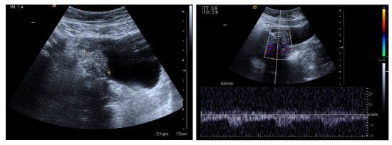
Figure 1: Sagittal transabdominal ultrasound image of the endometriotic mass located at the right posterolateral bladder wall. Power Doppler imaging reveals the presence of high amount of blood vessels inside the lesion.

Rocio Barrabino1 Antonio Fernández-Sánchez1 Hernani Gil-Julio2*
1Department of Urology, Complejo Hospitalario de Granada. Granada, Spain*Corresponding author: Hernani Gil Julio, Servicio de Urología, Hospital Don Benito, Ctra. Don Benito-Villanueva, Km 3 – 06400, Don Benito, Spain, Tel: +34 924 38 68 00; E-mail: mavangos@gmail.com
Endometriosis is a severely debilitating disease which affects primarily women of reproductive age. Endometriotic nodules involving the urinary tract is less common and can occur in 1–2% of all patients with endometriosis, and affects the bladder in 90% of these cases. In this article we report two cases of bladder endometriosis in two young females who were referred for macroscopic haematuria and the finding of exophytic intravesical lesions by transabdominal ultrasound imaging. In both cases a transurethral resection of the bladder lesions was performed. There was no evidence of symptoms or cystoscopic relapse in the follow-up period.
Deep Infiltrating Endometriosis; Interstitial Cystitis; Bladder Endometriosis; Haematuria; Transvaginal Ultrasound
Endometriosis is one of the most challenging gynaecological disorders [1], and rarely involves the bladder. Approximately 10% of all women in their reproductive phase develop endometriosis. Moreover, 30–40% of patients with this disorder are infertile [2]. Between 15–30% of women with endometriosis will have deep infiltrating disease [1]. Deep infiltrating endometriosis (DIE) is a particular form of this disorder which penetrates >5 mm under the peritoneal surface [3-5]. The most common sites for DIE are the recto-vaginal space and the recto-sigmoid [1,4,5]. Involvement of the urinary tract occurs in approximately 1–2% of patients, and involves the bladder in 90% of these cases [3,5]. When the bladder is affected the most common symptoms include suprapubic pain, menstrual cycle-related haematuria and cyclic urinary symptoms such as dysuria, vesical tenesmus, urinary urgency and frequency [5]. Transvaginal sonography (TVS) is recommended as the method of choice for the primary and preoperative assessment of pelvic pain patients with suspected endometriosis [1,3], and in cases with negative sonographic findings a magnetic resonance imaging scan can be useful [3]. A definitive diagnosis can only be achieved following surgery, ideally with histological confirmation [1].
The main purpose of endometriosis management is alleviating pain associated with the disease, which can be achieved surgically or medically, although in most women a combination of both is required [6]. Surgical treatment of this condition, which has traditionally been performed by laparotomy, consists of surgical ablation of endometriotic nodules [7] in order to offer long-term symptomatic relief; specially for those with severe or debilitating symptoms [8]. We suggest that an endoscopic surgery for these patients should be an option to consider for endometriosis affecting the bladder.
Case Reports
A 31-year-old female, with otherwise unremarkable health history, was admitted in our Hospital with a long-lasting history of cyclic macroscopic haematuria and pain with micturition during menstruation. No history of gynaecological disease. Physical and gynaecological examination was unremarkable. Urinary cytology was negative. On trans-abdominal doppler ultrasound imaging she had a 3 cm exophytic mass located at the right posterolateral bladder wall with vascularization (Figure 1). This was suspected to be transitional cell carcinoma. She underwent a complete transurethral resection of the bladder lesion using a bipolar resectoscope. Resection started from adjacent normal bladder mucosa through the lesion, until normal detrusor muscle at the base of the lesion was found. The base was fulgurated. Pathological examination revealed mixed glandular structures at the level of the lamina propia consistent with bladder endometriosis. Patient´s symptoms resolved and no medical therapy was needed. 21 months after the bladder surgery she underwent a preterm caesarean operation birth due to rupture of membranes. After 46 months follow-up there is no evidence of symptoms or cystoscopic relapse.
Figure 1: Sagittal transabdominal ultrasound image of the endometriotic mass located at the right posterolateral bladder wall. Power Doppler imaging reveals the presence of high amount of blood vessels inside the lesion.
A 35-year-old female with a previous history of endometriosis, a caesarean operation due to premature rupture of membranes, chronic anaemia and hyperthyroidism was referred to our Hospital due to macroscopic haematuria and the finding of a bladder exophytic lesion on transabdominal ultrasound imaging. The findings from physical examination, serum labs and urine cytology were unremarkable. Cystoscopic examination showed a haemorrhagic lesion at the right urinary bladder wall. A complete endoscopic resection of the lesion was performed using a bipolar resectoscope stopping at the level of normal detrusor muscle and fulgurating the base. Pathology was consistent with bladder endometriosis. No further treatment was needed. She remains with no symptoms after 17 months of follow-up.
Endometriosis is a severely debilitating disease which affects primarily women of reproductive age and is characterized by such symptoms as pelvic pain, dysmenorrhoea, dysuria and infertility [8]. The pathogenesis of endometriosis is not known. Approximately 10% of all women in their reproductive phase develop endometriosis [2,5]. Moreover, 30-40% of patients with this disorder are infertile [2]. There are two main theories to explain the pathogenesis of endometriosis. The first, the metaplasia theory, postulates that endometriotic cells originate from coelomic epithelium, which normally gives rise to the Mullerian duct in the embryo. By contrast, Gaetje et al. [2] support the transplantation theory in that endometrial cells leave their primary site, possibly by retrograde menstruation, implant at distant sites, penetrate into other tissues, and proliferate in the newly colonised organ.
Deep Infiltrating Endometriosis (DIE) refers to endometriotic lesions penetrating into the retroperitoneum to a depth of at least 5 mm, [3- 5]. The depth of the lesion reflects the severity of symptoms and guides therapeutic management [5]. The anatomical classification of DIE divides such lesions into two groups, defined by location in the anterior or posterior compartment. Anterior DIE corresponds to bladder endometriosis, in which the endometrial glands and stroma infiltrate the bladder muscularis propria and is a specific entity responsible for pelvic pain and dysuria [3]. Studies report that between 15% and 30% of women with endometriosis will have deep infiltrating disease [1]. The most common sites for DIE are the cul-de-sac, the rectovaginal space, the rectosigmoid and the muscular wall of pelvic organs [1,4,5].
Involvement of the urinary tract occurs in approximately 1–2% of patients with endometriosis and involves the bladder in 90% of these cases [3,5]. DIE impairs bladder function, causing peripheral damage of bladder and detrusor innervations in women with urinary symptoms [4]. The severity and frequency of urinary tract symptoms have a positive correlation with, respectively, the diameter and the presence of the lesion in base of the bladder [5]. Involuntary detrusor contractions are statistically more frequent in patients with DIE [4].
In patients in whom the bladder is affected, it is more frequent to find cyclic urinary symptoms such as suprapubic pain with a menstrual cycle-related flare in bladder pain, accompanied by polyuria, haematuria and dysuria. Recurrent urinary infections may also occur. In cases of ureteral involvement, it is common to find high incidence of severe or incapacitating dysmenorrhea and nonspecific urinary symptoms. Cyclic bowel symptoms may occur because most of the patients with bladder endometriosis also have lesions affecting the recto-sigmoid [5]. Bladder endometriosis is probably under diagnosed owing to non-specific symptoms, often mimicking interstitial cystitis/painful bladder syndrome (IC/BPS). Endometriosis may be present in the absence of clinically relevant pain [9]. The non-specific presentation and insidious onset can delay diagnosis, leading to increased morbidity and incorrect treatment [3].
Involuntary detrusor contractions are statistically more frequent in patients with a diagnosis of DIE due to an endometriotic or fibrotic neural involvement that may impair visceral and motor functions causing major problems such as chronic pelvic pain. The involvement of nerve fibers may play an important role in the mechanisms of pain generation since both the peritoneal and the deep infiltrating lesions have a high concentration of sensory, cholinergic and adrenergic nerve fibers, whose density appear to be significantly greater in DIE than in superficial peritoneal endometriosis [4].The organ-destructive growth of DIE may lead to longterm impairment of organ function, particularly in cases of intestinal and ureteral endometriosis [1].
Transvaginal ultrasonography (TVS) has been shown to be accurate at diagnosing endometriomas when carried out by experienced sonographers [1,3]. Vaginal examination alone may be insufficient to detect endometriosis prior to surgery [1]. Previous surgery for endometriosis increases the likelihood of detecting bladder endometriosis. Accurate preoperative mapping of all endometriotic lesions is required for planning complete excision. Bladder endometriotic nodules can be seen at TVS as discrete hypoechogenic lesions in the bladder wall [3]. TVS is recommended as the method of choice for the primary and preoperative assessment of pelvic pain patients with suspected endometriosis [1]. Given the fairly low sensitivity of TVS, especially in small endometriotic bladder implants, a magnetic resonance imaging scan can be useful in reaching a correct diagnosis [3].
A definitive diagnosis can only be achieved following surgery, ideally with histological confirmation. This need for a surgical diagnosis has contributed to an average diagnostic delay of between 6 and 8 years [1]. Hormonal treatments currently available are equally effective in the relief of endometriosis-associated pain, producing temporary relief of symptoms, but none has yet been shown to achieve a long-term cure [6,8]. Indeed interruption of medical treatment is associated with a high risk of recurrence [8]. Treatments approved and recommended for this purpose include hormonal contraceptives, progestogens and antiprogestogens, and GnRH agonists. Given that endometriosis is classified as an angiogenic disease, antiangiogenic factors could be important components of endometriosis therapy in the future [6].
Surgical treatment of this condition, which has traditionally been performed by laparotomy, consists of surgical ablation of endometriotic nodules [7]. A complete laparoscopic excision of endometriosis offers good long-term symptomatic relief, especially for those patients with severe or debilitating symptoms and results in a low rate of persistent/ recurrent disease [7,8]. For the endometriosis affecting the vesical dome, the success rate arises 100% of cases [7]. The limiting extension of the surgical resection around the nodule and the adenomyotic foci of the anterior uterine wall are the most important factors responsible for the higher recurrence rate associated with lesions of the vesical base.
Laparotomy has been the standard surgical approach when dealing with lesions of the vesical base as it allows manual exploration of the detrusor and of the involved myometrium. Extension of the deepest lesions and integrity of the ureters in their intravesical tract can also be ascertained manually. Such information cannot be obtained by any means through laparoscopy, even when the ureters have been previously catheterized [7].
Reviewing the literature, a partial cystectomy with maximum sparing of unaffected mucosa is the elective treatment for DIE affecting the bladder. An endoscopic surgical approach tailored to bladder endometriosis, as in our cases, may be beneficial for controlling the disease and does not necessarily imply poor results as has been previously described.
Injury to the detrusor branches of the pelvic splanchnic nerves can cause detrusor denervation and urinary retention [8]. Voiding dysfunction is a frequent postoperative complication [4,8], but these specific urinary symptoms and signs may also be present before surgery for endometriosis [4]. Considering the asymmetric distribution of the endometriosis, it seems unlikely that complete excision of endometriosis could lead to a bilateral damage to the nerves and hence to bladder function [8].
To conclude, endometriotic nodules involving the urinary tract are rare, and the most frequently affected site is the bladder. TVS should be the first-line investigation to detect DIE presurgically. Although partial cystectomy is the elective treatment, we suggest that an endoscopic surgery for these patients should be a feasible option. We need more time of follow-up to reach a positive conclusion.
Conflicts of Interest
The authors indicated no potential conflicts of interest.
Download Provisional PDF Here
Article Type: Case Report
Citation: Barrabino R, Fernández-Sánchez A, GilJulio H (2016) Bladder Endometriosis; An Under Diagnosed cause of Bladder Pain Syndrome: Report of Two New Cases Managed with Endoscopic Resection. J Surg Open Access 2(2): doi http://dx.doi.org/10.16966/2470-0991.114
Copyright: © 2016 Barrabino R, et al. This is an open-access article distributed under the terms of the Creative Commons Attribution License, which permits unrestricted use, distribution, and reproduction in any medium, provided the original author and source are credited.
Publication history:
All Sci Forschen Journals are Open Access