Introduction
From the late 1990s, Stem cells have been in speculation for their
potential to differentiate into multiple types of cells and their selfrenewal
[1]. However, at the time they were discovered, the only type
of stem cell available was Embryonic Stem Cells (ESCs). To obtain
viable ESCs, researchers had to complete many daunting tasks and mix
political perceptions with research, which made conducting research very
challenging [2]. Due to this, stem cell research was delayed and conflict of
interest rose. In later years, regenerative medicine came on to the scene.
This revived interest in stem cell research and knowledge of stem cells
grew rapidly [3,4].
Around the same time, additive manufacturing was also in demand,
with speculation on the ability to create industrial products out hardened
Polyactic Acid (PLA) [5]. What made it so appealing to clinical researchers
was its precision, which would open opportunities to reduce error in
creating three-dimensional scaffolds by hand. Researchers from across the
globe began testing stem cells and biomaterials together in an attempt to
develop one of the first artificial organs [6].
This effort was achieved in 2009 and soon after bioprinting was made
possible [7]. In short, the technique is simple: biomaterials are allotted
into a tubule, which is then pressurized and pushed out to form a tiny
droplet. All of these droplets are formed instantaneously upon contact
with the printer bed.
The mechanics of additive manufacturing work well, but there is one
problem: cells are living organisms. They are not stationary and have a
tendency to migrate naturally. They may also die if the printing method
destroys their microenvironment or if their microenvironment is not
stable. Additionally, if stem cells are not as responsive to work with the
biomaterial it is printed with, then the stem cells may migrate to certain
areas of the print and create abnormalities in larger scale tissues [8]. In
order to master bioprinting, understanding the properties and behaviors
of stem cells with biomaterials are necessary.
Stem Cell Types
There are now more than a couple different types of stem cells and
featured here is their ideal properties to be put to use in three-dimensional
printing methods to create living tissue and organs. Moreover, the most
essential part of printing cells is how they will react to it.
To start, it is necessary to look at what types of stem cells are capable
of undergoing the additive manufacturing process. Most stem cells are
capable of being extruded. The following stem cell types have been studied
in numerous labs and are displayed here figure 1 to show the variety and
difference of each one.
Embryotic stem cells
When scientists first discovered ESCs, they were astonished to find
they had the potential to solve certain ailments and diseases. In contrast,
ESCs would be difficult to harvest in mass quantities. These cells are only
available during embryonic fertilization where they differentiate into the
design of the human anatomy [9]. What makes these special, in particular,
is their ability to differentiate into almost every cell type [10]. Embryonic
stem cells need no former parent lineage to match the desired cell,
reducing the need for proteins or agents to morph the cell.
What makes ESCs unique and different from most other stem cells is
the ability to create three different germ layers: endoderm, mesoderm, and
ectoderm. Each one of these layers accompanies the stem cell and gives
the multipotency factor that makes the cell universal. Per layer, the germ
layers act as influencers to differentiate the cell into a separate lineage
[11,12].
Adult stem cells
Derived from adult tissue, adult stem cells (ASCs) come from certain
areas of tissue that serve as a niche and are released during an injury to
rebuild the tissue necessary to re-growth [13,14]. Also known as somatic
stem cells, each ASC can only differentiate into their parent lineage and
their respective cells. That does not put a limit on stem cells but it does
affect their ability to regenerate into their former cell type. Some of the
most common areas of ASCs are in epithelial tissue, cardiovascular tissue,
muscle tissue, and one of the most popular is bone marrow tissue [15]. For
bone marrow stem cells (BMSCs), they are widely regarded as one of the
easiest to use [16]. Many research groups have taken advantage of using
them to develop three-dimensional printers as bone is one of the simplest
tissue types in the body made of calcium, fat, and blood/plasma [17].
Neonatal stem cells
After childbirth, the remainder of the uterus contains the rest of the
umbilical cord and amniotic fluid. These are not hazardous wastes, as
most of the material is neonatal. A large portion of this waste contains
stem cells that are alive and culturing, in part of creating the child inside
the womb [12]. In fact, neonatal waste is donated to a public/private bank
for cryopreservation and examination to harvest the stem cells that have
been derived from the womb. The stem cells derived from it are almost a
sister to ESCs [12]. Neonatal stem cells (NSCs) have abilities similar to
ESCs, including regeneration and pluripotency. Yet, what limits their use
in the field of regenerative medicine is that they are autologous, meaning
they can only be used on the individual they came from [18].
Human-induced pluripotent stem cells
Another relative to ESCs, human-induced pluripotent stem cells
(hPSCs) which are derived from somatic, terminally ill cells that have
been reprogrammed to function as ESCs [19]. The current methods of
reprogramming are almost new, but the cells act as they are told to be.
The limitations on hPSCs are the limit in gene expression [2]. Research
has yet to detest the effects of reprogramming and how they act between
natural cells instead of reprogrammed cells [20].
There is one factor that matters the most and that is how to cultivate
stem cells into abundant quantities. As of yet, no efficient way has come to
light, since stem cells are capable of differentiating into an undesired cell
depending on the conditions they were raised in.
The same could be said about similar cell types as well, depending on
the multipotency of the cell itself. Some stem cells, including ASCs are
only capable of differentiating into their parent tissue [10]. If epithelial
adult stem cells were taken and cultured, they would regenerate only into
epithelial cells. In some cases, morphogenic proteins have been studied
with stem cells to influence them to grow into other cells unlike their
parent source [21].
Influencing a new protein into the cell culture has numerous effects. One
study on multipotent adult bone marrow-derived mesenchymal stem cells
(MSCs) is experimenting for use in develops different tissue types using
Bone Morphogenic Protein-2 (BMP2) [22]. In this case, BMP2 classified
the cells into three different lineages: cartilage, renal, and epithelial. All
three were situated in a certain environment that had materials in the
surrounding matrix with each respectively influencing the cells to the
appropriate lineage. The cells are able to know thanks to BMP2, as it serves
as a communicator for cell signaling and cell environments. This protein
gives the guidelines and knowledge for stem cells to differentiate into the
desired cell and develop tissue based on the experiment conducted.
It would be ideal to control cell signaling between stem cells, primarily
to grow in abundance for the use of additive manufacturing.
Stem Cell Microenvironments
Research has shown that four factors need to be addressed when
developing a stem cell microenvironment: cell migration and movement,
environment remodeling, change in phenotypic expression, and cell
viability. Each plays an important role in controlling a stem cell and the
reactions that it may have in an engineered microenvironment should not
be treated as a material, but a living organism [23,24].
Despite success with BMP2, stem cells cannot rely on cell signaling
alone to maintain homeostasis. In some cases, they may not find
the environment they are placed in suitable which could lead to cell
differentiation or cell destruction [22,25,26]. One way to reduce cell
destruction is in microencapsulation, which surrounds the cell in an
extra-cellular matrix (ECM) environment to provide proper nutrition,
hydration, and accessibility to communicate with other cells.
The most popular method is by encapsulating stem cells in hydrogels,
which contain all of the resources necessary to keep stem cells intact and
undifferentiated. Hydrogels themselves provide an atmosphere suitable
and ideal because they are porous, made of water, and biodegradable
[24]. Most hydrogels are made of organic material, some of which are
polysaccharides like alginate or proteins such as collagen and fibrin [27].
All of these become hydrogels by creating a rigid shape for the addition
of water molecules or a type of liquid to enter. Cells are then encapsulated
into the material and begin to retain homeostasis by adjusting to the new
environment [23,26].
However, if the stem cells cannot adjust to their new environment, they
will modify the environment. It comes down to what is needed inside of
the ECM they are introduced to and how it effects them. Hydrogels may
seem ideal, but it may not hold a rigid, mechanically robust structure. This
technique works well with for single stem cell printings on cells and tissue;
full-scale organs may not find them beneficial [25]. The intricacies of
organs themselves may make it complicated for hydrogels alone to create
a structure so precise. If all else fails, they will release particles into the
matrix to develop their own habitat, some of which are basic proteins for
cell survival. This is all for the stem cell to adapt to its new environment
and ensure its self-assurance for survival.
Biomaterials
Once stem cells are homeostatic in the environment they are placed
in, they have the potential to be used to develop tissues and even organs
[28,29]. Although hydrogels with encapsulated stem cells cannot create
tissue, there are biopolymers or biomaterials that serve as a compliment to
create a rigid, self-standing object. Biopolymers range from a wide variety
of materials [27].
For this to occur, specific qualifications for creating a supplemental
biomaterial need to be addressed; cell adherence, low toxicity,
biodegradable, and permeable. For example, when applying hydro gel
micro beads onto the biomaterial, it is good to ensure that the structure
itself will not fall apart. Some adhesives on biomaterials will connect to
the hydrogels using chemical properties or sometimes through the cells
themselves [23].
For cell scaffolding, this is an easy process. The cells attach themselves
to the biomaterial and then when implanted in vivo slowly take over and
expand across the material it is scaffold with. The process works for bone
and hard cartilage tissue, but as for fully-functional organs, there should
not be any scaffolding.
Currently, researchers are looking forward to creating scaffold-free
organs which would involve making the stem cells dominant and possibly
take over the entire structure to degrade it down [10]. That way, the
artificial organ would have only tissue around it and not biopolymers.
There are not too many drawbacks to taking this method. For one thing,
scaffold-free organs are similar to methods in tissue engineering [30]. In
tissue engineering, stem cells are cultivated and seeded onto a 3D scaffold
made by the researchers themselves although now it could be printed
using additive manufacturing. Once cell cultivation grows a sufficient
amount, they are seeded onto the scaffold and the cells attach themselves
with one another to engulf the scaffold. The cells adjust, decompose the
biomaterial and begin to form the desired shape of the organ and most
likely begin filling in the functions of the organ itself [31].
Polyactic acid
Polyactic acid (PLA) has been shown to assist industrial use than for
clinical. Yet there is a need to take notice that PLA is not a synthetically
manufactured, but rather naturally grown [5]. PLA is derived from
cornstarch and other such plants, purified and composited into a filament
for traditional extrusion [32].
Even though PLA is not generally used for biomaterials, it is for one
thing biomimetic. Its material is mechanically robust and is made of natural
resources which could hold hydrogels or similar microencapsulating
gels. The toxicity level on PLA is low as well [5]; bearing in mind that
cornstarch is not one to have too many toxins in it [2].
As far as permeability is concerned, it has recently come to limelight
[23]. As a cell, it must be able to transfer nutrients and protein synthesis
as well as waste materials across its semi permeable membrane [23].
Unfortunately, researchers cannot use PLA due to its porosity as the
membrane would inhibit the waste inside of the material and could
transfer over to the body.
Alginate
Formed from red algae, alginate is almost a gel but its sol-gel mechanism
creates it to be a mechanically robust structure. It also has a wide pore
distribution, allowing materials to go through a concentration gradient at
the stem cell’s discretion. Unfortunately, alginate is not biodegradable or
adhesive. The material itself does not have the chemical composition to
hold onto other materials or the ability to allow biodegradation.
Fortunately, there is a loophole around the methods of using alginate
with stem cells, which can explain why it has been used clinically for
so long [23,31]. Alginate is a rare biomaterial and to keep its properties
without compromise, agents can be used to add additional features. For
cell adhesion, collagen can be mixed with alginate. Collagen is another
natural biomaterial that is derived from ligaments and skin. Thanks to
its elasticity and triple helix structure, it connects with other soluble
materials to itself [23]. This links with the alginate and the hydrogel that is
applied onto it for stability.
As far as biodegradability, in vivo still remains as a problem. That
is not to say biodegradability in vitro will not be. An agent called
Ethylenediaminetetraacetic acid (EDTA) is applied onto the alginate
substrate. Over time, it begins to breakdown the internal structure that
formed the shape of the scaffold to allow stem cells to overwhelm and take
its shape [27]. By being able to find a biomaterial that supplements the
structure of the desired tissue, biomaterials have the ability to design fully
functional tissues and even organs.
Hyaluronic acid
Probably the most ideal, hyaluronic acid (HA) has what almost any
biomaterial would need. The polysaccharide is natural, stemming from
a variety of different organisms [23]. It is biodegradable and allows
other organisms to attach to it thanks to its RGD adhesive [23]. HA is a
compatible biomaterial and could be used over alginate as it serves to be
the opposite of what alginate does not have.
Unfortunately, there are a few things that make the material different.
The effects of HA in vivo and in vitro are a bit different [24]. The water
content in HA is higher than most biomaterials and could lyse some of the
cells upon attachment or encapsulation [23]. However, the degradation
rate is lower than most biomaterials, making it less susceptible of giving
dominance to stem cell cultures that would take over the biomaterial postprinting.
The effects of said properties would not be so drastic in vitro, but
may have long-term effects to the metabolic processes in the body.
Reactions of Stem Cells and Biomaterials
Knowing that biomaterials have the potential to create a temporary
scaffold and proper microenvironment, what matters the most is how
combining both a hydrogel and a biomaterial together would give the cells
freedom to develop tissue. Unfortunately, it all depends on the situation
alone. Stem cells can grow their own viable scaffold on their own if the
tissue is epithelial and injected in situ [15]. In demonstration, this almost
replicates the methods tissue engineers use to recreate epithelial tissue by
simple micropipetting and drop-on-demand [33,34].
Once the cells are placed onto the area, they begin to recognize the
environment they are placed on and begin differentiating into the
appropriate tissue. For example, the cells are simply injected, leaving their
new environment to do the rest. On the other hand, stem cells may be
combined with a heterogeneous mixture of biomaterials reminiscent to
a gel [27]. This provides an ideal ECM and encourages cellular growth.
When using this method, there was a significant advantage as opposed
to using stem cells alone [23,33]. In the same lab where stem cells were
injected in situ onto open wounds, stem cells and a couple gels (one of
them being alginate) had an increase in cell migration [15].
Although this was not expected as the purpose was to demonstrated the
gel’s mechanical robustness, it highlighted that cells injected in another
biomaterial increase cell migration, as the tissue treated went from 42%
damage to 3% damage within two weeks [15]. The cells formed a tight
bond with one another and then formed naturally into the epithelial layers.
Besides tissues, there is a need to develop three-dimensional and
fully functional organs. The method here proves that biomaterials work
well with stem cells and promote cell growth, but does not prove how
structures remained intact [35]. Efficient structure and rigidity are major
functions in developing artificial organs.
Some methods rely on structures embedded into the biomaterials
themselves for the cells to grow around18. That is a possibility and may
work for creating the intricacies of most organs like kidneys [14,36]. It is
also important to note cell positioning. There is not much consideration
where a cell is encapsulated into or where it is placed. Research has noted
that there are variations on where a cell might have the best or worst
results based on their location [21,37,38].
Primarily, the focus is on cell differentiation and spatial distribution to
get stem cells to their fullest potential. This mainly has to involve with the
cell culture that takes over the biomaterial-scaffold, which will have a later
effect on how the cells interact with each other. For example, if an organ
was to be bioprinted, there are different tissue types that will be used. Each
stem cell should not become the same cell type but rather a variety of cell
types.
In addition, the spatial distribution is dependent on the exact area on
where each tissue resides of that respective organ. Within the printing
process, the biomaterials may also contain agents or protein that would
influence particular differentiation. All of these factors are crucial to
making all of the working systems of the tissue synchronize together so
that the tissue itself performs seamlessly.
Bioprinting Methods
When investigating bioprinting, three factors that need to be addressed
when stem cells and biomaterials dispensed from three-dimensional
printers are: (1) forces acting on the materials and dispenser, (2) timing of
the dispenser, and (3) cell deposition. All of these are critical to the success
of building organic tissue or artificial organs. Yet, there are many ways
that have been tested to create tissue using modified printers, extruders,
or even lasers [28,29,35]. The following discusses more in-depth on the
advantages and disadvantages of printer methods that have been used for
experimentation (Figure 2).
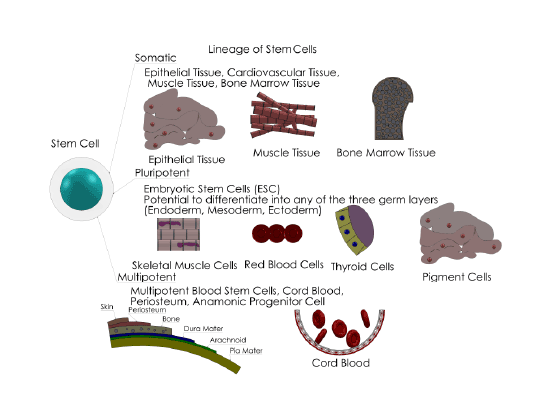
Figure 1: The most common stem cell types. Each one has their own unique pathway and includes several possible descriptions. A) Somatic Cells,
also known as adult stem cells, do not differentiate into a different type of cell. Rather, they stay among the cell groups inside of the niche it is held
in. It is capable of regenerating and reviving specific tissues it is grouped in. Somatic stem cells can be found in many places in the body, depending
on the niche location of each tissue. B) Pluripotent Stem Cells are stem cells that can differentiate into any type of cell and will thrive in any type of
tissue. Embryonic Stem Cells are the most common in this category. These cells have certain properties that can generate the three germ layers of the
cell. Most of these are found in neonatal material or embryos C) Multipotent Stem Cells are similar to pluripotent stem cells, but to renew itself it must
differentiate into a different type of cell. This specializes them and their self-renewing property is makes them capable of producing a few more cycles.
Multipotent stem cells are more commonly found in bone and cord blood.
Extrusion
One of the most common and made for industrial purposes, and most
preferred is three-dimensional extrusion [7,32,39]. By using a piezoelectric
pressure valve and a xyz-axis, extrusion accepts most biomaterials and
stem cells to create a layer-by-layer deposition. As the object is created,
the user has the ability to control the extrusion speed, thermodynamic
properties, and cell placement.
Creating the tissue itself is also efficient, too. Thanks to CAD/CAM
software [40], a tissue or organ can be scanned with computerized
tomography (CT) to generate the blueprints and target points to where
the cells will be positioned [32]. Timing is also appropriate, as the user is
able to control when the cells will be placed. This gives an advantage to
sol-gelation, if hydrogels are to be extruded through the material so that
they are not dehydrated when placed atop of the printer plate.
There is some debate in the extrusion process, as most extruders
are paired with a hot-end and plate is essentially a heat-bed [32]. With
client software, users are able to control if thermodynamics need to be
used or not. Should the materials be cryogenically frozen depends on
the experiment and the effects of cryo-freezing on stem cells. There is an
option to disable the thermodynamics to store cells and use them at a later
time (biomaterials won’t need to do so) [41].
In addition, there is also a concern on the shear forces of the piezoelectric
extruder. As a material is being dispositioned, it goes through the extruder
to form a viscous droplet. In doing so, there are forces that enact onto it
to make the materials rigid and precise so that it does not sputter onto
the plate [34,42]. However, this can be a risk as some cells are recorded to
decrease in viability after extrusion. Additionally, there should be enough
cells to engulf the structure the biomaterials formed, but that has yet to be
proven with more statistical data from other research groups.
In the end, there are modifications that can be done to the extruder,
some of which can dispense high viscosity materials. The newfound micro
extruder, which mimics micropipetting is able to generate more precise
prints and gives the advantage of recreating the intricacies of organ-specific
tissue functions [8,27] Scientists have also developed a piezoelectric
microfluidic chip [34], which borrows the idea of micropipetting and
reduces contact forces between the plate and extruder.
Inkjet printing
Similar to extrusion is the traditional inkjet printing. The mechanics of
a paper printer are the same, but instead of using ink and toner, bioinks
are in place of the cartridges [43].
Instead of using a plate, a solvent is sprayed onto the base where the
prints are to be dispensed on [41]. Inkjet printing has a small difference
from extrusion. Instead of releasing materials one at a time, the bioink is
sprayed over to a specific shape and pressed to form the geometric shape
designed. The solvent crosslink’s the bioink to the structure to form the
designed shape. Tissues and cells have been successfully printed from this
process.
Probably the most significant piece of inkjet technology is the bioink,
a heterogeneous fluid made of cells, proteins, and fluids to hydrate the
microenvironment. Inkjet technology was widely used in the mid-2000s,
before the age of three-dimensional printing extrusion. Before then, Dr.
Atala [44] developed a way to create heart valves using inkjet printing
technology, which inspired several scientists to try the same method.
The advantage of inkjet printing for biomaterials are similar to that of
extrusion, where prints can be controlled by will of the user and guided
by office software. Unlike extrusion, the prints avoid direct contact on
the plate, rather a solvent to guide the cells to migrate. The probable
disadvantage is cell lysing [35,41]. It does not have a dramatic effect on the
cells while being printed, but the concentration of fluid acting onto the
cell membrane can cause the cell to burst. Nevertheless, in a recent study,
approximately 10% of cells lysed [41]. The cause could be directly related
to the number of hydrogels that were in the bioink.
Laser-assisted printing
The most complex of all printing methods, laser-assisted printing is a
different realm of printing. The method start with are two glass slides, one
being the donor slide that has the encapsulated cells in hydrogels and the
collector slide which contains an additional hydrogel layer to reduce the
impact of the laser energy transfer [35,45]. When the laser is activated, it
shoots into a gold-film that covers the cells and prevents them from being
destroyed. This energy starts to absorb the hydrogels on the donor slide
and transfers them over to the collector slide. The cells are transferred to
their specific place based on the user’s CAD/CAM design and deposited.
Ironically, lasers may have no effect on the cell viability [11,45]. As the
cells are absorbed the gold plating acts as a protective shield for energy
absorbance and placed as designated [11]. It does introduce a new
technology and focuses more on cell absorbance through laser energy.
Photopolymerization
Similar to laser-assisted printing, photopolymerization utilizes light
energy to create objects [46]. The materials are placed into a resin bath
and covering it is the plate. The object is printed using UVA light that
photopolymerizes the materials depending on their cross-sections.
Wherever the UVA hits the material hardens and while UVA is flashed the
base plate lifts itself up to display the object [5].
The biggest advantage is the printer’s ability to absorb light energy and
harden cells fast and the process is quicker than most conventional threedimensional
printers [5,47]. The drawback to the printer’s method is the
UVA radiation. If the radiation is strong enough, stem cells inside may
differentiate and become carcinogenic. In one test, researchers measured
the affects of UVA radiation on cells using a cytotoxicity test in the course
of three days [46]. Over time, researchers found that most cells did
die over the process only to self-renew after the cells’ “lag phase.” [46].
Eventually stem cells started to culture and overpopulate the biomaterial
for degradation.
The biggest drawback was that this was designed only for a twodimensional
microenvironment. Cells are required to thrive in a threedimensional
microenvironment that would model in vivo conditions. As
it was not the case, this method would not be relevant towards the use of
creating tissues and organs (Figures 2 and 3).
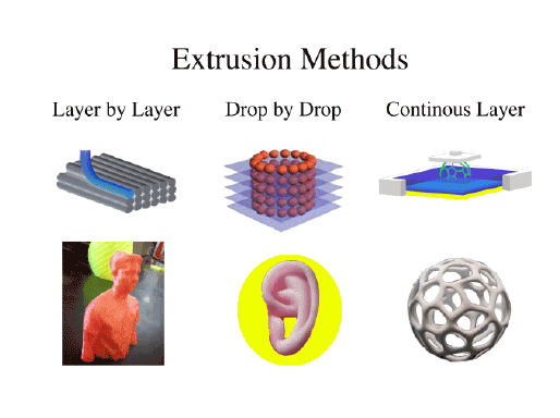
Figure 2: Extrusion three-dimensional printing methods and the prints that they have produced. Layer by Layer extrusion is a traditional approach to
creating a three-dimensional object. It divides the object into several cross sections and adds a layer into the appropriate cross-sectional area. Inkjet
printing and extrusion-based printing utilize this method and have the most precision. Drop by Drop extrusion is almost comparable to scaffolding with
cells. During the printing process, a scaffold is generated through a flexible biomaterial onto the printing surface. As it is printing, an additional nozzle
extrudes nanoparticles that contain cells. Once the biomaterial is placed onto the printer bed, the droplet nozzle carefully places cells onto the object
to create a cell culture. Continuous Layer Extrusion is a new method that has not been fully studied yet to its potential. Essentially, a plate slowly rises
above a viscous liquid. As it escalates, the material is photopolymerized by UVA light from beneath the plate. As photopolymerization occurs, crosssectional
layers are made and keep their rigidity, which keeps it stuck to the plate until the object is fully extruded.
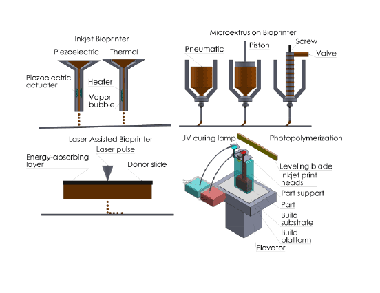
Figure 3: For each extrusion method, there are different forces and techniques applied per extruder. No extruder is deemed the same way, since
each one is specific to the biomaterial printed. Inkjet Bioprinters can use a piezoactuator to apply forces for fast extrusion or thermal heat to melt the
material as it drips down to the surface in a concise manner. Microextrusion is a more specific extruder than inkjet bioprinters. Since the materials are
much smaller, different forces can be used to dispense the material. These range from pneumatic, screw, and piston. Laser-Assisted Printing is unique
to those of the previous to extrusion methods. By using a laser, particles are guided from an energy absorbing layer to the donor slide. During this
transition, particles accumulate onto the slide and are not damaged by the laser’s
Considerations
Regardless of which method is chosen, it is safe to conduct a threedimensional
stem cell tissue or organ. Each method does have certain
advantages and drawbacks, but will give the best-produced result from
their system (Table 1). However, there are a couple points to consider
as to how these models are designed and created. When a stem cell
overpopulates the biomaterial, it degrades to give rise to the new tissues
naturally.
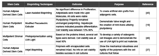
Table 1: Analysis of Bioprinting Methods with Biomaterials and Stem Cells
However, after the microenvironments the cells were once present in
are destroyed, how will the new tissues work as a system together? This
is what researchers call vascularization, the process in which vascular
systems are introduced into the system. This includes veins, capillaries,
and other blood pathways that filter and transfer materials into tissues.
It is not certain that a stem cell environment is capable of recreating a
vascular system, as the cells are designed to differentiate into the tissue
cell type.
So far, only a few methods have been published on the vascularization of
artificial tissues [31,47] with one of them includes artificially creating the
vascular system by hand. As the materials are deposited onto the printer
base, vascular grafts and capillaries are placed below the extruder to be
enveloped by the new material it will grow around. Once the biomaterials
degrade, the idea is to get the stem cells to differentiate not only into the
tissue cell type but into the vascular type as well. This may involve the
need to create a separate biomaterial that influences differentiation into
vascular grafts. Much has yet to be said about the subject.
Another consideration is the difference between the effects of in vitro
and in vivo environments for stem cells. On one hand the stem cells may
appear to differentiate as planned post-printing and may seem acceptable
for transplantation.
Yet, there are scarcely any findings on the effects of transplantation in
vivo. Dr. Atala [44] at Wake Forest University demonstrated it in 2011 on
a student named Luke, who received an artificial liver transplant created
in vitro by an inkjet printer. Luke is currently alive and well today, and
has not experience any terminal problems so far. Dr. Atala’s method is
briefly published, so it is not certain as to what materials or cells he did
use [6,7,10].
Modeling an environment that mimics the homeostatic functions of
the body is the best way to determine the functionality of artificial tissue.
It would dramatically reduce the risk of mammalian testing and risk of
implantation. A model has yet to be found, one that would stimulate
vascularization, metabolic processes, and immune reactions if the body
undergoes an infection. All things considered, these are just a few of what
would simulate the environment.
Lastly, there is still a need to consider the effects of stem cells and their
possibility to differentiate into unwanted cells. The printing methods
aforementioned can destroy cells, but in some cases stem cells can become
carcinogenic. For example, UVA radiation in photopolymerization
printing can create a tumorigenic cell type and stem cells could
overpopulate into a similar lineage, thus destroying the tissue and ruining
the model. The same could be said for extrusion, depending on the
user’s choice for thermodynamics. The hot end of the extruder can reach
temperatures of up to 200C which could cause a negative reaction in the
stem cells during extrusion. In the end, stem cell printing has a few things
to improve on. The methods used are sufficient, but some intricacies need
to be improved on.
Regardless of which method is chosen the most critical thing to
understand is the use of stem cells in a microenvironment and the
biomaterials that compliment them.
Acknowledgement
The authors would like to thank the National Institute of General
Medical Sciences of the National Institutes of Health under Award
Numbers; 8UL1GM118979-02; 8TL4GM118980-02; 8RL5GM118978-02,
and the California State University Long Beach’s College of Engineering
Faculty Startup Funds, Mini-Grant/Summer Stipend (MGSS) grant, and
seed grant.