
Full Text
Dr. Seyed Saeid Zamanieh Shahri, MD* Dr. Sonia Sayyedalhosseini, MD*
Faculty Members in California Northstate University, CNSU-University Professors in Losrios Community College District, USA*Corresponding author: Dr. Seyed Saeid Zamanieh Shahri, MD, Faculty Member in California Northstate University, CNSU-University Professor in Losrios Community College District, USA, Tel: (916)724-9485; E-mails: saeid.zamanieh@cnsu.edu, zamanis@flc.losrios.edu
Dr. Sonia Sayyedalhosseini, MD, Faculty Member in California Northstate University, CNSU-University Professor in Losrios Community College District, USA, E-mails: sonia.sayyedalhosseini@cnsu.edu, sayyeds@flc.losrios.edu
The domain of analysis of medical images has been developed increasingly over the years. With the increase in number of scan instruments and increase in size and volume of data, it is essential to develop the medical image analysis methods as much as they are developed. The advancements have caused development of extraction processes with high precision, which can lead to changing the images to analyzable data. The data are useful in decision making, diagnosis and treatment. Currently, there are many interests in field of understanding image from radiology images. In this study, it has been attempted to use image segmentation to detect the tissue in radiology images. For segmentation of images, thresholding is used, which is one of the most commonly used methods in field of image segmentation. This method is used in gray images to separate the pixels with higher brightness level from the pixels with lower brightness level. At the end, using this method, the lung is separated from other tissues in radiology images. The results of these segmentations are analyzed and the results showed high precision and accuracy of the proposed method.
Thresholding; Segmentation; Radiology images
Today, hospital environment is equipped to medical scanners, which provide valuable information to help diagnosis or treatment of a special disease. Computerized Tomography (CT), Magnetic Resonance Imaging (MRI), ultrasonography and Positron Emission Tomography (PET) are some examples of imaging methods, which are mostly used. Experienced radiologist can get lots of information with observation of the images produced by a scanner. To separate the image data to the components of that, data extraction or segmentation of a special area, radiologic image segmentation is required. Image segmentation can provide information such as size and volume about the desired area. Various methods are presented for radiology image segmentation, which are described here.
Artificial intelligence algorithms, especially deep learning, have shown considerable advancement in image diagnosis works. Methods such as circular neural networks have gained numerous programs in field of analysis of medical images and are taking the progress of the programs rapidly. In traditional form, in radiology, the educated physicians evaluate medical images for diagnosis, detection and monitoring diseases. Artificial intelligence have been used for automatic detection of complicated patterns in radiology images such as data extraction and presentation of quantitative evaluations in wide range.
Using Artificial Intelligence (AI) is powerful tool for image analysis, which is evaluated increasingly by radiology specialists. Segmentation of an image of regions with homogenous tissue, which is commonly recognized as image segmentation, is widely used as an important method for high level understanding and can decrease complexity of content analysis considerably. Image segmentation and high level methods have been widely designed to imitate specifications of visual understanding of human (e.g. object detection and image message understanding). One of the performance measurement methods can be segmentation of qualitative and quantitative comparison of the obtained results of method with human segmentation [1].
Liu C, et al., conducted an artificial intelligence project for automatic analysis of image in radiology to encourage running that in radiology units [2].
Mazurowski MA, et al., discussed generally on radiology and the opportunities of using deep learning algorithms. They provided a study on deep learning, which is applied on radiology. They organized types of special tasks they try to solve wide range of deep learning algorithms. Finally, they shortly discussed on the opportunities and challenges for inclusion of deep learning in future radiology operation [3].
Singh P studied on a neutrosophic-entropy based adaptive thresholding segmentation algorithm in MR images of Parkinson’s disease. Experimental results of this study showed that NEATSA can segment the main regions of MR images very clearly compared to the well-known methods of image segmentation available in literature of pattern recognition and computer vision domains [4].
Huang YP, Singh P, and Kuo H studied on a hybrid fuzzy clustering approach for the recognition and visualization of MRI images in Parkinson’s disease. Experimental results were provided by comparing PD patients’ segmented MRIs with seven well-known image segmentation methods. The performance showed an improvement in the average mean squared error, peak signal-to-noise ratio and Jaccard similarity coefficient compared to other image segmentation methods [5].
The other study by Singh P, presented a new method for MR image segmentation, which consisted of two main parts as: (a) NeutrosophicEntropy Based Clustering Algorithm (NEBCA), and (b) HSV color system. The proposed method showed effective segmentation differentiation regions of MR images and can clearly represent those segmented regions [6].
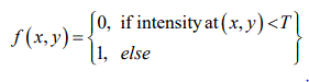
Image binarization is the process of separation of amounts of pixel to two black and white groups, in which black is the default and white is background. In this method, pixel division is done in comparison to the threshold value. Thresholding [7] is a basic important method in field of image segmentation, which is commonly used for gray images and is aimed at separating the pixels with higher brightness from those with lower brightness. Thresholds can be divided to two groups of local thresholds [8] and global thresholds [9]. In global threshold, the amount of threshold in whole image is considered fixed. Binary image is obtained by global threshold T for a gray image as follows:
Threshold T can be obtained using various methods. The most commonly used method is using histogram-based methods. If image includes an object and a default with homogenous brightness, then it has a bimodal histogram (Figure 1). The threshold value is equal to minimum local located in distance between two histogram peaks.
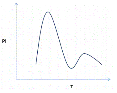
Figure 1: Histogram of a bimodal image.
Calculative complexity is low in the methods based on global threshold [10]; although this method can be used for image segmentation at the time that the intensity of image brightness has bimodal distribution. The better alternative method for global threshold can be using local threshold that segments the image to several sub-regions [11]. Threshold values in each sub-region can be different values and hence, threshold values can vary in whole image.
The first step in proposed algorithm is to obtain the histogram of interested region (that is close to central part of image). Minimum and maximum values of the histogram can specify the area of gray level in iterated thresholding. Then, in the gray area specified, algorithm should be iterated 7 times and the threshold in each selected step should be more than the previous step. Per iteration of algorithm, a binary image is created, in which only pixels with less gray levels than threshold remain light. 8-point connection map is used to make counters around the light and continuous pixels. Profile is made in mass center of each counter and this is then analyzed that whether the counter includes pixels belonged to lung or not. Pixels of counter that are out of lung are separated from the binary image that is made in later iterations. After seventh iteration, a set of answers of primary counters is obtained. To separate the lung completely, local thresholding method is used in first counter. The final counter set is obtained based on combined binary image made by means of thresholding certain local pixels of interested area. Then, the counter is normed and ball rolling algorithm [12] is run for that, so that the heterogeneity and problems of counter are resolved. Then, the process of determining costophrenic angles is done and exact segmentation of lung is obtained (Figure 2 and 3).
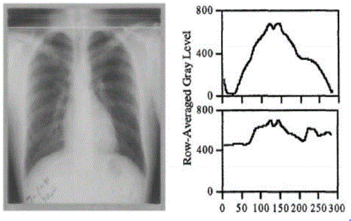
Figure 2: Schema of lung and determining the mid-line (computer has specified the beginning of lung and middle part with shine horizontal and vertical lines).
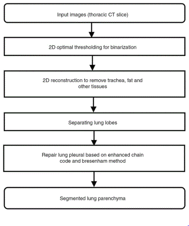
Figure 3: Flowchart diagram for segmentation method.
Global iterative thresholding algorithm
At the first, sobel filter [13] is convolved with lower ¼ and right side of the image to highlight the diaphragm lines and lighter edges [2]. Therefore, it prevents creation of large counters created because of unifying with intestine. Pixels of image ¼ that seem more powerful are filtered by image for suitable gradient and the pixels have desired value of 999. The value is large enough to go out of the realistic threshold limit in global histogram analysis. The increase in gray level acts as an assumed border, which prevents influence of lung counter to the area of stomach gases. Global histogram is the first step of segmentation that is illustrated in figure 1. To obtain sufficiently homogenous histograms, histogram estimations are restricted to 141*181 pixels (the early 140 pixels include the image). An area of image includes pixels with high density (low gray level) belonged to lungs and pixels with low density (high gray level) belonged to diaphragm, intercostal and chest edges.
Therefore, the histogram obtained from this area is bimodal and includes one peak to center of lower gray level (lungs) and another peak to upper gray level (intercostal area) (Figure 4).
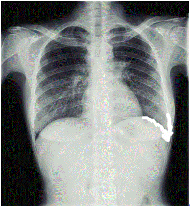
Figure 4: Applying Sobel filter for lower 1/4 and right side of image, gray level 999 with pixels filtered with value higher than threshold.
The purpose of global thresholding is to use histogram to find appropriate gray level to separate the gray levels belonged to pixels inside lung from the pixels out of that. It has been proved that only using a gray level can’t be useful to find suitable threshold. If the values of gray level are very small, the counters around lung are insufficient too and if the values of gray level are very large, the lung area or the outside area is placed in a single counter. Using iterative global thresholding solves the problem and an area is used for threshold instead of a value. The slope of global histogram is used to detect the gray levels, in which peak relevant to lung happens and the gray levels that happen minimum between lung and intercostal area (Figure 5). Then, iterative global thresholding technique used the gray level between the two points. The iterative process is defined with frequent thresholding in 7 points with equal distances in this limit. In first iteration of algorithm and by least gray level as threshold (from the existing 7 levels), binary image is created. If gray levels of image are less than threshold, the pixels are shown light in the binary image. As a result, a far counter is placed in the obtained binary image and is gradually improved. Some geometrical specifications of counter such as the center, compression and counter length and counter area can be estimated. The later iterations with larger thresholds can cause creation of additional binary images.
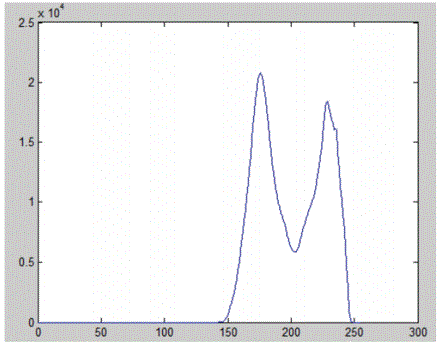
Figure 5: Global histogram that shows bimodal distribution (the arrows show pixels belonged to lung and intercostal area).
Counters are again created in areas containing continuous and light pixels and the geometric parameters of each counter are then estimated. The iterative process of the algorithm is vital for suitable lung segmentation. The position and minimum and maximum gray level and the center of pixels are used to determine that whether the counter has encompassed the lung area or not. Due to center situation, the center profile that is continued to the upper or lower part of image is analyzed to obtain the additional information on counter. If it is found with checking the center that counters is out of lung area, all pixels of image in counter are regarded as wrong pixels and the right counter is created in next iteration (Figure 6) and the outer areas are not again combined with lung area in next iterations.
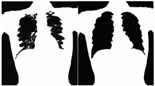
Figure 6: Global histogram that shows bimodal distribution (the arrows show pixels belonged to lung and intercostal area).
The binary image made in figure 6 has been created with checking central pixels in early iterations. In 7 iterations, the process of using threshold (to make binary image and to detect counter and to stop some pixels) is done through checking central pixel. Opening morphology operator [14,15] with 3*3 cornel is done after 3 iterations on binary image. Opening operator (erosion operator followed by dilation operator) can delete many artifacts of the image (Figure 7 and 8).
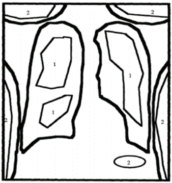
Figure 7: Global histogram that shows bimodal distribution (the arrows show pixels belonged to lung and intercostal area).
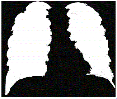
Figure 8: Global histogram that shows bimodal distribution (the arrows show pixels belonged to lung and intercostal area).
The final result of global thresholding algorithm is presented in the following and the lung area is separated by a counter (Figure 9).
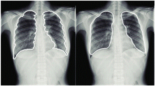
Figure 9: Global histogram that shows bimodal distribution (the arrows show pixels belonged to lung and intercostal area).
Local thresholding
Global thresholding can make problem with showing the real area of lung properly. To correct the position of points, after global thresholding, local thresholding algorithm is used (Figure 10).
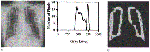
Figure 10: Global histogram that shows bimodal distribution (the arrows show pixels belonged to lung and intercostal area).
The size of interested area is selected to 31*31 pixels. Although all early counters are maintained, local thresholding is done only on two larger counters of the image. Then, if the counters take some different parts of chest, the interested area is allocated to one of them. Gray level analysis is done on pixels of each interested area and based on position of each interested area, good threshold is selected for them. After finding the counter, combined binary image is made to obtain final counter and then the counter is normed (as it was previously explained for the early counter).
Ball-Rolling algorithm
The final counter has sometimes some deviations appeared as protrusions or indentations. The deviations commonly happen in peak point of the area. For example, the shoulder area may be wrongly considered as a part of counter. To avoid such mistakes, rolling-ball algorithm is innovated. Ball-rolling has orbital structure that processes the image with moving on gray levels of image and using opening and closing morphology operators. Ball is an understanding of tubular object shown on 3-D screen showing gray levels as a function of distance. The main purpose of this algorithm is removing heterogeneity of image and removes them by the rolling ball and paves the curves. If the curve is not in contact with the ball, that point is shown and specified as the indentation point (like area 2 in Figure 11 and 12).
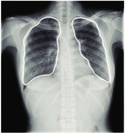
Figure 11: Global histogram that shows bimodal distribution (the arrows show pixels belonged to lung and intercostal area).
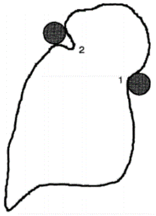
Figure 12: Global histogram that shows bimodal distribution (the arrows show pixels belonged to lung and intercostal area).
Indentation of point 1 is not to some extent that the area can be considered as a real indentation; although point 2 is considered as an indentation, because whole counter area is not in contact with ball (Figure 13 and 14).
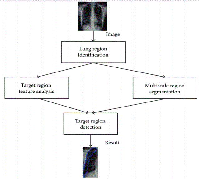
Figure 13: Global histogram that shows bimodal distribution (the arrows show pixels belonged to lung and intercostal area).
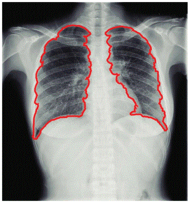
Figure 14: Global histogram that shows bimodal distribution (the arrows show pixels belonged to lung and intercostal area).
The main purpose of this study is segmentation of tissues in radiology images. In this regard, an image segmentation approach in radiology images is presented. In this segmentation method that has higher precision than the image segmentation by human, thresholding method is used for image segmentation. Segmentation of lung tissue was done in s\chest radiology images and the results of the segmentation were presented as figures in previous sections. The results obtained from this study showed that using the proposed method, one can separate the lung tissue with high precision and accuracy in radiology images. For further studies, it would be better to take the segmentation in other types of radiology images and it is also suggested to extract quantitative results such as volume, form and size of tissue. The problems with thresholding method can be inattentiveness to interaction of pixels. As the pixels belonged to a tissue in image may be discrete, segmentation error may be created in this method. High sensitivity to noise can be also another problem with this method, which may cause inaccurate segmentation. In this study, the innovative method based on area-based segmentation is used for lung image segmentation and has provided better answers than previous methods and the lung segmentation precision from other areas is very high in this method.
- Ramgopal K, Rahamatkar S (2019) Medical Image Segmentation: An Advanced Approach. In: Bit A, Bhattacharya P, Paul S (eds) Early Detection of Neurological Disorders Using Machine Learning Systems. IGI Global 292-321. [Ref.]
- Liu C, Chen L, Schroff F, Adam H, Hua W, et al. (2019) Auto-DeepLab: Hierarchical Neural Architecture Search for Semantic Image Segmentation. Proceedings of the IEEE Conference on Computer Vision and Pattern Recognition, Cornell University [Ref.]
- Mazurowski MA, Buda M, Saha A, Bashir MR (2019) Deep learning in radiology: An overview of the concepts and a survey of the state of the art with focus on MRI. J Magn Reson Imaging 49: 939-954. [Ref.]
- Singh P (2020) A neutrosophic-entropy based adaptive thresholding segmentation algorithm: A special application in MR images of Parkinson’s disease. Artif Intell Med 104: 101838. [Ref.]
- Huang YP, Singh P, Kuo H (2020) A Hybrid Fuzzy Clustering Approach for the Recognition and Visualization of MRI Images of Parkinson’s Disease. IEEE Access 8: 25041-25051. [Ref.]
- Singh P (2020) A neutrosophic-entropy based clustering algorithm (NEBCA) with HSV color system: A special application in segmentation of Parkinson’s disease (PD) MR images. Computer Methods and Programs in Biomedicine 189: 105317. [Ref.]
- Huang S, Yang S, Roberts J, Chen K (2011) Threshold Voltage Instability in Al2O3/GaN/AlGaN/GaN Metal? Insulator? Semiconductor HighElectron Mobility Transistors. Jpn J Appl Phys 50: 110202-110203. [Ref.]
- Lagger P, Reiner M, Pogany D, Ostermaier C (2014) Comprehensive Study of the Complex Dynamics of Forward Bias-Induced Threshold Voltage Drifts in GaN Based MIS-HEMTs by Stress/Recovery Experiments. IEEE Transactions on Electron Devices 61: 1022-1030. [Ref.]
- Yang S, Liu S, Liu C, Tang Z, Lu Y, et al. (2014) Thermally induced threshold voltage instability of III-Nitride MIS-HEMTs and MOSCHEMTs: Underlying mechanisms and optimization schemes. 2014 IEEE International Electron Devices Meeting. [Ref.]
- Lagger P, Schiffmann A, Pobegen G, Pogany D, Ostermaier C (2013) Very Fast Dynamics of Threshold Voltage Drifts in GaN-Based MISHEMTs. IEEE Electron Device Letters 34: 1112-1114. [Ref.]
- Johnson DW, Lee RTP, Hill RJW, Wong MH, Bersuker G, et al. (2013) Threshold Voltage Shift Due to Charge Trapping in Dielectric-Gated AlGaN/GaN High Electron Mobility Transistors Examined in Au-Free Technology. IEEE Transactions on Electron Devices 60: 3197- 3203. [Ref.]
- Joseph AB, Claude LB, William TC, Thomas ED, Wayne FE (2001) Rolling ball connector. United States Patent Application 20010002330. [Ref.]
- Gao W, Zhang X, Yang L, Liu H (2010) An improved Sobel edge detection. 3rd International Conference on Computer Science and Information Technology 5: 67-71. [Ref.]
- Schegloff EA, Sacks H (2009) Opening up closings. Semiotica 8: 289- 327. [Ref.]
- Seeman P, Ulpian C, Bergeron C, Riederer P, Jellinger K, et al. (1984) Bimodal distribution of dopamine receptor densities in brains of schizophrenics. Science 225: 728-731. [Ref.]
Download Provisional PDF Here
Article Type: REVIEW ARTICLE
Citation: Shahri SSZ, Sayyedalhosseini S (2020) Tissue Separation in Radiology Images with the Help of Image Segmentation. J Radiol Imaging Diagn 1(1): dx.doi.org/10.16966/JRID.101
Copyright: © 2020 Shahri SSZ, et al. This is an open-access article distributed under the terms of the Creative Commons Attribution License, which permits unrestricted use, distribution, and reproduction in any medium, provided the original author and source are credited.
Publication history:
SCI FORSCHEN JOURNALS
All Sci Forschen Journals are Open Access
New Journals
Best viewed in Google Chrome | Mozilla Firefox | Microsoft Edge
Copyright © 2024 Sci Forschen Inc., All Rights Reserved