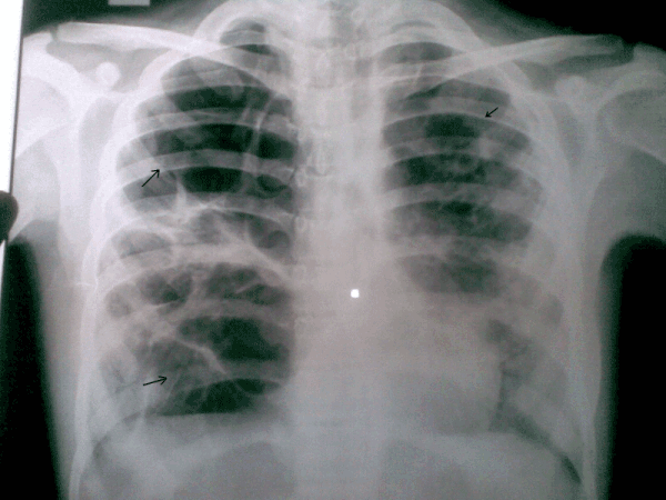
Figure 1: Plain chest radiograph (postero anterior view) revealing giant emphysematous bullae in both lung fields.


Indumathi CK* Savita Murthy
Department of Pediatrics, St. John’s Medical College Hospital, Sarjapur Road, Bangalore, India*Corresponding author: Dr. Indumathi CK, Associate Professor, Department of Pediatrics, St. John’s Medical College Hospital, Sarjapur Road, Bangalore - 560 034, Karnataka, India, Tel: 9180 - 2206 5456; 91 9448089891; Fax: 9180- 2553 0070; E-mail: ckindumathi@gmail.com
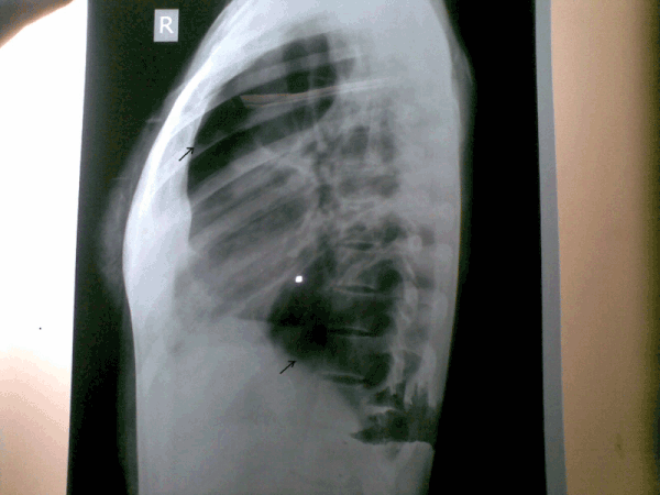
A 15 year old girl presented with history of fever and cough of 1 month duration. Chest radiograph revealed bilateral parenchymal infiltrations with cavities. Sputum Acid Fast Bacillus was positive. She was started on ATT (Anti Tubercular Therapy). At 6 months of treatment, she developed exertional dyspnoea. Repeat chest radiograph revealed giant emphysematous bullae in both lung fields (Figure 1 and Figure 2).

Figure 1: Plain chest radiograph (postero anterior view) revealing giant emphysematous bullae in both lung fields.

Figure 2: Plain chest radiograph (lateral view) revealing giant emphysematous bullae.
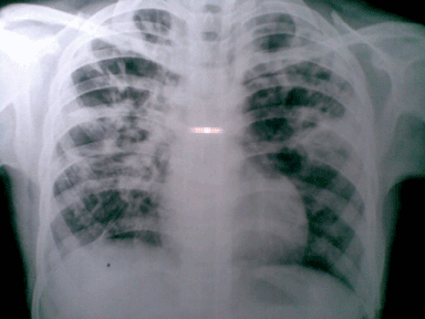
Development of emphysematous bullae during healing of Pulmonary Tuberculosis has been reported in 8 children, last being in 1967 [1-3]. Lesions appear soon after ATT (7-60 days) and reach maximum distension at 7 months. They disappear with time (6-15 months) and have favourable outcome [3]. One known complication is pneumothorax. Treatment is supportive. Excision has been advocated for persistent symptomatic lesions confined to localised areas. Our patient received supportive treatment. Currently she is well and chest X-ray at the end of 1 year revealed partial resolution of bullae (Figure 3).

Figure 3: Follow up plain chest radiograph (postero anterior view) revealing resolution of emphysematous bullae.
Download Provisional PDF Here
Article Type: Images in clinical practice
Citation: Indumathi CK, Murthy S (2015) Fifteen Year Old Girl with Giant Emphysematous Bullae. J Infect Pulm Dis 1(1): doi http://dx.doi. org/10.16966/2470-3176.101
Copyright:© 2015 Indumathi CK. This is an open-access article distributed under the terms of the Creative Commons Attribution License, which permits unrestricted use, distribution, and reproduction in any medium, provided the original author and source are credited.
Publication history:
All Sci Forschen Journals are Open Access