Abstract
To examine whether the clinical severity and antibiotic susceptibility of β-Lactamase-negative ampicillin-resistant Haemophilus influenza (BLNAR) causing adult pneumonia has changed with the times, we divided the study period into two periods from January 1, 2006 to May 31, 2011 and from June 1, 2011 to December 31, 2016. Then we compared the clinical severity and antibiotic susceptibility of BLNAR between those two periods. The result was that BLNAR holds large majority (65.5% to 75.5%) as pathogen of Haemophilus influenzae pneumonia in adult throughout whole period. Antibiotic susceptibility of BLNAR barely changed throughout the whole period. The pneumonia caused by BLNAR in adults during Period II has higher severity score and needed longer hospitalization than that during January 2006 to May 2011. Finally, our investigation suggests that the pneumonia caused by BLNAR gradually becomes more severe.
Keywords
BLNAR; Haemophilus influenza; Adult pneumonia; Drug resistance; Respiratory infection
Introduction
Haemophilus influenzae is one of the major pathogen in communityacquired pneumonia in adults [1]. Recently β-lactamase-negative ampicillin-resistant strain (BLNAR) has increased in the world especially in Japan [2], but the clinical impact of this finding in not completely understood. The aim of the study is confirm the change of the epidemiology data and evaluate the clinical impact of pneumonia due to H.influenzae.
Materials and Methods
The medical records of all patients who admitted to Department Respiratory Medicine and Allergology, Kindai University Nara hospital for pneumonia by H.influenzae were reviewed from January 1, 2006 to May 31, 2016. The study period was divided into two periods from January 1, 2006 to May 31, 2011 and from June 1, 2011 to December 31, 2016. We compared the number of patients, age, and severity of pneumonia, duration of hospitalization or drug resistance between the two study periods.
Patients of each period ware divided into three groups according to antimicrobial resistance of H.influenzae, such as, BLNAR group, β-lactamase negative ampicillin-sensitive strain (BLNAS) group and β-lactamase positive ampicillin-resistant strain (BLPAR) group.
Diagnostic criteria of H.influenzae pneumonia was defined by a new filtrate on radiograph and the presence of 1 or several of the following acute respiratory symptoms: productive cough, dyspnea, auscultate findings of crackles at breath, body temperature exceeding 37.5°C and quantitative cultures of good quality sputum containing >25 granulocytes and <10 epithelial cells per power field which was defined as H.influenzae.
Antibiotic susceptibility was defined according to guidelines of Clinical and laboratory Standards Institute by the disc diffusion method [3]. The nitrocefin disc method was used to detect whether the isolated strains produced β-lactamase. Susceptibility of BLNAR for each Ampicillin/ Sulbactam (ABPC/SBT), Cefepime (CPFN), Imipenem/Cilastatin (IPM), Azithromycin (AZM) or Levofloxacin (LVFX), each was compared between Period I and Period II.
Criteria of severity score of pneumonia at admission was defined by the criteria of Japanese Respiratory Society which is modified criteria of British Thoracic Society [4-6]. Diagnostic criteria that consists of five items includes the following topics: age (70 years of age or older male or 75 years of age or older female), dehydration or urea nitrogen concentration in the blood more than 21 mg/dl, 90% or less transcutaneous oxygen saturation, confusion, 90 mmHg or less systolic blood pressure. It represents the severity in total points as 1 point per corresponding criterion of each five criteria. If none of the five criterion was present, total points was 0.
All data were expressed as median and means ± SD. By dispersion test each two groups to be compared turned out not having homogeneity of variance. The data of each group was examined by Chi-square test or Shapiro-Wilk test to determine whether it is a normal distribution and all ware judged to be non-normal distribution. From these results differences between qualitative variables were examined for statistical significance using Brunner-Munzel test. A P-value less than 0.05 was considered to be a statistically significant difference. This retrospective study was conducted in accordance with the principles expressed in the Declaration of Helsinki.
Results
The total number of patients in this study was 82. In the period from Period I we found that β-lactamase-negative ampicillin-sensitive strain (BLNAS) accounted for 24.1%, BLNAR accounted for 65.5%, β-lactamasenegative ampicillin resistant strain (BLPAR) accounted for 10.3%. On the other hand, for the period from Period II, we found that BLNAS accounted for 18.9%, BLNAR accounted for 75.5%, BLPAR accounted for 5.6% (Table 1). Mean age of BLNAS cases was 73.9 ± 15.0 years old and median was 76.0 years old, that of BLNAR cases is 63.3 ± 20.5 years old and median was 64.0 years old, that of BLPAR cases is 63.0 ± 11.0 [7] years old and median was 73.0 years old in the period from January 2006 to May 2011,in the other hand, mean age of BLNAS cases was 72.2 ± 9.38 years old and median was 72.5 years old, that of BLNAR cases is 71.6 ± 11.4 years old and median was 71.0 years old, that of BLPAR cases is 75.0 ± 9.59 years old and median was 77.0 years old in the period from Period II (Table 1 and Figure 1). In comparison Period I and June 2011 to December 2016, there was no statistical significant difference in patient’s age of each BLNAS, BLNAR, BLPAR cases. Mean duration of hospitalization of BLNAS cases was 16.7 ± 12.2 days and median was 15.0 days, that of BLNAR cases was 12.8 ± 6.27 days and median was 11.0 days, that of BLPAR cases was 10.0 ± 2.83 days and median was 10.0 days in the period from January 2006 to May 2011. In the other hand, average duration of hospitalization of BLNAS cases was 21.8 ± 16.6 days and median was 15.0 days, that of BLNAR cases was 17.2 ± 9.48 days and median was 15.0 days, that of BLPAR cases was 13.0 ± 1.83 days and median was 13.0 days in the period from Period II (Table 1 and Figure 2). In comparison Period I and June 2011 to December 2016, there was statistical significant difference in duration of hospitalization of BLNAR cases (P=0.008). The duration of hospitalization of BLNAR cases in Period II is statistically longer than that in January 2006 to May 2011. In comparison January 2006 to May 2011and June 2011 to December 2016, there was no statistical significant difference in each BLNAS, BLPAR cases. There were six patients, three in BLNAR cases and three in BLNAS cases, those duration of hospitalization was over 30 days. Table 2 shows the details of the patients. Regardless of BLNAR or BLNAS, most were elderly people. There was also a tendency for the severity score to be high and COPD was the majority of underlying disease.
| |
January 2006 to
May 2011 |
June 2011 to
December 2016 |
P
value |
| BLNAS |
|
|
|
| Total number |
7 (male 5, female 2) |
10 (male 3, female 7) |
|
| % of total H.Influenzae pneumonia |
24.1 |
18.9 |
|
| Median (mean) age |
76.0 (73.9) ± 15.0 |
72.5 (72.2) ± 9.38 |
0.33 |
| Median (mean) duration of hospitalization |
15.0 (16.7) ± 12.2 |
15.0 (21.8) ± 16.6 |
0.247 |
| Median (mean) severity score of pneumonia |
2.00 (1.83) ± 1.27 |
1.00 (1.10) ± 1.20 |
0.525 |
| BLNAR |
|
|
|
| Total number |
19 (male 9, female 10) |
40 (male 24, female 16 ) |
|
| % of total H.Influenzae pneumonia |
65.5 |
75.5 |
|
| Median (mean) age |
64.0 (63.3) ± 20.5 |
71.0 (71.6) ± 11.4 |
0.101 |
| Median (mean) duration of hospitalization |
11.0 (12.8) ± 6.27 |
15.0 (17.2) ± 9.48 |
0.008 |
| Median (mean) severity score of pneumonia |
1.00 (0.890) ± 0.99 |
1.00 (1.50) ± 1.09 |
0.017 |
| BLPAR |
|
|
|
| Total number |
2 (male 1, female 1) |
4 (male 1, female 3) |
|
| % of total H.Influenzae pneumonia |
10.3 |
5.6 |
|
| Median (mean) age |
73.0 (63.0) ± 11.3 |
77.0 (75.0) ± 9.59 |
0.334 |
| Median (mean) duration of hospitalization |
10.0 (10.0) ± 2.83 |
13.0 (13.0) ± 1.83 |
0.15 |
| Median (mean) severity score of pneumonia |
0.00 (0.00) ± 0.00 |
1.00 (1.00) ± 0.83 |
0.029 |
Table 1: BLNAS, BLNAR and BLPAR groups.
P value was calculated by Brunner-Munzel test.
- BLNAS; β -lactamase negative ampicillin-sensitive strain group
- BLNAR; β -lactamase-negative ampicillin-resistant strain group
- BLPAR; β -lactamase positive ampicillin-resistant strain group
| Duration (days) |
Age (years old) |
Sex |
Severity score |
Underlying disease |
Pathogen |
| 36 |
71 |
Male |
2 |
COPD |
BLANR |
| 47 |
64 |
Male |
0 |
COPD |
BLANR |
| 56 |
78 |
Male |
3 |
COPD |
BLANR |
| 60 |
83 |
Male |
2 |
COPD |
BLNAS |
| 42 |
76 |
Female |
3 |
none |
BLNAS |
| 43 |
84 |
Male |
3 |
COPD, Diabetes |
BLNAS |
Table 2: Details of the patients those duration of hospitalization were over 30 days.
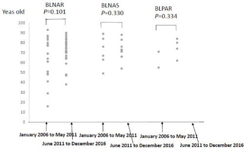
Figure 1: Age of the patients.
P value was calculated by Brunner-Munzel test.
BLNAS; β -lactamase negative ampicillin-sensitive strain group
BLNAR; β -lactamase-negative ampicillin-resistant strain group
BLPAR; β -lactamase positive ampicillin-resistant strain group
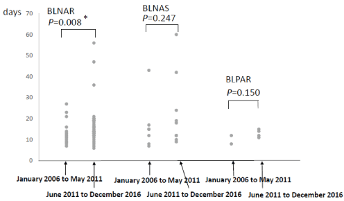
Figure 2: Duration of the hospitalization.
P value was calculated by Brunner-Munzel test.
BLNAS; β -lactamase negative ampicillin-sensitive strain group
BLNAR; β -lactamase-negative ampicillin-resistant strain group
BLPAR; β -lactamase positive ampicillin-resistant strain group
Mean severity score of pneumonia of BLNAS was 1.83 ± 1.27 and median was 2.00, that of BLNAR was 0.89 ± 0.99 and median was 1.00, that of BLPAR was 0.00 ± 0.00 and median was 0.00 in the period from January 2006 to May 2011, in the other hand average severity score of pneumonia of BLNAS was 1.10 ± 1.20 and median was 1.00, that of BLNAR was 1.50 ± 1.09 and median was 1.00, that of BLPAR was 1.00 ± 0.83 and median was 1.00 in the period from Period II (Table 1 and Figure 3). In comparison Period I and June 2011 to December 2016, there was statistical significant difference in severity points of BLNAR cases (P=0.017). There was statistical significant difference in BLPAR cases either (P=0.029). The severity points of pneumonia of BLNAR cases or BLPAR cases in Period II was statistically higher than that in January 2006 to May 2011. In the other hand, there was no statistical significant difference in BLNAS cases in comparison Period I and June 2011 to December 2016.
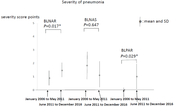
Figure 3: Severity of pneumonia.
P value was calculated by Brunner-Munzel test.
BLNAS; β -lactamase negative ampicillin-sensitive strain group
BLNAR; β -lactamase-negative ampicillin-resistant strain group
BLPAR; β -lactamase positive ampicillin-resistant strain group
The susceptibility of BLNAR for each ABPC/SBT, CFPN, IPM, IPM, AZM and LVFX was presented in figure 4. In comparison between Period I and January 2011 to May 2016, there was no change in susceptibility of BLNAR for ABPC/SBT, AZM, LVFX. Further, in comparison between Period I and June 2011 to December 2016, the susceptibility of BLNAR for CFPM and IPM seems to have a tendency of improving (Figure 4).
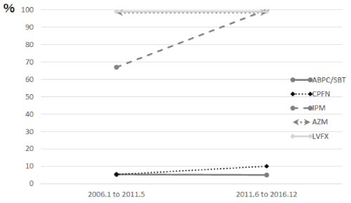
Figure 4: Antibiotic susceptibility of BLNAR.
Discussion
This is the first study that evaluate modification of severity of pneumonia due to change of epidemiology of subgroup of H.influenzae. There is the limitation that the results are applicable to hospitalized patients and not to patients managed at home. In this report we confirm that BLNAR holds large majority as the pathogen of H.influenzae pneumonia in adult throughout January 2006 to December 2016. Further, distribution of BLNAR in Period II is larger than that in January 2006 to May 2011. The most interesting point of our investigation is that the pneumonia caused by BLNAR in adult in Period II had higher severity score and needed longer hospitalization than that in January 2006 to May 2011.This difference suggests that the pneumonia caused by BLNAR in adult has become more severe. Because there was no significant difference in age of patients between two periods, the tendency of the pneumonia by BLNAR becoming more severe has no relation to age. Further, because of better antibiotic susceptibility of BLNAR in Period II compared to January 2006 to May 2011, antibiotic susceptibility seems to be no relation to the tendency of the pneumonia by BLNAR becoming severer It can be speculated that certain clinical pathogenic factor of BLNAR in adult pneumonia in Period II became stronger than in January 2006 to May 2011. There are many reports about antibiotic susceptibility of H.influenzae, but few about clinical pathogenic factor yet. One report suggests the biofilm formation is a pathogenic factor of H.influenzae in vitro [8]. Another report suggests the Lic B in Lic 1 operon on Phosphorylcholine decoration of lipopolysaccharide of H.influenzae contributes to lung damage in an aged mice co-infection model [9]. One report suggests that host-pathogen cross talk during human bronchial epithelial cell infection in non typeable H.influenzae provides important insight into non typeable H.influenzae pathogenesis in vitro [10]. There were no reports about change of clinical pathogenesis or clinical severity of BLNAR. We think the pneumonia caused by BLNAR in adult becomes gradually severe. Our report could be useful to design future prospective study.
Article Information
Article Type: Research Article
Citation: Sawaguchi H, Gose K, Wada S, Muraki M (2017) The Trend of Clinical Severity and Antibiotic Susceptibility of β-Lactamase-negative Ampicillinresistant Haemophilus influenzae causing Adult Pneumonia from June 2011 until May 2016 compared with those from June 2006 until May 2011. J Infect Pulm Dis 3(1): doi http://dx.doi. org/10.16966/2470-3176.123
Copyright: © 2017 Sawaguchi H, et al. This is an open-access article distributed under the terms of the Creative Commons Attribution License, which permits unrestricted use, distribution, and reproduction in any medium, provided the original author and source are credited.
Publication history:
Received date: 11 Nov 2016
Accepted date: 27 Feb 2017
Published date: 04 Mar 2017