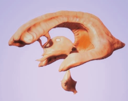
Figure 1: Drawing of the cerebral ventricles. The shape of the ventricles seems to envelop at least some of the most important structures of the Papez Circuit, or at least those that requires more oxygen and hydrogen (energy).


Arturo Solis Herrera*
Human Photosynthesis™ Research Center, Aguascalientes, Mexico*Corresponding author: Arturo Solis Herrera, MD, PhD, Human Photosynthesis™ Research Center, Aguascalientes 2000, Mexico, Tel: 014499160048, 014499150042; E-mail: comagua2000@yahoo.com
Since the seventeenth century began to glimpse the existence of oxygen in small amounts in the atmosphere, and yet, inside our body the amount of oxygen is almost 5 times greater than in the air around us.
It seems obvious that, somehow, when breathing, our body concentrates atmospheric oxygen and carries it into the blood. But after 200 years, no mechanism has been found that concentrates oxygen from the atmosphere and raises it almost 5 times inside the body.
So, the mystery remains to this day: where do the CNS’s high oxygen levels come from? And, circumstantially, we find the answer: our body does not take oxygen from the air around us, but from the water it contains inside, in each of the cells that make us up.
Papez; Limbic; Oxygen; Hydrogen; Metabolism; Memory; CNS
The Papez circuit was described in 1937 by Papez JW in his paper “A Proposed Mechanism of Emotion” [1]. This work was a different understanding of how the limbic lobe directed emotions and memory through its connections with the hypothalamus.
Papez presented a description of the hippocampus, hypothalamus (mammillary bodies), anterior thalamus, cingulate gyrus, and their interconnections as the “anatomic basis of the emotions”.Although it has subsequently emerged as more important in the function of memory than in emotional processing [2].
Anatomical dissection-based studies and neuroimaging techniques have consistently described the precise white matter interconnections between circuit hubs including the cingulum [3], perforate pathway [4], fornix [5], and mammillothalamic tract [6].
The circuit of Papez included cortical and sub cortical structures such as the hippocampus, fornix, mammillary bodies, mammillothalamic tract, and anterior nuclei of the thalamus, cingulate gyrus, and entorhinal cortex.
Clinically the anterior thalamic nucleus has a role in episodic memory, as it and its afferent connections implicated in anterograde memory deficits, e.g., in Korsakoff ’s syndrome [7]. Additionally, the nucleus may be affected by prion diseases such as fatal familial insomnia [8], and Creutzfeldt-Jakob disease [9]. The anterior thalamus may also have a role in seizure propagation, as demonstrated by the success of deep brain stimulation of the nucleus in patients with refractory epilepsy [10].
The cingulum is a distinctive white matter tract that forms a nearcomplete ring underneath the cingulate gyrus from the orbitofrontal cortex anteriorly to the temporal pole posteriorly [11]. The sizeable connectivity of the cingulum facilitates its involvement in diverse cortical processes such as executive function, attention, emotional and behavioral regulation, memory, and visuospatial processing [12]. The cingulum bundle has been implicated in many neuropsychiatric diseases including schizophrenia [13], depression [14], posttraumatic stress disorder [15], obsessive compulsive disorder [16], autism [17], and Alzheimer’s disease [18].
Connections between the Anterior Thalamic Nucleus (ATN) and the cingulum were first described in the rat brain by Clark WE, et al.[19] and Waller WH [20]. Papez described his emotional circuit with the connection between the anterior thalamus and cingulum as a fundamental component.
The anatomical course of the thalamocingulate tract remains uncertain despite 80 years since Papez’s original work [21].
The anatomy of the limbic system has been studied exhaustively for almost a century, for its implications on transcendent brain functions. It is remarkable the time, money and effort that have been invested both by clinicians, researchers, as well as by the patients themselves.
And the relative closeness of the Papez circuit to the ventricles and thereby to (Cephalic Spinal Fluid) CSF does not seem to have given greater importance (Figures 1 and 2).

Figure 1: Drawing of the cerebral ventricles. The shape of the ventricles seems to envelop at least some of the most important structures of the Papez Circuit, or at least those that requires more oxygen and hydrogen (energy).
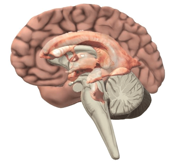
Figure 2: The limbic system is surrounded by the cerebral ventricles, so the water of CSF is quite close to the anatomical structures that make it up. Structures with high metabolic activity seem as requiring the water of CSF close.
It is possible that the careful proximity of the CSF with structures of the limbic system is fundamental since the water of the CSF constitutes its source of oxygen and hydrogen [22]. It is a long-lasting mistake to believe that the oxygen found within the central nervous system comes from the atmosphere. The air around us contains very little oxygen, a knowledge that began to be glimpsed since the seventeenth century by Boyle and Mariotte, and as the knowledge of biology progressed, 100 years later, it was found that there is almost five times more oxygen in our body than in the atmosphere.
But such a dogma has been difficult to banish because it seems obvious that respiratory movements introduce oxygen into the lung, where it is supposedly concentrated and then passed into the blood. But inspiration and expiration have as their fundamental purpose the expulsion of CO2 that continuously forms inside our body.
No living being takes oxygen from the atmosphere but extracts it from the water it contains [23].
The oxygen and hydrogen in the gaseous state that comes from the dissociation of water into neuromelanin diffuses through tissues in the form of increasing spheres that follow the laws of simple diffusion. In their journey, hydrogen and oxygen are captured by the different tissues and molecules that integrate them in a very precise way.
Melanin or neuromelanin, a hybrid electronic/ionic conductor have the potential to split the water molecule into molecular hydrogen and molecular oxygen. Molecular hydrogen is an antioxidant and may be instrumental in preventing the excessive oxidation leading to Parkinson’s disease.
Melanin, located in the SubstantiaNigra, locus coeruleus, ependyma tissues, deteriorates in Parkinson’s disease so may be related to the development and progression of the disease, since molecular hydrogen would no longer be generated as it deteriorates. Environmental toxins, thought to be related to development of Parkinson’s disease, may cause deterioration of intrinsic chemistry of melanin, since it is a chelator which would collect such environmental contaminants, but its function of splitting the water molecule into molecular hydrogen and oxygen could be affected, therefore, restoring melanin function or providing supplemental molecular hydrogen might be potential treatments for Parkinson’s disease [24].
Neuromelanin is one of several molecules that the body possesses, which can split the molecule from water. The reaction can be described as follows:
2H2O(liq)→2H2(gas)+O2(gas)→2H2O(liq)+4e-
Neuromelanin can dissociate and reform water. For every two water molecules that are reformed, 4 high-energy electrons are generated. The molecular oxygen and molecular hydrogen that are released by breaking the water molecule are distributed in the tissues following the laws of simple diffusion. In the diagram in figure 3, they are represented as increasing circles, which expand in all directions and exemplifies the oxygen and hydrogen source of the Papez circuit.
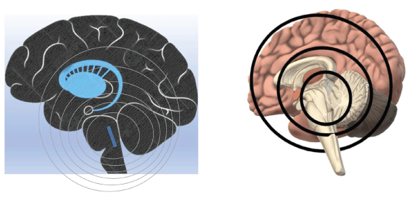
Figure 3: The abundant oxygen that we find inside the body, for example in the CNS, does not come from the atmosphere, but from the water that the body contains in each of its cells, like plants. The blue slash means the approximate location of the substantia nigra.
The proximity of the limbic system to the CSF ensures the source of hydrogen and oxygen that the system requires to function properly. It is common to find in neurodegenerative diseases the increase in volume of the ventricles, which can be explained by the fact that the dissociation of water is not happening with the celerity adequate, thereby water begins to accumulate, therefore the ventricles dilate, and the surrounding tissues tend to retract at low levels of hydrogen and oxygen.
Tissues require a constant supply of hydrogen and oxygen, and when the process that extracts oxygen from the water is disturbed, then the affected tissues tend to become disorganized, and in the case of the limbic system, the manifestations are varied since there is no single or specific symptomatology in the case of water dissociation. Any disease begins when the rate of turnover of water dissociation is significantly altered.
Female, date of birth: 09/06/2012. Refractory Epilepsy diagnosed in August 2017 after several studies, but no one drug controls it. She has 10 to 30 crises a day, more in the night. She no longer talks; she can’t walk anymore (Figures 4-10).

Figure 4: Photography taken during the first consultation. The patient had to be charged all the time. Notice the facial expression of the paternal grandmother (left).

Figure 5: The patient cannot wander without help because constant falls and cannot even hold her head.
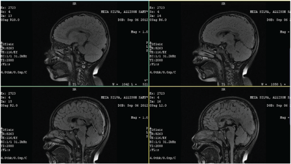
Figure 6: The patient cannot wander without help because constant falls and cannot even hold her head.
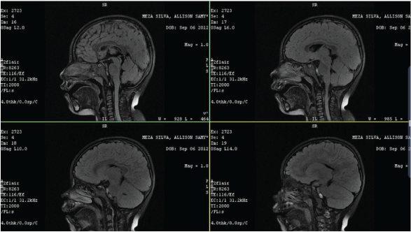
Figure 7: The structures do not present significant alterations. The ventricles are slightly increased in volume.
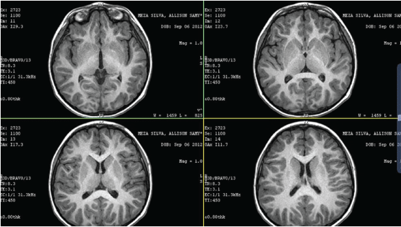
Figure 8: The neuroanatomy is preserved, only the ventricles are discreetly increased in volume, which suggests that the dissociation of water is not happening with the proper turnover rate, and therefore water is accumulating.
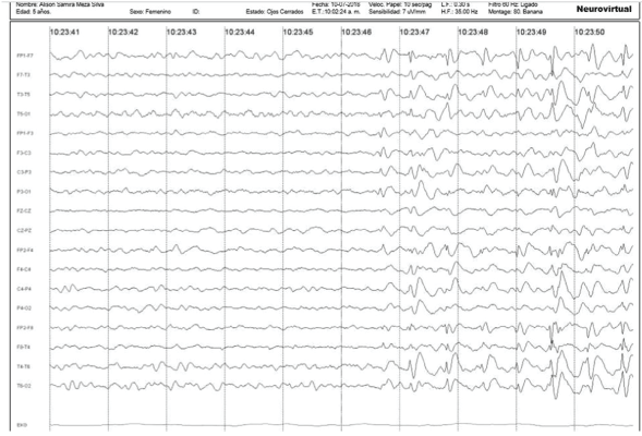
Figure 9: Example of the patient’s Electroencephalogram (EEG).

Figure 10: The patient required support for all kinds of activities. The date of the first consultation was 17/07/2018.(EEG).
The same day, treatment with QIAPI 1® (in patent proceedings), 17 July 2018, was started. The other treatments were suspended. Next photographs were taken on July 31, 2018 (15 days later) (Figures 11-14).

Figure 11: The improvement of the picture, with only 15 days of treatment, is impressive. The patient’s smile says it all.

Figure 12: The patient is already walking again, already talking, she can already sit alone, and of course she is happy.
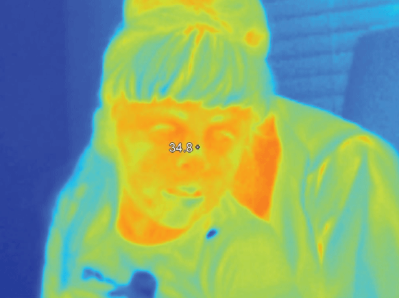
Figure 13: Body temperature shows 34.8° Celsius, pre-treatment.
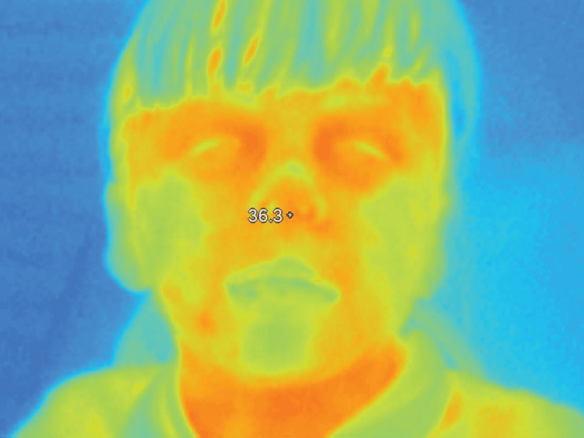
Figure 14: The body temperature 15 days after starting the treatment shows 36.3º Celsius.
The responses of the human body to the pharmacological restoration of oxygen levels are impressive. They demonstrate without a doubt the enormous physiological importance of a process that was only known in plants and that we circumstantially detected in the retina. It wasdetected during an observational study that was carried out from 1990 to 2002 and included ophthalmological studies of 6000 patients.
It was a huge effort, but fortunately we were able to achieve a useful result that allows us to improve the level of health and quality of life of patients. The oxygen levels inside the body are essential for the proper functioning of the body. Even the body temperature is improved. This case of refractory epilepsy is just one example of the benefits of the new neurophysiology that we propose.
Many disorders, including neurological and psychiatric, are strongly influenced by genetic variation altering gene function [25]. But in turn, genes are not independent structures, they also require oxygen and hydrogen (energy) and in quantized quantities, i.e., exact. And genes (and cells and neurons) require them constantly, day and night, throughout life.
The unsuspected ability of the human neuronal cell to take oxygen from water is the very first reaction in life. This allows neurons to get oxygen and hydrogen (energy) at the same time. The energy that is released by separating the molecule from water into its gaseous components is captured and transported by hydrogen, the main carrier of energy in the entire universe, so our body can be no different.
And when the amazing ability of the eukaryotic cell to break down the molecule of water is disturbed, mainly by environmental factors related with water, then the availability of oxygen and hydrogen decreases. And in any system, when the energy decreases, the mass tends to disappear.
That is why we think that when the dissociation of water is altered, then liquid water tends to accumulate abnormally in the ventricles, fissures, and subarachnoid space. This is followed by what we call brain atrophy, in which the tissue becomes rarer and loses anatomical, physiological, and functional qualities. including the appearance of abnormal metabolites such as amyloid β, alpha-synuclein aggregation, hyperphosphorylated tau accumulation, neuron loss, neurofibrillary tangles, senile plaques, etc. [26], which, although could be normal in physiological amounts, when the availability of oxygen and hydrogen in the tissue is inadequate, then the failure is widespread and various substances appear in abnormally high amounts, but the explanation is that low levels of oxygen and hydrogen (energy) do not allow the cell to properly carry out the functions it has done thousands or millions of years.
The signs and symptoms of an extra pyramidal movement disorder are often associated with increasing SN pathology. It is confirmed that pathological lesions in the SN are a common feature of AD and an uncommon feature in normal aging [27].
Written informed consent was obtained from the patient for the publication of this case report.
This work was supported by an unrestricted grant of the Human Photosynthesis™ Research Centre, Aguascalientes 20000, Mexico.
Download PDF Here
Article Type: CASE REPORT
Citation: Herrera AS (2022) The Circuit of Papez Needs Oxygen to Work Properly, Where Do the CNS’s High Oxygen Levels come from? J Psychiatry Ment Health 7(1): dx.doi.org/10.16966/2474-7769.149
Copyright: © 2022 Herrera AS. This is an open-access article distributed under the terms of the Creative Commons Attribution License, which permits unrestricted use, distribution, and reproduction in any medium, provided the original author and source are credited.
Publication history:
All Sci Forschen Journals are Open Access