Introduction
Despite advances in neonatal nutritional management, somatic
and tissue-specific growth deficiencies remain a common finding in
premature infants diagnosed with bronchopulmonary dysplasia (BPD),
a form of neonatal chronic lung disease characterized by a reduction in
alveolar surface area and aberrant capillary development [1]. The altered
pulmonary architecture in BPD leads to diminished pulmonary function,
which may persist into childhood and potentially throughout adult life
[2]. During the treatment of respiratory distress syndrome in preterm
newborns, exposure to high inspired oxygen concentrations is potentially
life-saving. Nevertheless, hyperoxia, the administration of oxygen at a
higher partial pressure than is necessary to meet tissues and organs needs,
is well-established for its capacity to hinder lung growth in newborn
animals and for its etiologic role in the development of BPD [3,4]. Free
radicals and reactive species generated by hyperoxia have the capacity
to modulate vital cellular metabolic functions which influence growth,
including the translational control of gene expression. Within the adult
rodent lung, hyperoxia reduces the fractional rate of protein synthesis
(FSR) and the efficiency of protein synthesis without altering total lung
RNA or RNA/protein content [5]. In the newborn rat lung, hyperoxia
suppresses global mRNA translation at the level of initiation and alters
the activity of eukaryotic initiation factors (eIF) that regulate mRNA/
ribosome binding [6].
Translation of mRNA into protein begins with initiation, which
entails binding of the mRNA to 40S ribosomal subunit to form the 43S
pre-initiation complex, scanning of the ribosome complex to the start
codon, and binding of the Met-tRNAiMet (initiator methionyl-tRNA) to
the 40S ribosomal subunit. The 7-methyl-GTP cap at the 5’ terminus of
all nuclear-encoded eukaryotic mRNAs binds the 40S ribosomal subunit
to the mRNA via the eIF4F complex, a heterotrimeric aggregation of
the cap-binding protein, eIF4E, the scaffolding protein, eIF4G, and the
RNA helicase, eIF4A [7]. Regulation of binding of eIF4E to the 5’-cap is
repressed by eIF4E-binding protein-1 (4E-BP1). Phosphorylation of 4EBP1
by the nutrient-sensitive kinase, mammalian target of rapamycin
(mTOR), releases eIF4E, allowing for the formation of active eIF4G:eIF4E
complexes [8]. Binding of the Met-tRNAiMet to the 40S ribosomal subunit
is mediated by eIF2 [7]. In order to attract Met-tRNAiMet, eIF2 must
generate GTP, a step catalyzed by the guanylate nucleotide exchange factor
eIF2B [9]. Phosphorylation of the α subunit of eIF2 on Ser51 stabilizes the
inactive, GDP-bound, form of eIF2B [10]. The activity of eIF2B is also
mediated by its catalytic ε subunit, which is regulated by expression and
glycogen synthase kinase-3-mediated phosphorylation [11]. In summary,
translational regulation of initiation involves both the assembly of the
eIF4F complex on the 5’-mRNA cap and the successful recruitment of the Met-tRNAiMet by eIF2.
During the newborn period, rapid growth corresponds to high rates of
protein synthesis. Accretion of skeletal muscle mass in neonates directly
relates to the heightened protein synthetic response to feeding [12].
Feeding also increases the FSR in the newborn lung, but to a lesser extent
than in muscle [13]. Mature milk, colostrum, and formula feedings nearly
double FSR in the lung of colostrum-deprived, 4-6 day-old piglets [13].
In skeletal muscle, meal-feeding stimulates polysome assembly, eIF4F
assembly (increased eIF4G:eIF4E binding), and the phosphorylation of
4E-BP1and ribosomal protein S6 (S6Rp) within 30 minutes [14]. Pathologic
conditions have also been shown to negatively influence the translational
regulation of protein synthesis. Lipopolysaccharide decreases FSR in
glycolytic muscle concomitant with decreases in eIF4G:eIF4E binding and
the phosphorylation mTOR substrates, 4E-BP1 and ribosomal protein S6
kinase-1 (S6K1), suggesting that a reduction in mTOR activity is at least
partially responsible for the reductions in protein synthesis [15].
We have previously shown that exposure to 95% O2 reduces protein
synthesis and polysome aggregation while simultaneously increasing
eIF2α phosphorylation in the lungs of continuously nursing neonatal rats
[6]. To directly evaluate the ability of hyperoxia to alter feeding-induced
protein synthesis, we designed the current study to test the hypothesis that
hyperoxia does not alter feeding-induced initiation complex assembly
or mTOR-mediated regulation of mRNA translation in the lungs of
newborn rat pups.
Methods
Animal model and conditions
Timed-pregnant, Sprague Dawley rats at day 14 of gestation, (Charles
River Laboratories, Boston, MA) were housed in standard rat cages until
day 4 after delivery. Littermates were scrambled between dams and the total
number of pups culled to 12/ litter. On day-of-life 4, pups were weighed
and litters placed into Plexiglas chambers circulated with either room
air (RA) or 95% O2 (Ox) as previously described [6]. Delivery of 100%
O2 was continually adjusted using a computerized system (BioSpherix
Ltd., OxyCycler A). In both chambers, the atmosphere was circulated
via a small fan and CO2 maintained at <0.5%. Room air and O2
-exposed
animals were studied concurrently. Dams were supplied with standard rat
chow and water ad libitum, exposed toroutine day/light cycles of 12 hours,
and maintained at 26°C and 75-80% humidity.
After 16 hours of exposure to either RA or Ox, pups were weighed and
separated into 2 groups. In group one, animals were returned to the dam
for a total of 24 hours in either RA or Ox (dam-fed, n=3 per atmosphere).
The remaining pups were separated from the dam to induce fasting and
placed into separate cages (n=9 per atmosphere) lined with standard rodent
bedding covering a water-jacketed warming blanket set at 37° C (Gaymar,
Orchard Park, NY). Preliminary studies using the phosphorylation of
S6K1 in the lungs as an indicator of mTOR activity revealed that 8 hours
of separation was required to reduce S6K1 phosphorylation. After fasting,
pups were fed 0.4 ml of sterile water (water-fed, n=3 per atmosphere) or
human infant premature formula (24 kcal/oz) using a 24 gauge feeding
needle attached to a syringe (formula-fed, n=6 per atmosphere). The 0.4
ml volume was determined as the approximate volume of the newborn
stomach based upon analysis of dam-fed pups. After feeding, pups were
placed back into their respective chamber. Water-fed animals were studied
after 30 minutes while half of the formula-fed pups were studied at 30
minutes and half at 60 minutes. At these intervals, pups were sacrificed by
decapitation and lungs harvested as described [6]. In a subset of litters, the
right gastrocnemius muscle and the entire liver were also harvested. To
examine residual mTOR activity after fasting, a group of water-fed pups
were treated with rapamycin [4 mg/kg, i.p. (LC Laboratories (Woburn,
MA)] in 5% DMSO or an equivalent volume of DMSO vehicle 1 hour
prior to placement in their respective atmosphere. Animal protocols were
approved by the Institutional Animal Care and Use Committee at the
Pennsylvania State University College of Medicine.
eIF4F assembly
Affinity chromatography was used to separate eIF4E containing protein
complexes [6]. Frozen left lungs were pulverized using an ice-cooled,
Dounce tissue homogenizer and dissolved in CHAPS lysis buffer (40 mM
HEPES PH 7.5, 120 mM NaCl, 1 mM EDTA, 10 mM pyrophosphate, 10
mM β-glycerophosphate, 40 mMNaF, 1.5 mM sodium orthovanadate,
0.1 mM PMSF, 1 mM benzamidine, 1 mM DTT, 0.3% CHAPS). One
hundred micrograms of total lung protein was combined with washed
m7
-GTP Sepharose beads (GE Scientific, Piscataway, NJ) and incubated
for 2 hours at 4°C. Following incubation, beads were pelleted, washed,
and the protein complexes removed by boiling. Affinity purified protein
complexes were separated by electrophoresis and immunoblotted with
antibodies to eIF4E, eIF4G, and 4E-BP1. Immunoblots were visualized
via chemiluminescence and quantified using a Gene Gnome Bio-Imaging
System (Syngene Incorporated, Fredericksburg, MD) with blots of eIF4E,
eIF4G, and 4E-BP1 developed under identical conditions within each litter
set (RA and Ox). For immunoblot figures, the large number of animals
and conditions necessitated “pasting” together lanes from the same gel [6].
Immunoblotting
Equal amounts of protein in CHAPS lysis buffer were electrophoretically
separated on 10% Bis-Tris gels (Invitrogen/Biosource, Carlsbad, CA) and
transferred to PVDF membranes [6]. Membranes were incubated with
following antibodies: S6K1 (Thr389), S6rp (Thr235/236), 4E-BP1 (Thr70),
eIF4E, eIF4G, eIF2B, and eIF2α (all 1:1000, except S6Rp (Thr235/236) at
1:2000; Cell Signaling Technologies, Beverly, MA); S6K1 and 4E-BP1
(1:2500 and 1:10,000, respectively, Bethyl Laboratories, Montgomery,
TX); eIF2α (Ser51) and eIF2Bε (Ser535) (1:1000; Invitrogen); β-actin
(1:5000; Sigma Chemical, St. Louis, MO); and species-specific horseradish
peroxidase-conjugated secondary antibodies (1:5000; GE Scientific).
Protein expression was normalized to β-actin on each gel and the ratio of
phosphorylated to total proteins calculated.
Polysome analysis
Sucrose gradient centrifugation was employed to analyze lung
polysome aggregation as previously described [6]. Briefly, lung tissue was
homogenized in resuspension buffer [50 mM HEPES, pH 7.4; 75 mM
KCl; 5 mM Mg2
Cl2
; 250 mM sucrose; 100 µg/ml cyclohexamide; 2 mM
DTT; 1% Triton X-100; 1.3% deoxycholate; 10 µl/ml Super-asin (Ambion,
Austin, TX)], cleared by centrifugation, and the supernatants layered onto
20-47% linear sucrose gradients. Gradients were separated at 90,000 x g
for 4 hours at 4°C in an ultracentrifuge. Each gradient was then upwardly
displaced using a Fluorinet fractionator and the optical density at 254 nm
recorded on a chart recorder.
Statistical analysis
Exposures were conducted on a minimum of 2 liters per atmosphere,
with the exception of the rapamycin experiment which represents a single
trial using two litters. Normally distributed data were analyzed using
t-tests or analysis of variance (ANOVA, one- or two-way) with Tukey
testing to identify individual differences when appropriate. Non-normally
distributed data were analyzed using Mann-Whitney or Kruskal-Wallis
ANOVA on ranks with testing for multiple comparisons by Dunn’s
method when appropriate. For simplicity, data in figures is listed as mean
± SEM regardless of testing method and the level of significance set at p<0.05.
Results
Fasting suppresses mTOR activity in the lungs RA and Oxexposed
rat pups
To delineate the effect of hyperoxia on eIF4F assembly, we used maternal
separation and newborn fasting followed by re-feeding with the aim of
first suppressing, then stimulating, mTOR activity and eIF4F assembly
in the presence of hyperoxia. Using S6K1 phosphorylation as marker of
mTOR activation, preliminary studies revealed that 8 hours of fasting was
necessary to reduce S6K1 phosphorylation on Thr389 (not shown). Even
with this period of fasting, mTOR activity was not completely suppressed,
as intraperitoneal injection of rapamycin further suppressed S6K1 and
4E-BP1 phosphorylation below that of fasting levels (Figure 1). During
the 8-hour fast, serum glucose levels remained unchanged independent of
whether the pups were reared in RA or Ox (RA: non-fasted, 92 ± 5; fasted
105 ± 15; Ox: non-fasted, 126 ± 23; fasted 95 ± 6 mg/dl, NS). In agreement
with our previous findings, rat pups exposed to 24 hours of 95% O2
gained
a similar amount of weight to those reared in room air (RA: time 0-11.2 ±
0.3 g, 24 hrs-13.2 ± 0.4 g; Ox: time 0-11.3 ± 0.2 g, 24 hrs-13.1 ± 0.2 g, NS)
[6]. Weights were unaltered over the 8-hour period of fasting in either RA
or Ox animals.
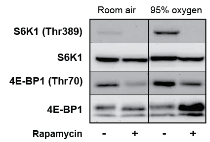
Figure 1: Fasting fails to completely suppress pulmonary mTOR
activity. Rat pups received i.p. injection of rapamycin (4 mg/kg) or an
equivalent volume of DMSO prior to exposure to room air or 95% O2
for
24 hrs. After 24 hrs, pups were fasted for 8 hrs by maternal separation,
gavage fed sterile H2
O, and lungs collected 30 min later. Figure depicts
representative immunoblots of the phosphorylation of downstream
mTOR substrates, S6K1 and 4E-BP1 (n=3 pups/condition)
As illustrated in figure 2, 8 hours of fasting (Dam-fed vs. Water-fed)
reduced the phosphorylation of S6K1 on Thr389 in lung tissue from both RA
and Ox exposed pups, but had no effect on the phosphorylation of 4E-BP1
at Thr70. Fasting also reduced the phosphorylation of the S6K1 substrate,
S6Rp, though only in the RA animals (Figure 2). Formula feeding,
however, failed to stimulate S6K1, S6Rp, or 4E-BP1 phosphorylation
above fasted levels after 30 or 60 minutes (Figure 2). These studies suggest
that while fasting is capable of suppressing mTOR activity in the lung,
formula feeding-induced stimulation in the lung is rather insensitive.
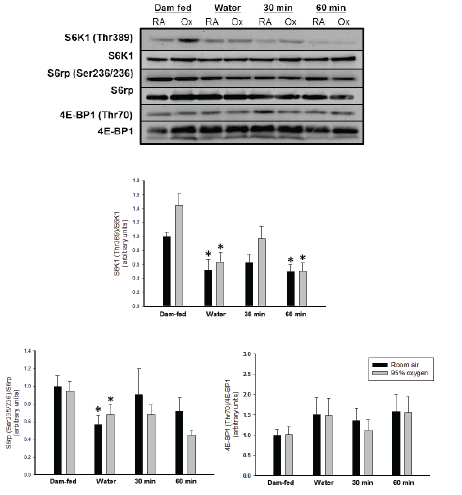
Figure 2: Feeding fails to stimulate mTOR activity in RA and Ox-exposed
rat pups. A) Immunoblots are representative of thephosphorylation
of S6K1, S6Rp, and 4E-BP1. Histograms (B-D) show the mean
phosphorylation in the lungs of room air (black) and 95% O2
-exposed
(gray) pups. Columns represent mean (n=11-12) and bars SEM. “*”
denotes a significant difference (p<0.05) compared to dam-fed pups.
Protein synthetic rates generally directly correlate with formation of
active eIF4F complexes. The association of eIF4G with eIF4E (eIF4G:eIF4E)
represents a surrogate for the assembly of active eIF4F complexes. Affinity
purification of eIF4E from lung homogenates permits the identification
of the relative association of eIF4E with both eIF4G and 4E-BP1. Using
this technique, we found that fasting and re-feeding significantly altered
eIF4G:eIF4E in RA and Ox animals (p<0.05) (Figure 3). In RA-exposed
pups, eIFG:eIF4E doubled 60 minutes post-feeding (Water-fed: 0.91 ± 0.22
vs. 60 min-fed: 1.88 ± 0.2, p<0.05-t test). By comparison, in Ox animals
maximal eIF4G:eIF4E occurred 30 minutes post-feeding (Water-fed: 1.00
± 0.20 vs. 30 min-fed: 1.99 ± 0.21, NS – Mann-Whitney). Availability of
eIF4E is regulated by the release from 4E-BPs, the phosphorylation of
which is mTOR-dependent. Assessing of the association of 4E-BP1:eIF4E
in the same samples revealed that fasting increased 4E-BP1 binding in
both RA and Ox-exposed animals (RA: Dam-fed, 1.0 ± 0.0 vs. Water-fed,
2.6 ± 0.5; Ox: Dam-fed, 1.2 ± 0.1 vs. Water-fed, 3.2 ± 0.6, both p <0.001).
Neither feeding nor hyperoxia, however, altered 4E-BP1:eIF4E.
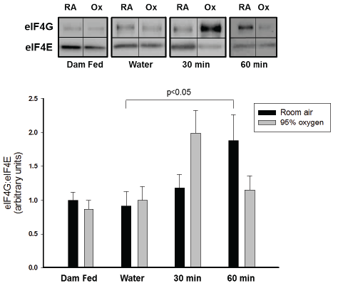
Figure 3: Hyperoxia does not suppress meal-stimulated eIF4F assembly.
Affinity purified lung extracts were separated by electrophoresis and
the relative amount of eIF4G co-precipitating with eIF4E identified by
immunoblotting. A) Representative immunoblots depict a single animal
in each group. Lines of separation represent non-adjacent lanes. B)
Histograms display relative association of eIF4G with eIF4E. Columns
(black – room air; gray – 95% O2
) represent means (n=11-12) and
bars SEM. Analysis found a positive effect of feeding on eIF4G:eIF4E
binding in RA pups (ANOVA, p=0.038) with a difference identified
between water- and formula-fed pups at 60 min (p<0.05). For Ox pups,
testing revealed an effect of feeding (Kruskal-Wallis, p = 0.041), but no
individual differences.
The relative insensitivity of the lung to meal-feeding raised concern
a cow’s milk-based formula may not stimulate translational regulatory
pathways in the newborn rat. As a control, we studied meal-stimulated
eIF4F assembly in skeletal muscle and liver. Figure 4 demonstrates that
formula feeding following an 8-hour fast dramatically stimulates active
eIF4F complex assembly in liver but has little effect in gastrocnemius
muscle. The combined results from this series of experiments indicate that
cow’s-milk feedings can increase translational activity in a tissue-specific
manner and that hyperoxia does not blunt this response in the lung.
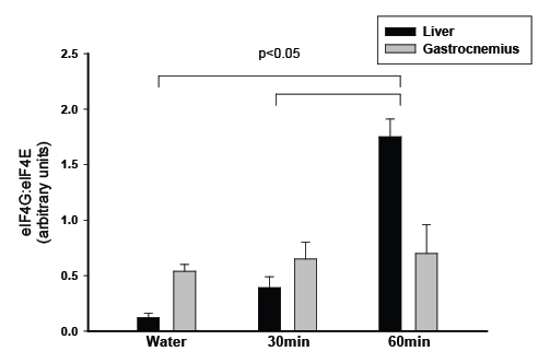
Figure 4: Feeding induces eIF4F assembly in liver. Affinity-purified liver
and gastrocnemius extracts were separated by electrophoresis and
the amount of eIF4G co-precipitating with eIF4E determined. Panel A
shows representative immunoblots from a single animal for each tissue.
B) Histogram shows the relative binding of eIF4G with eIF4E. Columns
represent means ± SEM (n=3) and bars SEM. Statistical analysis
revealed that 60 min after feeding eIF4G:eIF4E binding increased
relative to water-fed or meal-fed pups at 30 min (p<0.05, t test)
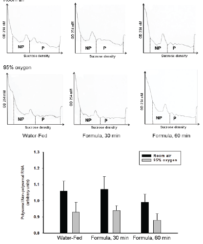
Figure 5: Hyperoxia suppresses polysome aggregation irrespective of
feeding. Total lung RNA from pups exposed to RA or Ox was subjected
to sucrose density gradient centrifugation as described under Methods.
A) Tracings represent the quantity of RNA at specific sucrose density
as measured by OD 254 nm. The dashed lines separate non-polysomal
(NP) and polysomal (P) RNA. B) The mean ratio of P/NP for RA (black)
and Ox (gray) animals is represented by columns (n=6 per feeding
condition/atmosphere). Bars represent SEM. Two-way ANOVA revealed
a significant effect of Ox on polysome aggregation (p=0.012), but no
feeding effect in RA or Ox animals
Hyperoxia suppresses polysome aggregation irrespective of
feeding
An increase in the polysomal to non-polysomal RNA ratio is indicative
of a relative enhancement of initiation events. Figure 5 illustrates that
polysome aggregation was not responsive to formula feeding in either
group, a finding consistent with the lack of change in S6K1 and 4E-BP1
phosphorylation. Hyperoxia, however, decreased the ratio of polysomal to
non-polysomal RNA in response to both water and formula feeding (2-
way ANOVA, p<0.05). Interpretation of this finding suggests non-eIF4F
events inhibit mRNA translation at the level of initiation.
Hyperoxia induces sustained phosphorylation of eIF2α
Similar to our previous observations, we found that hyperoxia tends to
increase eIF2α phosphorylation on Ser51 during continuous feeding (RA:
1.0 ± 0.1 vs. Ox: 3.5 ± 1.0, p=0.08, figure 6) [6]. In comparison to water,
formula feeding neither increased nor decreased eIF2α phosphorylation
in either atmospheric group. Among the formula fed animals, the
phosphorylation of eIF2α was greater 60 minutes post-feeding in Oxcompared
to RA-exposed pups (RA: 1.2 ± 0.3 vs. Ox: 2.9 ± 1.0, p<0.05,
figure 6). Analysis of eIF2Bε, on the other hand, failed to identify an effect
of feeding on eIF2Bε expression or phosphorylation on Ser535 (2-way
ANOVA).
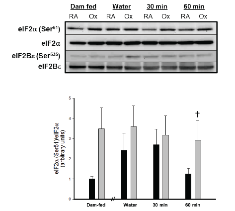
Figure 6: Effect of 95% O2
on phosphorylation of eIF2α.A) Immunoblots
represent the relative phosphorylation of eIF2α and eIF2Bε. B)
Histograms show the mean phosphorylation of eIF2α in RA (black) and
Ox (gray) pups. Columns represent mean (n=12) and bars SEM. In damfed
animals, eIF2α phosphorylation tended to be higher in Ox than in RA
pups (p=0.08). Formula-feeding failed to alter eIF2α phosphorylation in
animals reared in either atmosphere. “†” denotes a significant difference
(p<0.05) between RA and OX at 60 min.
Discussion
Aggressive nutritional support of premature infants at risk for the
development of BPD is standard practice in neonatal intensive care.
Despite these efforts, nutritional deficiencies persist in this population
illustrated by the high incidence of growth failure relative to gestational
age-matched controls [16]. Animal studies provide convincing evidence
for a global impact of under-nutrition, encompassing reductions in lung
weight, alveolar number, elastin and hydroxyproline content, and type
II cell maturation [17-19]. Growth failure in preterm infants is further
confounded by risk factors that contribute to the development of BPD
[20,21]. In newborn guinea pigs, restriction of nutrient intake enhances
the toxicity of hyperoxia, leading to increased mortality without altering
pulmonary inflammation or antioxidant capacity [22]. Whether hyperoxia
alters nutritional signaling regulating mRNA translation in the lung and
whether such changes influence lung growth remains to be established.
Exposure to95% O2
attenuates pulmonary protein synthesis and transiently
augments the activity of mTOR in continually nursing term, newborn rats
[6]. The current study expands upon these earlier findings by illustrating
that hyperoxia does not alter feeding-induced initiation complex
assembly or major signaling events regulating translation initiation and by
reaffirming that hyperoxia-induced reductions in polysome aggregation
correlate with eIF2α phosphorylation.
Within the lung, colostrum, mature porcine milk, and casein-based
formula all increase the FSR following fasting, while only colostrum
enhances protein synthetic capacity (RNA/protein ratio) [13]. The
stimulatory effect of feeding in the lung is responsive to changes in serum
amino acid levels, not insulin levels, denoting nutritional protein as a
major contributor to the growth [23]. Based upon methodology designed
to investigate meal-stimulated effects in skeletal muscle from newborn
pigs, the present study examined the impact of hyperoxia on nutritional
signaling at 30 and 60 minutes post-feeding [14]. Although an 8-hour fast
was sufficient to reduce S6K1 and S6Rp phosphorylation and increase
4E-BP1:eIF4E association, it did not produce the anticipated reductions
in 4E-BP1 phosphorylation or eIF4G:eIF4E assembly in either the RA or
Ox exposed animals. The disparate response to fasting may be explained
by the insensitivity of Thr70 to fasting relative to other phosphorylation
sites or the presence of fasting resistance eIF4F factors. Differences in the
molecular weights of fetal and adult eIF4G1 and in eIF4A1/2 expression
have been proposed to explain fetal hepatic rapamycin resistance, an
effect independent of mTOR substrate phosphorylation [24]. Although
the current study was not designed to detect such changes, the results are
entirely compatible with a developmental paradigm which fosters eIF4F
assembly even in the presence of hyperoxia.
Activation of translational signaling components by feeding has been
extensively investigated in skeletal muscle and liver of newborn piglets.
In skeletal muscle, dietary protein stimulates Akt, S6K1, and 4E-BP1
phosphorylation and the assembly of active eIF4F complexes [25]. In stark
contrast, complete meal-feeding in the present study did not enhance
the phosphorylation of S6K1, S6Rp, or 4E-BP1, but still stimulated the
formation of eIF4G:eIF4E complexes in both RA and Ox treated pups.
Phosphorylation of eIF4G on Ser1108 is directly correlated with active
eIF4G:eIF4E assembly in several studies, including investigations of
feeding-induced protein synthesis in the skeletal muscle [26]. Rapamycinindependent
eIF4G phosphorylation has been linked to PKCε activation
following feeding raising the possibility that mTOR-independent eIF4F
formation might also be involved in the lung [26].
Phosphorylation of eIF2α increases the affinity of eIF2-GDP for eIF2B
150-fold, indicating that relatively minor increases in phosphorylation
markedly inhibit cap-dependent mRNA translation [27]. In RA exposed
newborn rats, fasting and re-feeding gradually, though not significantly,
enhance eIF2α phosphorylation until 30 minutes at which point
phosphorylation declines toward the dam-fed state. This temporal pattern
of eIF2α phosphorylation is consistent with activation of GCN2 (General
Control Non-derepressible-2), the eIF2α kinase responsive to amino
acid deprivation [28]. While the phosphorylation of eIF2α tends to be
greater in hyperoxia, the effect does not reach statistical significance until
60 minutes post-prandial. Within the Ox group, eIF2α phosphorylation
parallels polysome aggregation more closely than eIF4F assembly,
suggesting that eIF2α, alone or in tandem with other signaling events
regulating initiation, inhibits feeding-stimulated initiation events. The lack
of association between polysome aggregation and eIF4F assembly in RA
pups may reflect equal changes in initiation and elongation rates during
fasting, resulting in a consistently higher polysomal:non-polysomal RNA
(P/NP) ratio.
Although the findings presented herein suggest that hyperoxia has
little effect on the mTOR signaling pathways regulating initiation, some
concerns remain. The use of a human whey/casein-based formula may
have been insufficient to stimulate mTOR signaling. Mature rat milk or a
casein formula based upon the composition of rat rather than human milk
may have produced different results. Ideally, assessment of feeding induced
translational effects would also include measuring protein synthetic
rates. This approach was consciously omitted to avoid introduction of an
additional stressor in the immediate post-prandial period and to avoid
prolonged removal from hyperoxia. Even without this potentially useful
data, the selected methodology was capable of stimulating eIF4F assembly
and discerning differences between RA and Ox animals.
In summary, the results from this study show that hyperoxia reduces
the ability of feeding to globally stimulate translation initiation in the lung
possibly through mechanisms involving stress-induced phosphorylation
of eIF2α. Given that whole body protein synthetic rates in mammals are
higher in the immediate postnatal period than during any subsequent
phase of life, brief reductions in polysomal aggregation have the potential
to alter the expression of growth factors integral to critical developmental
windows [29]. The rapid alveolarization following birth renders the lung
uniquely vulnerable to the negative influence of hyperoxia on protein
synthesis, with the potential to adversely influence life-long lung function
Statement of Financial Support
This work was funded in part by a grant from the Children’s Miracle
Network at the Penn State Children’s Hospital.


