
Figure 1 (OD) and Figure 2 (OS): Farnsworth Munsell-100 hue test shows normal colour findings.

John Maggiano Millie Mei Liu* Timothy You Sanford Chen Raj Rathod
Orange County Retina Medical Group, Santa Ana, California, USA*Corresponding author: Millie Mei Liu, Orange County Retina Medical Group, Santa Ana, California, USA, Tel: 714 972- 8432; Fax: 714 560- 8402.
Purpose: To report the rare visual consequence of palinopsia seen after short use of Clomiphene Citrate (Clomid) medication and lingering effects seen even after discontinuation.
Methods: A 29-year-old Caucasian female presented with long-lasting visual disturbances after taking Clomiphene. Clinical examination, high resolution spectral domain optical coherence tomography, Humphrey visual fields, Contrast sensitivity, Farnsworth Munsell 100 Hue test, Electroretinogram, Multifocal Electroretinogram, Macular Integrity Assessment and Visual Evoked Potential test were performed.
Results: Generally negative findings in all physiological testing and clinical exam.
Conclusion: This case is to our knowledge the fourth repeated case of Clomiphene-induced palinopsia even after cessation of the drug. The negative result of our testing does not support other diseases in the differential diagnosis of palinopsia. This case demonstrates infertility treatment with Clomiphene can cause prolonged visual disturbances years after discontinuation.
This is a case of a rare visual consequence of palinopsia seen after short use of infertility treatment with Clomiphene Citrate (Clomid) medication. Negative findings in all physiological testing demonstrate healthy eyes with prolonged visual disturbances years after discontinuation.
Clomid; Clomiphene citrate; Palinopsia
Palinopsia describes a visual disturbance involving the persistence or recurrence of a visual image after the stimulus object has been removed [1]. Although normal after images are generally understood to be a common retinal adaptation phenomenon with benign physiological origins, palinopsia is thought to be a pathological symptom, a brain-related disorder and not a retina-related disorder. Therefore, it is imperative to differentiate the two, as palinopsia has been associated with brain neoplasia, epilepsy, trauma, systemic disease, psychiatric illness, and illicit as well as prescribed drug use [2].
A comprehensive review of all palinopsia cases in the literature was published in 2015 by Gersztenkorn and Lee [3]. They categorized palinopsia into two clinically relevant subdivisions comprising of illusory and hallucinatory palinopsia. They described illusory palinopsia as a visual disturbance that is low resolution, indistinct and unformed [3]. Hallucinatory palinopsia on the hand is high resolution, long-lasting, formed and distinct, often due to posterior cortical lesions (e.g., tumors, abscess, infarction, aneurysm) [3]. The pathology of lesions that cause hallucinatory palinopsia is due to focal cortical hyper excitability resulting in persistent activation of the visual memory circuit [3]. Illusory palinopsia, often caused by prescription medications, is considered a dysfunction of visual perception resulting from alterations in neuronal excitability that affect physiological mechanisms of light or motion perception [3]. This categorization gives us a better idea of the nature, prognosis, urgency and treatment of patients with palinopsia.
A 29-year old Caucasian female presented for a comprehensive exam to our retina practice on a referral from a local ophthalmologist. After taking Clomiphene in May 2013, the patient had the following symptoms, occurring approximate a week after beginning Clomiphene:
The onset of these symptoms started two and a half years ago immediately a week after she started taking Clomid pills for 1 month (2 rounds of 5pills/day) in May 2013. The patient consulted with four other eye doctors including optometrists, ophthalmologist and retinal specialists prior to this exam regarding her unusual visual complaints. She reports no trauma and underwent an MRI/MRA 2 years ago which she reports was normal.
Patient denies any history of other medical conditions or diseases. She reported no past surgeries. Family history is positive for Glaucoma (father, aunt and grandparent), macular degeneration (grandparents) and cancer (aunt). Social history is negative for tobacco, alcohol, or recreational drug abuse. She has no known drug allergies and is not currently taking any medications or eye drops. Patient does not work outside the home. There is no antecedent psychiatric or neurological history. Patient denies headache and negative family history of migraines.
Her corrected visual acuity OU (Both Eyes) was 20/20 at distance and near OU with spectacle prescription of -0.25-2.25 X 092 OD (Right eye) and -0.25-2.25 X 087 OS (Left eye). Intraocular pressure measured 15 mmHg OD and 16 mmHg OS by Tono-Pen. Pupils were equally round and reactive to light without afferent pupil defect OU. Extraocular muscles were unrestricted in all gazes. Confrontation fields were full to finger count OU. Amsler grid testing was normal OU.
Slit lamp bio-microscopy revealed normal adnexa, lids, lashes, puncta, quiet bulbar and palpebral conjunctivae OU. The cornea was intact and clear OU. Irises were flat and blue OU. Anterior chambers appeared quiet without evidence of cell or flare with normal AC depth.
Pupils were dilated using one drop 1% Tropicamide and one drop 2.5% phenylepherine. Evaluation of the posterior segment revealed optically clear lens OU. Fundus assessment with 90D lens and by Binocular Indirect Opthalmoloscope revealed optic nerves with a cupto-disc ratio of 0.30 Round OU with good color and healthy rim. The vitreous was optically clear in both eyes. The vasculature was normal with an arterial venous ratio of 2/3 noted OU, the macula was flat OU and the retinal periphery was intact without breaks and no pathology in both eyes.
Our patient underwent an intensive work-up in the Physiology laboratory. All testing showed typical findings in healthy eyes. Normal color findings (Figures 1 and 2), normal contrast sensitivity values (Figure 3), normal retinal layers (Figure 4a and 4b), normal retinal nerve fiber layer (Figure 4c), normal visual fields (Figure 5a and 5b), normal retinal functioning on multifocal ERG (Figure 6a), normal photopic and scoptopic responses on ERG (Figure 6b and 6c), normal macular sensitivity (Figure 7a and 7b), and normal occipital response on pattern visual evoke potential (Figure 8a and 8b).
Figure 1 (OD) and Figure 2 (OS): Farnsworth Munsell-100 hue test shows normal colour findings.
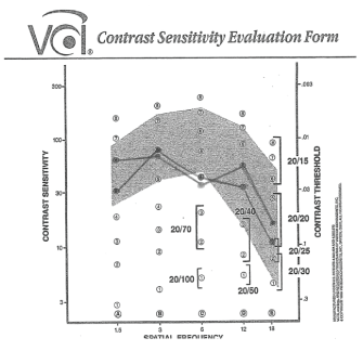
Figure 3: Contrast Sensitivity results shows normal values with a minimal non-significant decrease at spatial frequency of 6 cpd.
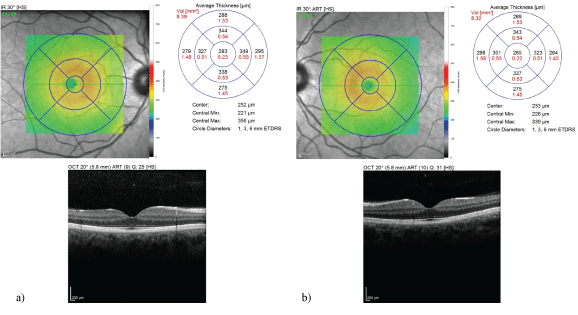
Figure 4a (OD) and Figure 4b (OS): Optical Coherence Tomography (OCT) findings portrays adequate average retinal thickness OD and OS. It shows typical curvature of the macula with all layers intact and no abnormalities.
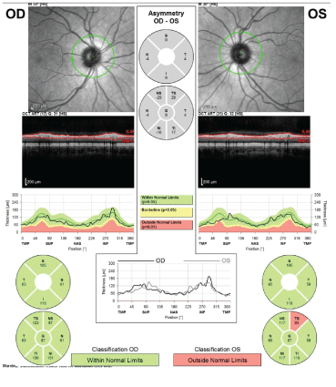
Figure 4c: Optical Coherence Tomography of retinal Nerve Fiber Layer is within normal limits OD. There is possible minimal rim thinning superior temporal of the optic nerve.
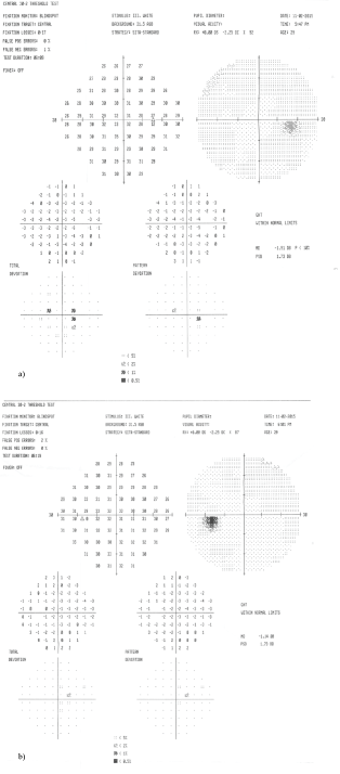
Figure 5a (OD) and Figure 5b (OS): Humphrey visual fields shows a full field with no defects, no fixation losses and minimal 1-2% false positive and negative errors OU.
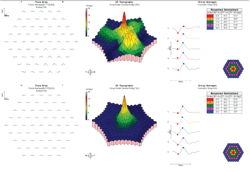
Figure 6a: Multifocal Electroretinography waveforms shows normal retinal function OS and increased paracentral waveform likely due to testing error. No retinal loss is seen OU.
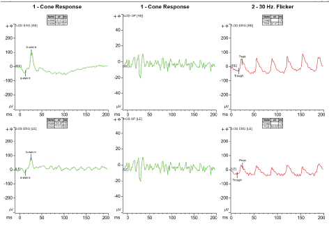
Figure 6b: Electroretinography shows normal photopic responses.
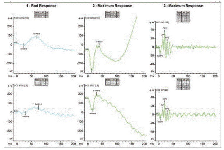
Figure 6c: Electroretinography shows normal scoptopic responses.
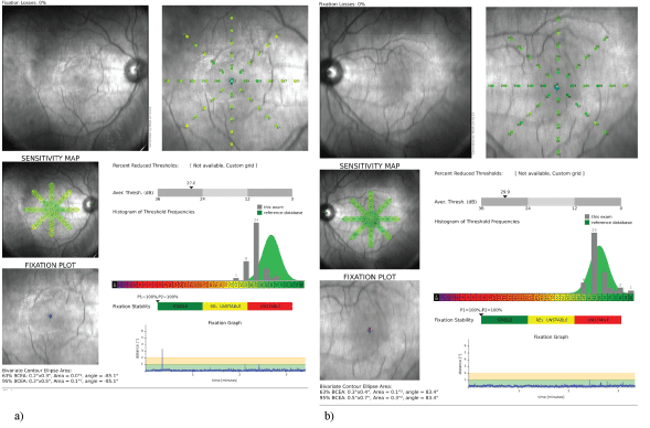
Figure 7a (OD) and 7b (OS): Macular Integrity Assessment test shows normal sensitivity and matches normal population values of her age OD and OS.
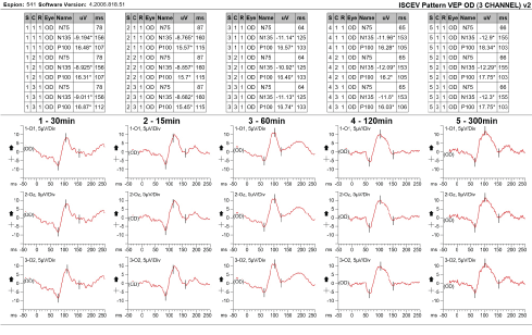
Figure 8a: Pattern Visual Evoked Potential test OD shows normal occipital responses.
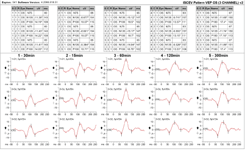
Figure 8b: Pattern Visual Evoked Potential test OS shows normal occipital responses.
The patient was lost to follow-up as she transferred her care to yet a different eye doctor.
Differential diagnosis includes those of organic and supratentorial origins.
In malingering patients, one might find abnormalities without a specific pattern or disparity between subjective and objective testing. Other clinical diagnosis to take into consideration is another visual phenomenon like synaesthesia which may involve similar abstract visual movements. However, this is often present from childhood and rarely acquired unless from drugs or brain lesions in parietooccipital areas. Negative findings on all tests including visual fields, MRI & MRA do not support a serious brain lesion including strokes, tumors, abscess or vascular malformations. However, we do not have her MRI/MRA results, we do not know the radiological sequences and purely trusting her report of the MRI/MRA was normal. Also, her symptoms matched that of illusory palinopsia which is often not caused by posterior cortical lesions. We can also rule out migraine aura due to absence of a headache, the short duration of the episodes and unilaterality. The testing was able rule out all of the differentials except for toxicity.
Literature review showed eight adult female patients taking clomiphene citrate who also experienced similar visual disturbances [4]. However, the visual disturbances were generally reversible in all eight patients and a bilateral reduction in flicker sensitivity was found with negative findings in all other tests [4].
We found analogous rare cases of three women aged 32-36 years old with visual disturbances secondary to clomiphene treatment [5]. They experienced similar symptoms of prolonged afterimages, shimmering of peripheral field and photophobia with normal neuroophthalmologic examination and electrophysiologic findings [5]. Similar to our patient, these women’s visual symptoms did not resolve on cessation of treatment [5]. They remained symptomatic from 2 to 7 years after discontinuing treatment with the medication [5].
Our research confirms our suspicious of clomiphene toxicity in our patient. A flicker fusion test might prove useful in similar patients.
This case along with our research demonstrates treatment with Clomiphene can cause prolonged visual disturbance even years after discontinuation of drug administration. Women with characteristic visual disturbances of palinopsia should be questioned about past use of clomiphene. It is important to identify the symptoms of palinopsia that may be a manifestation of a more serious underlying systemic condition that would require treatment. In addition, the clinician who is able to recognize palinopsia symptoms can help calm and educate the patient.
All authors declare no support from any organization for the submitted work; no financial relationships with any organizations that might have an interest in the submitted; no other relationships or activities that could appear to have influenced the submitted work.
Download Provisional PDF Here
Article Type: CASE REPORT
Citation: Maggiano J, Liu MM, You T, Chen S, Rathod R (2020) A Clinical Case of Clomiphene-Induced Palinopsia. J Ophthalmic Stud 2(2): dx.doi. org/10.16966/2639-152X.116
Copyright: © 2020 Maggiano J, et al. This is an open-access article distributed under the terms of the Creative Commons Attribution License, which permits unrestricted use, distribution, and reproduction in any medium, provided the original author and source are credited.
Publication history:
All Sci Forschen Journals are Open Access