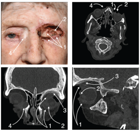
Neglected basal cell carcinomas can cause devastating localised tissue destruction. Clinical and radiological images demonstrate the erosion of the eyelids and perforation of the globe following 15 years of growth.
Basal cell carcinomas (BCC) account for 80% of all skin cancers, and due to their association with UV exposure, are commonly found on the face and eyelids. BCCs make up a large portion of the oculoplastic workload. Increased awareness and good outcomes with interventions means that we rarely see the aggressive localised impact of these tumours. In these cases, they are referred to as locally advanced BCCs (LABCC).
This frail 88-year-old female developed an eyelid growth 15 years ago. An extreme phobia of hospitals prevented any review until she was admitted with an episode of confusion and the near-complete erosion of the eyelids and globe perforation were noted. Ophthalmology was promptly involved and intervention commenced with mapping biopsies obtained under local anaesthetic via punch biopsies and small excisional biopsies of the residual eyelid tissue and conjunctival tissue respectively. Specimens were sent for histopathology, which confirmed infiltrative BCC.
CT orbits (Figure 1) showed tumour in the minimal residual lid tissue, medial and inferior orbital walls and extraocular muscles. The globe was perforated with intraocular tumour invasion evident. Ill-defined cortex and hyperostosis of the left frontal bone also suggested tumour involvement. A lacrimal sac mucocele with associated epiphora was present on the right, but intervention declined.

Figure 1: Locally advanced BCC: colour photograph and axial, coronal and sagittal CT sections respectively.
Note:
- Residual tumour in the lid tissue, medial and inferior orbital walls and extraocular muscles.
- Perforated globe.
- Hyperostosis of the frontal bone.
- Lacrimal sac mucocele.
Treatment options were discussed in a skin cancer multidisciplinary team meeting and the options of radiotherapy, vismodegib and surgical exenteration were discussed with the patient and her sons. Although exenteration is often preferred in cases of extensive orbital involvement, the patient was extremely phobic to the prospect of surgery and was a high anaesthetic risk. Radiotherapy was likely to cause breakdown of the tumour with increased infection risk and therefore could only be offered in a palliative fashion for pain management. Thankfully no pain was present despite the phthsical perforated globe. Vismodegib was ruled out due to her poor nutritional state and low weight of 39kg, which can be exacerbated with the drug. Other treatments used for BCCs include Mohs micrographic surgery, wide surgical excision, cryotherapy, photodynamic therapy or imiquimod but could not all be considered in this case.
The joint decision for palliative management was reached given the extensive nature of the disease and the frail state of the patient. Wound care and antibiotics in response to episodes of recurrent socket infection would be provided. The potentially devastating destructive nature of these lesions is a reminder of the importance of not only prevention, but also early recognition and intervention.
Download Provisional PDF Here
Article Information
Article Type: OPINION ARTICLE
Citation: McVeigh K, Vahdani K, Garrott H, Ford R (2018) A Case of a Locally Advanced Periorbital Basal Cell Carcinoma. J Ophthalmic Stud1 (1): dx.doi.org/10.16966/2639-152X.105
Copyright: © 2018 McVeigh K, et al. This is an open-access article distributed under the terms of the Creative Commons Attribution License, which permits unrestricted use, distribution, and reproduction in any medium, provided the original author and source are credited.
Publication history:
Received date: 25 Sep, 2017
Accepted date: 31 Jan, 2018
Published date: 07 Feb, 2018


