Figure 1: Schematic of Data Flow through the Brain and the Autonomic Nervous System.

Graham Wilfred Ewing*
Chief Executive, Mimex Montague Healthcare Limited, Mulberry House, United Kingdom*Corresponding author: Graham Wilfred Ewing, Chief Executive, Mimex Montague Healthcare Limited, Mimex Montague Healthcare Limited, Mulberry House, 6 Vine Farm Close, Cotgrave, Nottingham NG12 3TU, United Kingdom, E-mail: graham.ewing@mmhcl.co.uk
The first precise mathematical model of the relationship between sense perception, brain structure and/or function, the autonomic nervous system and/or physiological systems, and cellular and molecular biology was developed by Dr. Igor Gennadyevich Grakov in the period 1981-1997.
The aim of this article is to illustrate how such knowledge can be applied to better comprehend how the whole body functions and, in particular, how this can be applied to understand the process (es) responsible for aging and to understand why we ultimately expire. It supports the conclusion that the aging process is associated with the many and various factors which materially influence the stable function of the autonomic nervous system and physiological systems and that our ultimate demise is associated with the inability of the brain to sustain such functional relationships.
The author presents this knowledge in the ‘Autonomic Hypothesis’ outlined in this report, discusses the significance of the many and various factors which influence the stable and coherent function of the autonomic nervous system, which sustains the body’s function and thereby influences the aging process. It incorporates case reports to justify such hypothesis, in particular how this knowledge can be used to screen patient health and optimize longevity.
Genotype; Phenotype; Physiological systems; Autonomic Nervous System
ROS: Reactive Oxygen Species/oxidative stress; NDE: Near Death Event
Why do we age and why, ultimately, do we die? It is an issue which concerns us all, increasingly so as we age.
All aspects of the body’s function, without exception, involve an understanding of how the body functions, in particular how pathological onset is associated with (i) changes of sense perception, (ii) changes of brain structure and/or function, (iii) how this influences the stable and coherent function of the autonomic nervous system and/or physiological systems, (iv) leads to the changes of cellular and molecular biology which accompany pathological onset, and (v) influences how we behave. It is upon such relationship(s) that biomedicine and, more generally, healthcare is based.
The first precise and sophisticated simulation in the form of a mathematical model of this relationship between sense perception, brain structure and/or function, the autonomic nervous system and/ or physiological systems, and cellular and molecular biology [1,2] takes into account changes of gender, weight, and age. It illustrates that these parameters are pathologically significant. Within this model, the nature of the body’s physiological systems is clearly defined [3].
This article considers how such knowledge can be applied to better comprehend how the body functions and how this can be applied to understand the process (es) responsible for aging and to understand why we ultimately expire.
We are born from the DNA, genes, and chromosomes which have been inherited from our natural parents. Thereafter our lives evolve taking into account the myriad of interactions with our environment which influence our genotype, in particular viruses [4-6] and viruslike particles [7-9], and the many and various non-genetic factors which we experience in our lives which influence how we function i.e., our phenotype.
The genes are part of a complex mechanism which is part of a greater design however, despite the hype, it is rare that a single gene does anything. In almost all cases it takes a spectrum of genes, including many non-coding genes, to express a particular protein. The non-coding genes are not directly associated with genetic expression of proteins but they nevertheless have an indirect role in this process of genetic expression. In addition it is often overlooked that genetic expression is a chemical reaction therefore the expression of proteins and/or substrates by this reaction must be influenced by prevailing the reaction conditions.
Often an environmental trigger cultivates pathological onset e.g. stress leads to inflammation and/or oxidative stress; thereby making the management of a genetic condition more difficult [10]. The spectrum of genes producing the same protein may differ [11] e.g., between people with differing racial sub-types; therefore a single gene can only part of the complex process by which one or more proteins are expressed. The evidence suggests the existence of a mechanism which is not directly genetic but which is not independent of genotype i.e., involving changes to gene conformation [12].
Each gene or spectrum of genes, including associated mutations, and epigenetic and chromatin modifications, plays a significant role in molecular and cellular biology, organ function and system function but this is where the biochemical paradigm struggles to elucidate the mechanisms by which the body functions. So how are these genes regulated?
“We now have a parts list. That’s what the Human Genome Project is about is getting a parts list. If you want to understand the plane, you have to have the parts list, but that’s not enough to understand why it flies. Of course you’d be crazy not to start with the parts list.” Lander E [13].
The process of genetic expression yields a variety of proteins which collectively contribute to sustain the function of the autonomic nervous system i.e., the organs, blood and blood vessels, and/or which are functional components of the healing process following injury e.g. stem cells, levels of immunochemicals, etc. There must, therefore, be a hierarchical process which defines and/or prioritises how proteins react with their reactive substrates and subsequently influences how the body is assembled and how it functions.
Contemporary biomedicine has hitherto considered that these two processes-genotype and phenotype-proceed independently however an emerging body of evidence illustrates that genotype and phenotype are comorbidities [14] e.g. diabetes is characterized by a reduction of the level of insulin (type 1 diabetes) and the inability of insulin to react with its receptor protein IRP2 (type 2 diabetes); but also that problems of blood glucose regulation invariably follow hysterectomy and/or endocrine problems, type 1 and type 2 diabetes occur as comorbidities, and that the administration of insulin often does not prevent the progression of advanced diabetic comorbidities [15] in other organs and systems. Just administering insulin addresses the shortage of insulin and raise blood glucose levels but does not prevent the progression of diabetic comorbidities. This illustrates the existence of a more complex regulatory mechanism than has hitherto been considered.
‘Patching up the phenotype is what medicine is all about’. Brenner S, 2008[16]
Moreover, it may have the perverse effect of suppressing or overcoming the brain’s ability to optimise systemic stability e.g. to regulate blood glucose levels; and indicates the presence of a biodynamic relationship between the function of the brain and the visceral organs [17] in which the brain regulates, in a best-fit manner, the stable and coherent function of the autonomic nervous system, the physiological systems and organs, and cellular and molecular biology; and that, conversely, changes of molecular biology influence brain function which is the case, for example, when we administer psychotropic medication.
In essence, the genotype is a measure of physiological capacity whilst the phenotype is a measure of physiological demand -comparable to the supply of fuel and air to an internal combustion engineboth reducing the levels of protein bioavailability and physiological sustainability. This body of evidence has been building since the 1980s, perhaps earlier, when beta-blockers were first administered in combination with a diuretic-the introduction of beta-blockers was intended to treat high blood pressure by slowing heartbeat and thereby reducing blood pressure however by doing so it slows metabolic rate which leads to increased weight which in turn leads to elevated blood pressure [18]. The beta-blocker and diuretic act upon pathologies in different physiological systems. It illustrates that pathological onset is invariably multi-systemic, multi-pathological and polygenomic; and that each and every medical condition is influenced by its genotype and/or its phenotype.
In years passed the cause of death would often be declared by the doctor to be due to ‘old age’ however the progression of medical research has reached the stage when this is no longer considered to be viable. Instead, we die of a particular medical condition so it is reasonable, indeed logical within this text, to comment upon such observations and introduce into this text how pathological onset leads to our demise but how do such pathological changes occur and how do they influence the quality of our lives and our longevity?
Our physiology is genetically predetermined and is manifest from the point of conception. Many of us will develop medical conditions which appear to have been inherited from parent(s) who have the same condition and many of us will develop psychological and/or personality characteristics which are similar to a parent’s psychological traits.
There are various stages of growth and development which are physiologically significant: (i) from birth until puberty, (ii) from puberty until the menopause whilst (iii) sports physiologists will attest the existence of an intermediate second phase which occurs at ca 25- 27 years [19] when peak physical capabilities declines (arguably at ca 30 years) until (iv) we reach a further period of decline following the menopause from ca 50 years onwards. As each period involves biological changes then so too must our psychology and behaviour change.
Around age 50, women’s ovaries produce lower levels of oestrogen and progesterone and the pituitary gland increases levels of folliclestimulating hormone (FSH). For men, arguably, it is not about the onset of a male menopause although about 20% of men over age 60 and 30-50% of men over age 80 will experience a significant decline in testosterone production. Our ability to produce and/or fertilise and/or bring a fertilised egg to maturity/birth, which is free of birth defects, declines as we get older i.e. beyond 40 years [20].
More generally, our ability to express proteins declines with age e.g. our 70/80’s our ability to genetically express insulin declines, conceivably to as little as 10% of the levels which we were capable of expressing when we were at our peak years until our insulin levels are unable to sustain the body’s function and we expire [21].
Age is pathologically significant.
Unsurprisingly, and excluding the trauma of the birthing process, most of us are at our healthiest in the initial months of our lives before we experience the complex range of pathogenic factors which adversely influence our health. When nourished by the mother through breast milk this period of protection can continue for several months/years until breast feeding is discontinued and/or later.
Moreover, as outlined above, if we live long enough and live a happy and healthy life, we will likely expire due to a lack of insulin (and/ or perhaps other physiologically significant moieties). If we encounter a shortage of insulin (either as a shortage of insulin, or that the available level of insulin is unreactive or unable to react) we will likely develop the consequences thereof e.g. of diabetic comorbidities incl. Alzheimer’s Disease in which 60-80% of Alzheimer‘s patients have ‘diabetes’ [22].
We may be living longer than our parents, but the quality of our lives in our latter years remains relatively poor. See Note 1. Our longevity may have increased, (perhaps due to diet, better housing, antibiotics, etc), but the overall limit of our lives remains below 100 years i.e. the average lifespan, depending upon where you live, is ca 80-85years, 95% expire before 100 years, and few live beyond 110 years. Moreover, our latter years are of suffering and illness. It is inevitable that the quality of our lives declines as we age until we finally expire. Nevertheless, contemporary biomedicine and science more generally, continues to grasp at ‘evidence’ which suggests that some miracle component could enhance the quality of our lives and, in particular, our longevity.
Note 1: The drug Manoplax was very well received by patients. It enabled patients with chronic heart failure to have a better quality of life however the drug increased the risk of death in patients who ceased the treatment and it was, therefore, withdrawn from the market.
There is evidence that people live longer in some regions e.g. in Okinawa (Japan); some who live at altitude in Georgia; Ikaria(Gr); Nikoya (Costa Rica);and in Sardinia(I) where M26 marker, a genetic variant linked to extreme longevity, has been passed down through generations. This increase in longevity is invariably linked to regular exercise, diet (seafood/Mediterranean diet) and social links to family and friends [23] however many with those with such variants have lives which compensate for their genetic inheritance and, consequently, they expire earlier. Having the M26 marker is not a guarantee that you will live longer! It highlights the immense complexity of the aging process. Nevertheless, and irrespective of whether someone lives a longer or shorter life, their body remains regulated in an extraordinary level of precision until they expire their last breath. For some who have expired, perhaps as a result of injury, surgeons and/or doctors are occasionally able to resuscitate them and hence allow them to live for many more years. We can reach a stage when the body is unable to continue to function but medicine allows us to patch-up the problem and then resume our lives. See note 2.
Note 2: At age 27 years the author suffered a sub-dural haematoma whilst playing rugby. The surgery conducted at Newcastle-upon-Tyne General Hospital was able to deal with the damaged blood vessel and enabled the author to resume his life.
Despite the evidence for a complex multi-systemic paradigm biomedical researchers continue to focus upon particular biological components e.g. mTor, Telomerase, Vitamin D, etc, etc; although the research tends to be based upon the reductionist approach in which particular chemicals are considered as targets which can lead to the development of drugs, which mitigate against the effects of a particular ailment, and thereby extend our lives rather considering how such chemicals fit into the wider scheme, in particular an understanding of how the whole body functions e.g. too much vitamin D is toxic.
Many eminent researchers have considered, and continue to consider, why we age. Theories have been advanced which contribute to this debate including (i) the free radical theory of aging [24,25]; (ii) the evolutionary hypothesis or antagonistic pleiotropy hypothesis [26]; (iii) the Hyper function theory of ageing [27]; (iv) the damage accumulation theory [28]. Such theories are interesting because they link different aspects of the body’s physiology and function. Each has particular merits. This paper differs from previous research by considering the aging process from the neural perspective-how the brain interprets changes of sense perception and regulates the body’s function.
Weisman’s ‘wear and tear’ hypothesis considered that the accumulation of cellular damage progressively overcomes the body’s reparative abilities whilst Gerschman, Gilbert, & Harman considered the cumulative effect of degeneration caused by free radicals, in particular ROS, and showed how this altered the lifespan of yeast, worms, flies.
Williams GC [26] considered the genetic basis for disease in particular how genetic mutations may have a positive and/or negative effect e.g. that mutations which cause Huntington’s disease improve fertility and decrease the risk of cancer; or that mutations which cause sickle cell disease protect against malaria; and/or that mutations associated with cystic fibrosis also improve fertility. This is intriguing because genetic changes arising from exposure to viruses or virus-like particles are recognised to have pathological significance e.g. Jenner’s recognition that milkmaids exposed to cowpox were immune to smallpox illustrated that the genetic change arising from exposure to the cowpox virus creates the changes to genetic function and structure which protects against the smallpox virus; or that the administration of the MMR vaccine in Finland was accompanied by a 57% increase in the numbers with type 1 diabetes over a period of 20 years [29]; or that each and every genetic change appears to alter our predisposition to particular disease(s) or infection(s) [9].
During 1965 to 1984 the incidence of type 1 diabetes increased by 57% or by ca 2.4% annually. Tuomilehto et al, 1991 [29].
Blagosklonny MV [27] suggested proteins (and the genes responsible for making them), are the cause of the aging process. Some of these proteins are enzymes which assist chemical reactions to happen in our body. Among them is an enzyme (TOR) which initiates cell growth, in particular during the early part of our lives however high levels of TOR can be pathogenic in later life.
More recently, Chatelain M, Drobniak SM & Szulkin M [30] highlighted the significance of physiological stress upon DNA, in particular how telomere length declines during successive cell divisions. They associated the gradual shortening of telomere length with exposure to free radical reactions and thereafter a gradual decline of the quality and quantity of our lives.
In 1962 Hayflick L and Moorehead PS [31], demonstrated the senescence of cultured human cells. Hayflick L, in particular, hypothesised that the aging process was controlled by a biological clock contained within each living cell. Their studies concluded that various human cells (in the lung, skin, muscle, heart) have a limited life span i.e. they divided approximately 50 times over a period and then suddenly stopped and that nutrition influenced the rate of cell division: (i) overfed cells made up to 40-60 divisions before entering a period of senescence, (ii) underfed cells took up to three times longer to divide, and (iii) alterations and degenerations occurred within some cells before they reached their growth limit. The most evident changes took place in the cell organelles, membranes, and genetic material. This improper functioning of cells, due to the shortening of telomeres (cell division ceases when the telomeres reach a critical length, and loss of cells in organs and tissues) is partially associated with the onset of aging i.e. the biological clock envisaged by Hayflick & Moorhead is associated with the rate at which intercellular processes proceed i.e. the reaction kinetics, involving the genes and the intercellular environment, in which cell division was taking place.
In 1985, Grakov IG [1-3] recognised that that the ‘Accumulation of errors in the neural control programme’ leads to problems with cell-division. Grakov’s research which commenced around 1981/2 led to the development of a mathematical model of the autonomic nervous system and physiological systems. It included a mathematical computation of the ‘Hayflick Limit’ which defines the extent of cell division and also a comparison between calendar and biological age.
Genotype and phenotype are two physiologically distinct processes. The first defining the extent of genetic expression of a protein and the second defining the rate/level of protein reactivity i.e. how well the genetically expressed protein reacts with its reactive substrate. This is particularly evident in diabetes where the two components are the type 1 (genotype) and the type 2 (phenotype) however it is not necessarily the case that you will inherit such condition(s). Changes to our DNA occur as a result of exposure to viruses [32,33] and viruslike particles [29,34-37]; our exposure to stress which influences the epigenetic profile [38] and associated intercellular environment; and subsequent genetic changes which occur as a result of exposure to free radical reactions [39-41]. Such genetic changes may have a positive life-extending significance [42], whilst others clearly have a negative life-shortening significance [43,44].
In general, free radical/oxidative reactions in the body do not occur in patients with intercellular pH of >7.35. They occur as intercellular acidity increases- in those who are mainly overweight and/or physically inactive -in which the levels of essential minerals (Ca, Mg, Zn, Cr, and Se) declines, and the levels of transition minerals (Fe, Al, Mn, Pb, Hg, etc) increase. This is significant because transition minerals catalyse free radical reactions [41] which are associated with genetic changes and pathological onset e.g. the production of complex lipids which characterise diabetes [45], and atherosclerosis [46] i.e. where there is a substrate (of glucose and/or the glycation process) which can be metabolised into complex lipid(s). Moreover, antioxidants often appear to have a moderating effect upon such ROS reactions [39,40] which confirms that ROS are directly implicated in such oxidative process(es).
Intercellular pH influences the onset of free radical reactions and is pathologically significant.
In addition, (i) telomere shortening accompanies a wide range of pathological indications and is implicated in the aging process [47] i.e. it influences genetic expression and results in reduced levels of protein expression of for example, insulin [48] and hence its subsequent manifestation in a wide range of indications including cardiovascular indications [49,50], Alzheimer’s disease [51], age-related life stresses [52], etc ; and (ii) over 191 genes have been implicated in the risk of contracting breast cancer [53] and thereby highlights the existence of a metabolic/pathogenic pathway(s) which is not unique to a single gene i.e. which disrupts normal metabolic processes and leads to the onset of breast and other cancers.
The level of physical injury-whether at the systemic, organ, cellular and/or molecular level - influences whether the body’s natural healing mechanism is able to recover normal function e.g. (i) if someone has broken their leg and the leg does not heal correctly this will influence their ability to recover their ability to run (and/or associated activities) which will in turn have a knock-on effect upon how they resume and/or conduct their lives; (ii) if the patient has broken their back the re-alignment of their bones will influence how they resume and/ or conduct their lives e.g. the incorrect or abnormal alignment as a result of the injury may leave them with less mobility and susceptible to become diabetic and obese or disposed to have migraines as a result of restricted supply of oxygen via the spinal cord to the brain; (iii) if the female recovers normal BMI following childbirth; etc.
There are many vitamins and/or cofactors which have an antioxidant effect. Some are fat-soluble and others are water soluble which illustrates the need for a balanced diet. Vitamin D is significant [54] because it is produced by the action of sunlight, in particular UV-B, upon the skin but there are many other reactions which occur as a result of exposure to sunlight. Vitamin D is only one of many chemicals which effectively function as an antioxidant e.g. Melatonin, Serotonin, Superoxide Dismutase, Vitamin C, etc.
Proteins are characterised by having -NH2 and -COOH group(s) in their chemical structure which confers polarity, solubility and conformation. Moreover proteins are visually active i.e. they absorb and emit biophotons of light during reaction with their reactive substrate(s). They absorb energy in order to reach their reactive state and then release energy following reaction as the protein decays to its base state. The energy released (often referred to as bioluminescence or autofluorescence), which becomes most intense in the retina, has colour and intensity which is a measure of the genotype and phenotype for each pathological reaction and/or medical indication [55,56].
Proteins may be coiled when in their reactive state and uncoiled when in their unreactive state hence the use of the term ‘resistance’ to describe how a protein e.g. leptin (leptin-resistance), insulin (insulinresistance) and ghrelin (ghrelin-resistance); is not able to react with their reactive substrate. It is what proteins DO which is significant, not what they ARE.
“Proteins do the work. Genes are the instruction sets for those proteins. Actually, only a few percent of all of your DNA is devoted to writing down the instructions for those proteins”. Lander E [13].
It is an innate property of proteins that they are visually active i.e. they require energy in order to reach their reactive state when they react with their reactive substrates. At this point they release energy, often in the form of light, as they decay to their unreactive base state. As this release of biophotons occurs in the bloodstream, the light released influences colour perception in the retina. This is a direct and precise measure of pathological onset and provides the data sets upon which to base a mathematical model of the autonomic nervous system.
This phenomenon is often known as autofluorescence or bioluminescence and is widely recognised for its value in analytical chemistry and in astronomy e.g. the company BioAstral, prior to its demise, (https://www.le.ac.uk/biology/phh4/bioastral/home. htm) was researching advances in space-science imaging technology i.e., to measure the release of biophotons from blood (quote: ‘Our STJ cryogenic detector is 1000 times more powerful at detecting fluorescence in biological assays than current technology, and is unique in giving the colours of photons detected without requiring filters, gratings or other techniques’).
Changes of sense perception, in particular of colour perception (but also of odour/smell, taste, touch, and the vocal spectrum), accompany almost every known medical indication [56], drug, vaccine, virus, etc; so using measures of colour perception (light is considered to be responsible for ca 85% of sensory input; the eyes respond to as little as 7*10^2 biophotons per second) we can, in principle, relate changes of colour perception to the onset and progression of every medical indication.
Perhaps the most obvious example of such changes of colour perception occurs in the diabetic patient where the condition is accompanied by changes of blue/yellow colour perception [56]. It illustrates how changes of sense perception have pathological significance but also that changes of colour perception, in particular, influence brain function, in particular the amygdala [57], hippocampus [58], lateral geniculate nucleus, [59] hypothalamus [60], etc; and thereby influence the function of the autonomic nervous system [61].
Light comprises ca 85% of sensory input and is the primary mechanism by which we engage with family, friends and colleagues. It influences the immune response. Light, in particular through changes to sense/colour perception, has pathological significance.
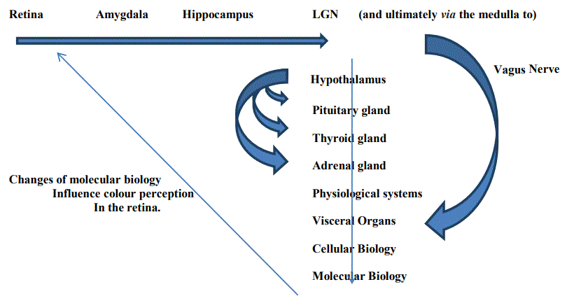
The eye is the part of the brain which conveys information into the limbic system. It initiates our function and/or behaviours. The sequence of data receipt and/or processing, as described in figure 1 below, illustrates how data is delivered as electrical impulses by the vagus nerve [62] from the medulla oblongata to the heart, lungs, liver, stomach, etc; but also as biochemical changes in the blood and peripheral blood vessels.
Figure 1: Schematic of Data Flow through the Brain and the Autonomic Nervous System.
The term ‘the Hypothalamic-Pituitary-Adrenal (HPA) axis’ has been coined to describe the interaction between the hypothalamus and primary endocrine glands and how they respond to stress however psychological stress is primarily experienced through the visual pathways i.e. through the eyes; but is only interpreted by the body as a stress following comparison and/or interaction with memories (of events which influence our ability to express proteins, which reduce protein reactivity, or otherwise influence the body’s function) which must therefore involve the hippocampus and its relationship with longterm memories stored throughout the cortex. The key endocrines are the pituitary, thyroid and adrenal glands are essential and fundamental components in the body’s physiological systems.
It illustrates the existence of two distinct mechanisms and/or systems (i) which supply the components required by the autonomic nervous system i.e., a relationship involving the hypothalamus, thyroid, pituitary and adrenal glands (often called ‘the HPA axis’); and (ii) which regulates the supply of these components. The lack of a precise understanding of techniques which are intended to stimulate the vagus nerve leads to the onset of side-effects e.g. altered heart rate, difficulty breathing, digestive issues. Despite this lack of understanding vagus nerve stimulation has been approved in the US to treat depression, epilepsy, and other indications.
Different senses influence different parts of the brain via differing sensory pathways. This illustrates that brain function is influenced at different levels by multi-sensory input and/or influences autonomic output i.e. that there is a predictable pattern of response based upon the levels of multi-sensory input and pathological onset; and there is a functional connection between the brain, the autonomic nervous system and physiological systems, which incorporates the endocrine glands/organs, and which leads to a spectrum of changes of both genotype and phenotype in each organ i.e. pathological onset.
Note 3: It is conceivable that altered and/or uncoordinated flow of sensory data through different sensory pathways would lead to sensory synaesthesia.
All behavioural functions involve precise and specific patterns of autonomic nerve activity [56-63] which regulate the various patterns of organs in each physiological system. These patterns of response to sensory challenges require specific anatomical connections, structures, neurons, etc.
The hypothalamus acts as a modulator of autonomic function. It stimulates the pituitary gland-acting in a concerted fashion with the thyroid gland-to feed a variety of hormones into the blood stream e.g., adrenocorticotropic hormone, and stimulate the adrenal glands [63] to secrete steroid hormones e.g. cortisol; human growth hormone which regulates growth and body composition; luteinising hormone; follicle-stimulating hormone; prolactin; thyroid-stimulating hormone, which stimulates the thyroid gland to secrete thyroid hormones; the hormones vasopressin and oxytocin which are stored in the posterior pituitary gland prior to their release into the bloodstream; a melanocytestimulating hormone which is produced in the intermediate pituitary gland and acts upon skin cells to stimulate the production of melanin; sustain immune function; etc.
Neural components respond in a manner akin to smooth muscle i.e. according to demand. What we do in our lives influences the brain’s functional size, plasticity, and capabilities e.g. the birth control pill provides hormones which alter fertility and thereby prevent birth however recent research has now illustrated that these pharmacological substances reduce the size of the hypothalamus by ca 5-10% [64]. Conversely, the size of specific neural components will often increase in response to intense and specific types of activity e.g. the size of the hippocampus is often larger in drivers of London taxicabs [65], there is higher microstructural diffusion in the corpus callosum of longterm drummers [66], and those with greater social circles have greater amygdala volume [67].
The pituitary gland responds to light e.g. (i) exposure to bright light, acting through the pineal gland [68] stimulates melatonin production, influences the secretion of pituitary hormones [69]; (ii) melanocytestimulating hormone, produced in the intermediate pituitary gland, acts on skin cells to stimulate the production of melanin [70] which conceivably acts as an antioxidant [71]. The hypothalamus is linked with each of the primary endocrine glands and each of the body’s physiological systems (the networks of organs which act coherently in order to sustain a particular function) [60,61].
Over 100 medical indications appear to respond to the therapeutic effect of light e.g. multiple sclerosis [72], seasonal affective disorder and/or depression [73-76], lupus [77], neonatal jaundice [78,79], psoriasis [80], Alzheimer’s Disease [81], etc. Light, in particular exposure to normal levels of summer sunlight, has a neuro stimulating mode of action which has the potential to improve metabolic function, in particular in those who are diabetic and obese [82,83] and extend our longevity [84]. Accordingly, exposure to the blue light on the screens of mobile phones, computer screens, and televisions must inevitably, depending upon the extent of exposure, influence the body’s regulation and health.
Light is physiologically significant.
Longevity varies between the genders. In the UK the male currently lives until ca 79 years whilst the female lives to ca 83 years. In Japan, females live until ca 90 years whilst the males live until ca 85 years. It is clearly evident that females live longer i.e. that their unique biological and/or genetic profile predisposes them to live longer whilst the unique biological and/or genetic profile of the male predisposes them to live ca 3-5 years less. This implicates the genetic profiles, which differ between male and females [85], and the subsequent relationship between molecular and cellular biology, organ function and the coherent function of the organ networks (more commonly known as the physiological systems) e.g. chromosomes (female XX, male XY), reproductive organs (ovaries, testes), hormones (oestrogen, progesterone, testosterone) [86,87].
To illustrate the point: the SRY gene, in particular, initiates a spectrum of genes which are activated in male embryos or inactivated in female embryos. Consequently, genetic changes [88,89] influence gender and predisposition to disease e.g. (i) diseases which occur in only one sex e.g. autoimmune diseases, (ii) diseases which are more usual in one sex but which may manifest differently in each sex e.g. 90% of primary biliary cirrhosis cases occur in the female whilst primary sclerosing cholangitis is more common in 60-70% of males [90].
Gender is pathologically significant.
The body is a highly regulated entity [91-99] i.e. there is a precise and sophisticated mechanism which continuously regulates and micro manages the body’s physiological stability throughout every second of our lives in a best-fit manner in order to optimise and/or maintain the body’s key physiological parameters i.e. the brain regulates the autonomic nervous system. It does so by continuously modulating the coherent function of the various physiological systems. These systems comprise networks of organs which are neurally regulated and act coherently to perform a significant physiological function.
Each system performs a vital executive and/or regulatory function however biomedicine remains unable to explain how these networks of organs function in a coherent manner. The terms hyper-function and hypo-function are routinely used to describe pathological onset which illustrates a general acceptance of the phenomena. Thereafter histopathology tests are used to define the nature of pathological onset i.e. to identify specific changes of molecular biology which occur in response to changes of brain function.
The histopathology tests used in biomedicine identify many of the biological or biochemical consequences arising from the function or dysfunction of these systems but not how and why these systems function and are regulated. Moreover the measured parameters e.g. of a protein or other biochemical; are often unable to delineate between the coiled and reactive component versus the uncoiled and unreactive component [100]; whether the measured parameter is a systemic parameter or a molecular parameter; and/or whether it is a reliable indicator of pathological onset and/or progression.
Failure of the brain to regulate the coherent function of these systems- of sleep, body temperature, blood pressure, blood volume, intercellular pH, etc- is accompanied by changes of molecular biology and of pathological onset depending upon the origins of the autonomic instability as discussed in this text e.g.
Sports physiologists recognise that the brain functions as a complex neuromodulator which continuously regulates the function of the body’s physiological systems [105-107] e.g. during extremes of athletic pursuits. For them, the issue is no longer whether the brain continuously regulates the autonomic nervous system but instead what are the mechanisms by which it does so.
How can we explain that (i) a child’s exposure to stressful experiences can influence the time when puberty takes place [108- 110]; (ii) alcoholic intake during pregnancy increases the risk of abortion or birth defects [111,112]; (iii) altered systemic stability during pregnancy e.g. of intercellular pH, elevated blood pressure, levels of blood glucose(diabetes); influences childbirth outcomes [113- 115]; (iv)an inadequate diet during childhood influences our growth and development [116]; (v) smoking during pregnancy can result in abortion or birth defects [117]?
How can we explain that when stressed, specific organs may become dysfunctional e.g.
There must therefore be a hierarchical process which regulates how proteins react with their reactive substrates and subsequently influence how the body functions e.g. (i) some physiological processes vary between the day and night periods, between the summer and winter periods [124], and/or during extremes of altitude and temperature; (ii) some biochemicals are stored in readiness for particular events e.g. insulin, magnesium, zinc, vitamin D; and (iii) how organs and organ systems function and/or are regulated.
There can be several outcomes involving both genotype and phenotype :
Note 4: low levels of magnesium and other essential minerals is a characteristic of both type 1 and type 2 diabetes.
Modern society, through its association with, and adoption of, anything which alters perceived taste(s) experiments with a wide range of chemicals and factors which alter the function of the sensory organs to give a sensory experience. This is perhaps most evident in products containing salt, flavour enhancers, phosphoric acid, acetic acid (vinegar), ethyl alcohol (alcoholic beverages), etc. Each has a physiological effect on the tongue and the brain.
The most basic studies of inorganic chemistry focus upon the solubility and insolubility of inorganic materials. This study of essential minerals is vitally important in biology because of the interplay between acidity and alkalinity and, in particular, how acids react with essential and/or transition metals. If essential minerals are not present in the intercellular medium-in particular Magnesium, Zinc and Chromium-the body’s metabolic processes are unable to proceed e.g. to metabolise blood glucose and generate energy, and results in weight gain.
There is, therefore a need to maintain a balance between the levels of essential and transition metals. This is sustained by keeping the body’s pH at ca 7.35. If levels of acidity increase and the pH declines, perhaps to 6.75 or below, this favours increased levels of transition metals and reduced levels of essential minerals; and vice versa. This creates an environment which favours the creation of reactive oxygen species [39-41] which react with available substrates e.g. glucose, LDL cholesterol, proteins, DNA; to create complex lipids and glycated proteins which are characteristic of diabetes and heart disease(s).
High levels of Salt are linked to hypertension [125]; high levels of acetic and, in particular, phosphoric acid are linked to osteoporosis [126] i.e. the acids react with essential minerals to form insoluble mineral salts; and the consumption of ethyl alcohol (which metabolises to acetaldehyde and acetic acid) in alcoholic beverages is linked to the progression of pathologies [127] in the pancreas, heart, liver, kidneys, peripheral blood vessels, brain: which present as diabetes, cardiac arrhythmia, cardiomyopathy, hypertension, cancer(s), liver cirrhosis, etc. Phosphoric Acid reacts with various minerals and has a demineralising effect. Without adequate levels of Magnesium, Zinc, and Chromium the body is unable to maintain normal levels of metabolic function [126]: (i) the reaction of insulin with its receptor protein is a magnesium-dependent reaction, (ii) the storage and supply of insulin is zinc dependent (zinc is required to store insulin as the zinc-hexamer), and (iii) the metabolism of blood glucose into energy in the smooth muscles is a chromium dependent reaction.
Ethyl Alcohol metabolises into acetic acid which also depletes the body of essential minerals [127] (forming insoluble or less soluble mineral acetates) and also creates the circumstances necessary to sustain oxidative stress reactions.
The prevailing level of Intercellular pH is physiologically significant.
The physiological systems are consistent with the body’s basic and most fundamental behaviours without which it could not continue to function and that the autonomic responses involve interaction between the brain and associated structures which are mediated by neurons and nerves which stimulate the body’s systems and organs i.e. the organisational principle of the autonomic nervous system.
A pattern of response to a particular sensory challenge requires specific functional and anatomical connections from modality-specific sources. Janig W, 2006 [128].
Memory is significant because the brain, primarily via the prefrontal cortex and hippocampus, is able to store and compare short-term memories with long-term memories and thereby establish whether a pattern of sensory response is characteristic of stress and/ or a pathological state. Our memory of stressful experiences alters the epigenetic profile [129,130,38] and influences (i) the rate and/or levels of protein expression, (ii) alters the phenotype and/or the shape/ conformation of proteins [131-133] (Figure 2), and (iii) the reactivity of the proteins which have been expressed.
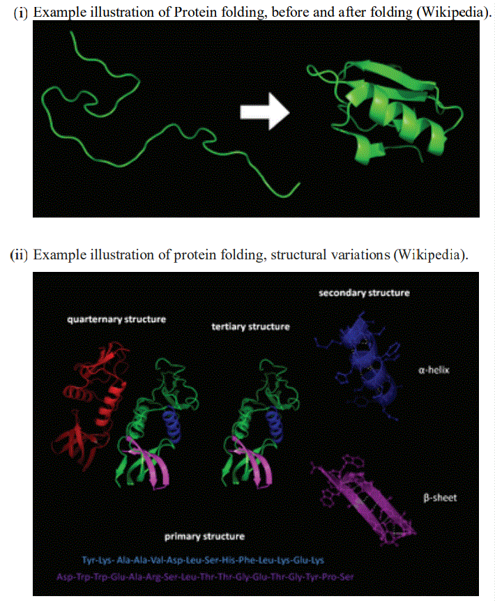
Figure 2: Protein folding.
In addition, the brain accumulates memories throughout our lives yet, as we age, the body becomes steadily less able to sustain the brain’s structures and function. Many of the essential hormones, which influence brain function, are produced by visceral organs i.e. there is a dynamic relationship between biological processes in the neural and visceral matrices. Accordingly pathological onset must inevitably lead to changes of brain function and/or cognitive decline e.g. as in Alzheimer’s Disease; and/or the ability of the brain to sustain the regulated function of the autonomic nervous system and thereby maintain the stable and coherent function of the physiological systems. Moreover the level of a stress and/or the time of exposure to a stress influence our long-term memory [134,135]. If the longterm memory is of stress we steadily become accustomed to the stress (stress becomes habitual) and also to the pathological onset which accompanies the stress.
The Stress response is more often defined as the relationship between the sympathetic and parasympathetic nervous system (the autonomic nervous system) or the ‘fight or flight’ response.
It influences the brain’s ability to regulate the autonomic nervous system and results in changes of cellular and molecular biology, and leads to pathological onset. This subsequently influences brain function i.e. there is a biodynamic relationship between the brain and visceral organs (Figure 3).
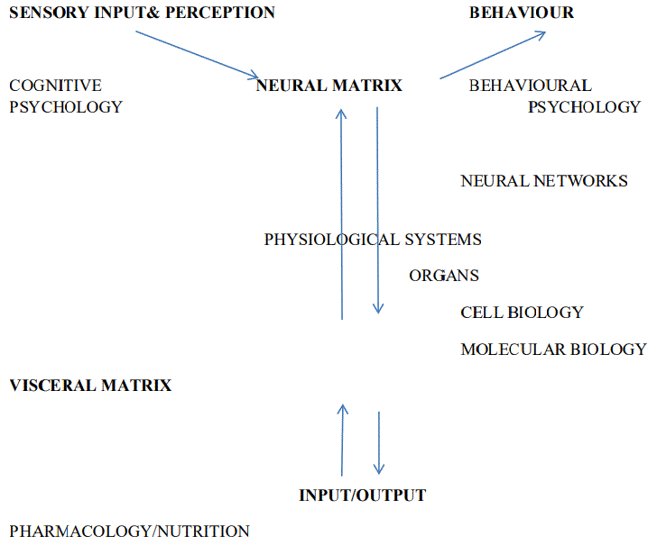
Figure 3: The Structural nature of the Autonomic Nervous System.
It influences the quality and quantity of sleep, the beneficial effect which sleep performs, including the ability of the neural structures to regulate the autonomic nervous system and the coherent function of the physiological systems.
Stress is experienced through the senses and influences the prevailing levels of intercellular pH. It influences brain function, and the coherent function of the autonomic nervous system including the coherent function of the digestive tract (retaining digestive acids within the stomach and duodenum), cellular biology, levels of essential minerals, protein conformation, etc; and leads to a wide range of problems including heart problems, digestive tract ailments, the quality and duration of sleep, inadequate supply of oxygen to the brain, elevated body temperature, elevated sympathetic response, poor appetite, etc.
Pharmacological substances are usually administered via the gut which enables the drug substance to reach the brain (in the case of psychotropic medications) and other visceral organs.
Stress can be characterised as psychological and/or psychoemotional stress (the level of hormones are often linked to specific emotional responses or medical indications) but also as psychophysiological stress due to the effect of excess body weight upon the body’s physiology.
It is experienced as a neurological response via the vagus nerve [136] and a biological response via the endocrine glands [137] which secrete their various hormones into the bloodstream [138,139] i.e. stress comprises both neurological/vagal and biochemical components.
Pathological onset rarely occurs when intercellular pH is maintained at ca 7.35 i.e. pathological onset arises mainly as a result of the onset and/or progression of innately acidifying processes in the brain and in the viscera and is the fundamental basis for the ‘inflammatory response’. Stress influences the ability of the digestive tract to retain acidity with the result that acidity levels increase in the intestines.
Different types of psychological/psychoemotional stress influence the body in subtly different ways [140] e.g. Some of these stressful responses can lead to endocrine disorders like Graves’ disease, gonadal dysfunction, dwarfism, obesity and cancer [141]. Stress can also alter the clinical status of many pre-existing endocrine disorders [142] thereby influencing the function of the adrenal and thyroid glands. It influences the phenotype of pathologies in all organs throughout the body. Stress is not a single phenomena.
Stress is pathologically significant.
Perhaps the most difficult factor to explain, and certainly one of the most controversial, is that of body weight. An immense amount of research illustrates that excess weight is pathologically significant. The following brief discussion is intended only to illustrate the general nature of this relationship.
In modern societies there is a reluctance to accept that lack of exercise [143] and consequently excess body weight [144,145] is fundamentally responsible for our poor health including the associated burden of cancer [146]. We are in denial i.e. we become diabetic, obese, lose our mobility and general capabilities yet deny that the excess weight is the cause of our poor health. Moreover being called ‘fat’ is often considered to be an insult so we are socially pressurised to accept people being overweight!
The male will often gain weight until abt. 55 years, and thereafter will start to lose weight whereas the female will often gain weight until abt. 65 years, and thereafter should begin to lose weight. This occurs, due to declining metabolic rate and, arguably, because fat (which weighs less than lean muscle tissue) replaces lean muscle tissue, however the body has an innate level of tolerance to excess weight however the body is designed to be physically active. Despite the epidemic of hyper indulgence most of us have relatively normal weight (BMI). Without adequate levels of exercise there is an accumulation of CO2 in the intercellular medium, the level of intercellular acidity increases as we consume excess levels of carbohydrates, the levels of essential minerals in the intercellular medium decline which alters Zinc levels and the ability to eliminate CO2, the smooth muscles become progressively less able to metabolise excess levels of blood glucose, body fat accumulates in the less active regions of the body, inflammatory processes become established, metabolic rate declines, etc.
Apart from the physical consequences of excess weight upon the joints which influences our agility and mobility excess weight has pathological consequences. The fatty acids in body fat are often highly acidic [147,148]. This acidity, directly or indirectly, influences intercellular pH (which at its extreme can decline to below pH 6.5), levels of essential minerals, and is pathologically significant and leads to a range of pathological indications e.g. the onset of oxidative (ROS) reactions which lead to the glycation of essential proteins [149], increased levels of complex lipids [150], increased blood viscosity [151], elevated heart rate and blood pressure [152], the functional capability of the liver and kidney declines, circulatory/ulcerative problems [153], development of cataracts and other visual problems [154], prematurely degraded joints/onset of osteoporosis [155], cerebrovascular disease/stroke [156], chronic obstructive pulmonary disease [157], asthma [158], polycystic ovarian syndrome [159], kidney stones, depression [160], cancer [161], etc.
Weight is pathologically significant.
Although the body is an intensely regulated moiety, and although the brain manages the body’s executive functions and has immense processing capability, and despite the arguments summarised and/or outlined in this text, it is not yet accepted that the brain regulates the body’s function. It is however accepted by sports physiologists who recognise that the body’s systemic stability is neurally regulated [105-107].
The identification of the multi-level nature of brain function is one of the primary objectives of the EC’s Human Brain Project however there is not, as yet, a fundamental understanding or agreement of such phenomena. Nevertheless there is widespread interest in using such techniques [162] (biofeedback, neurofeedback, neuromodulation, brainwave coherence, transcranial magnetic stimulation, and deep brain stimulation use electrical stimuli to stimulate the basal ganglia) to treat Multiple Sclerosis [163,164], Parkinsonism [165], Alzheimer ’s disease [166-168], Migraine [169], Premenstrual Syndrome [170], etc.
The EEG states are particularly significant because we require typically 8 hours of sleep each day and we sleep for the majority of this period in the delta state. Without sleep the quality of our lives declines significantly. It is a physiologically important and indispensable aspect of our lives. In normal periods of activity the brain functions continuously in the delta and theta states throughout the 24 hour cycle. This is augmented by activity in the gamma, beta and alpha states, during the non-sleep period i.e. when we are awake. In the dead patient, all EEG activity ceases whereas in the comatose patient the brain may be locked in the delta state.
It is increasingly apparent that the EEG states reflect different levels of neurological function. Different stimuli act upon the function of different EEG states e.g. EFT (emotional freedom technique), CBT (cognitive behavioural therapy), reiki, hypnosis, meditation, music, biofeedback techniques, sleep, neuromodulation techniques (SLT), etc. Accordingly, the issue is no longer whether such techniques can be effective, but instead, why do these techniques have an effect and to what extent can these techniques be expected to be effective i.e. the precise selection of applied parameters influences the desired outcomes.
Although the body is a biological entity its biology does not directly ‘regulate’ the body’s function e.g. if we administer psychotropic medication (i) the drug will often influence systemic stability often leading to the onset of diabetes and/or obesity, however (ii) over a period the drug will become increasingly less effective as the brain learns and/or adapts to compensate for the effect of the medication [171-173].
The brain uses the EEG frequencies to regulate the body’s multilevel function i.e. the brain functions as a neuromodulator yet biological changes are responsible for the autonomic dysfunction which influences systemic stability and which alters the perceived stability of the EEG frequencies. This is apparent if we consider the EEG-based mechanisms which sustain the body’s function and existence.
The body is a biological entity which fuels a biophysical entity-the brain-therefore pathological onset influences the ability of the brain to perform its essential function of maintaining the body’s physiological stability.
This dysregulation or imbalance is responsible for what we experience as pathologies and morbidities. Accordingly, an understanding of this mechanism would enable the provision of healthcare to treat the fundamental causal mechanisms and the symptomatic presentation of the person’s unique medical conditions.
The brain uses frequency to regulate the coherent function of the autonomic nervous system and physiological systems [174,175]. It explains why, for example, the selection of the wrong frequencies can cause photosensitive events [176] e.g. by stimulating reductions in blood flow and/or the flow of oxygen to the brain, as occurs in complex medical indications such as migraine or epilepsy which often have complex multi-systemic and multi-pathological origins.
The onset of molecular and cellular pathologies are the consequence of this process however biological input (nutrition, air, water, drugs) influences brain function i.e. the neural and visceral data matrices operate dynamically. Significant variations to what we eat, drink, breathe and body temperature influences the body’s function. Stress (the stress response or phenotype) influences this dynamic and is manifest as a spectrum of pathologies which influences heart function, breathing, kidney function, pancreatic function, sleep, etc. The outcome of this process is changes of how we behave-of memory(s), speed of movement, smoothness of movement; how we organise our lives and our priorities throughout the day, weeks and months ahead; and our effectiveness i.e. our ability to start and complete the many tasks which we require to complete each day.
Pathological onset influences the brain’s ability to regulate the coherent function of the organ networks. In order to do so the brain functions as a neuromodulator this uses the EEG frequencies to modulate the coherent function of the organ networks. Such a phenomena involves the body’s chemistry but this is unable to explain the entirety of this phenomena. Pathological onset influences brainwave coherence and vice versa. Different components work at different frequencies i.e. the precise nature and degree of pathological onset in the patient influences the selection of modulating frequencies, dose responses, etc.
If eat and drink too much of the wrong things we will become diabetic and/or obese. The eating and drinking are the fundamental neurological cause. The diabetes and obesity is the consequence of this process. By the same token if we are stressed, perhaps as a result of a bereavement, divorce or as a result of stress in the workplace we can develop a range of pathological indications to various organs and physiological systems e.g. we might sleep poorly, develop a cardiac problem (the term ‘dying of a broken heart’ was not coined in ignorance), pains in the low back, digestive issues, etc. The stress is the cause whilst the medical indications are the consequences. Bearing this in mind it is clear that if we are overweight a drug is unlikely to address this problem. Only exercise and dieting will reduce weight and resolve the problem. A drug acts upon the consequences and masks or otherwise ameliorates the symptoms. It is therefore appropriate to question whether the drug is acting on the fundamental cause of the condition e.g. as an antibiotic would act to kill an invading bacterium; or whether is acting upon the consequences of the condition; as is the case with many life-style related drugs e.g. as outlined earlier regarding the use of beta-blockers to treat high blood pressure [18]. The drug lowered heart rate which had the effect of reducing metabolic rate and increasing weight: the exact opposite of what was claimed, and/or expected, and/or claimed in the medical literature.
Some drugs may be teratogenic or pathogenic [177,178] thereby illustrating that genetic changes caused by drugs can be deployed with positive and negative effects, and that such effects may be shortlasting but can also persist long after exposure to the drug has ceased. Many drugs influence the genome and/or epigenome by altering the underlying genetic sequence or by creating epigenetic [179] or chromatin modifications [180] which influence the ability of the gene to express a protein.
Drugs may alter epigenetic homeostasis by direct or indirect mechanisms. Direct effects may be caused by drugs which affect chromatin architecture or DNA methylation e.g. the antihypertensive hydralazine [181] inhibits DNA methylation; whereas the indirectly acting drug isotretinoin [182-189], influences the process of transcription.
The epigenetic side-effects of pharmaceuticals may be involved in the etiology of diabetes, obesity, heart disease, cancer, neurological and cognitive disorders, infertility, sexual dysfunction, etc. For example:
Association of safety concerns/and side-effects are wide and varied e.g. (i) low neuropsychologic performance following cancer treatments [203,204]; (ii) sexual dysfunction [205,206]; (iii) the occurrence of cancer [207,208]; (iv) side-effects from antibiotics [209-211]; (v) etc.
Under certain circumstances, some pharmaceutical products may have pathological significance which results in an unwelcome range of side-effects.
Strannik is the first technology to be based upon a mathematical model of the relationship between sense perception, brain function, the autonomic nervous system and physiological systems, and cellular and molecular biology.
By determining the neural mechanisms which regulate the function of physiological systems and inherent visceral organs, using the Strannik Virtual Scanning test, it is possible to determine the extent of pathological onset in different degrees of complexity and severity; and to determine the unique parameters of the Strannik Neuromodulation Therapy (SLT). It provides a validated scientific hypothesis [1-3] against which most other techniques can be compared.
As outlined earlier, neuromodulation techniques have been developed which illustrate the existence of a medical principle, in particular, that it is possible to manipulate brain function using applied impulses e.g. electrical impulses in the case of DBS, magnetic impulses in the case of TCMS, resonant frequencies in the case of EEG, and sensory impulses using sound and colour; however most such research is experiential.
Nevertheless it is important to recognise that all hypotheses, whether proven or unproven, are based upon a set of assumptions however the data presented in a programme of ca 80 peer-reviewed medical papers by the author suggests that the nature of these assumptions does not introduce errors which invalidate the scientific hypothesis but instead strengthens and validates the hypothesis presented by the author. Moreover, if the doctor cannot make an accurate diagnosis, which is often the case, how can they define the parameters of the drug or neuromodulation therapy?
This technology, known by the brand name Strannik, has been evaluated in an extensive series of clinical studies and case studies which illustrate how it can be applied to screen and/or treat the health of the patient. The clinical studies illustrate that the technology performs 2-23% more precisely than the entire range of diagnostic tests against which it was compared [2,212,213] and which were in use in the various test clinics and ca. 75-96% effectively as a therapeutic/ neuromodulation technique [2,212] i.e. treating ca. 30 categories of common medical indications. The extensive list of case studies illustrate how it performs as a diagnostic technology re the diagnosis of specific medical indications e.g. diabetes [214,215], cardiovascular disease [216], migraine [217], Raynaud’s phenomenon [218], dementia/ Alzheimer’s Disease [219], cancer [220], etc; and as a therapeutic modality treating diabetes and related diabetic comorbidities [214], mental health indications [221], migraine [176], sleep disorders [221], dysarthria [222], dyslexia [223], etc.
Such data indicates that the technology is based upon an advanced level of understanding of how the body functions- a more advanced understanding than currently prevails. Indeed, as our failing health is considered to be a measure of our declining longevity it follows that techniques which are able to measure our declining health must also, to some extent (and depending upon what is being measured), be a measure of our declining longevity (Figure 4 and Figure 5).
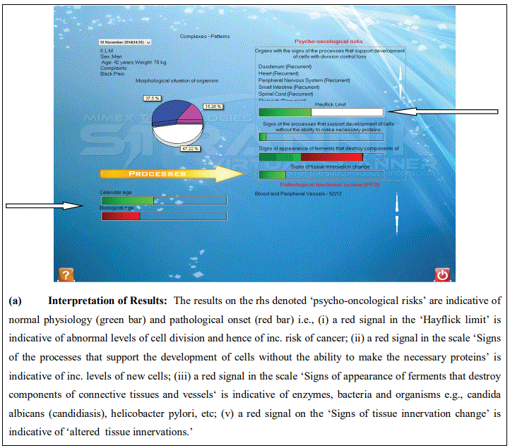
Figure 4: Hayflick report.
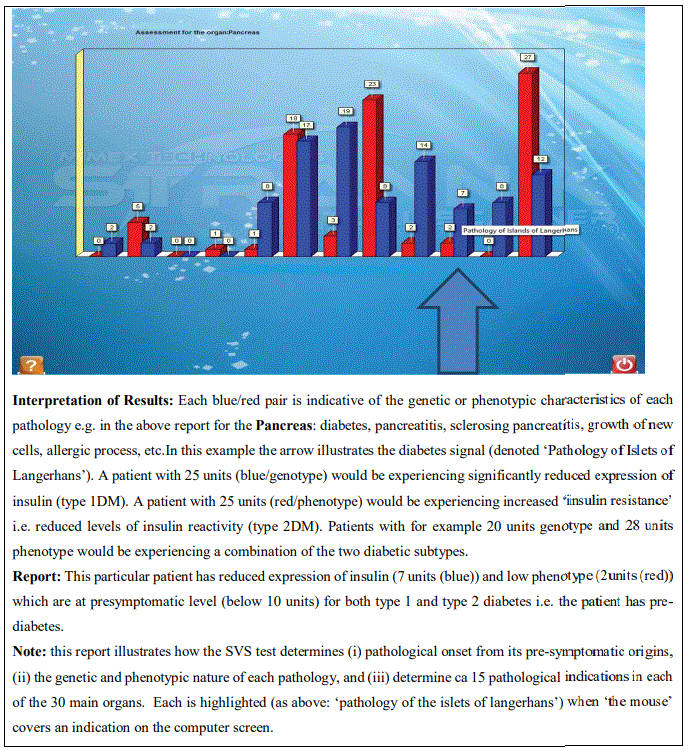
Figure 5: Strannik Virtual Scanning Test Report.
The SVS test was first placed on the market in ca 1997. Since then over 1M tests have been conducted, mainly in the Russian market. The results of ca 15-20 clinical studies conducted in the Russian market in the period 1997-2003 (comparing the results of the SVS test with that obtained by the GP using available histopathology tests was reported as 2-23% more accurate. This was corroborated by further studies conducted in South Africa, Spain and ca 500 tests/case studies which were conducted in the UK market during the period 2003-2017.
On one occasion the test was used to make a diagnosis of mesenteric insufficiency- a condition affecting 1 in 250,000 patients. This patient was considered by her consultant to have mental problems! Her discomfort was considered to have psychosomatic origins! In another case a patient was given a diagnosis of duodenal ulcer which was only confirmed by her GP 1-2 weeks later - when she started coughing up blood! Almost all of these ca 500 tests were patients who had unresolved health issues i.e. they were seeking something better than could be offered by contemporary biomedicine however despite the many reports which highlighted very significant ailments we refrained from giving alarmist comments to these patients.
In ca 2016 a reputable medical company wished to know whether SVS could determine whether a patient was likely to experience a heart attack, stroke, or other severe life-threatening event. They offered medical consultations to their clients, which included a barrage of histopathology tests (tests which are widely and commonly used in healthcare to monitor patient health), in order to assess their health and wellbeing however they had come to recognise the limitations of the tests upon which they based this service i.e. that many patients, who had been given an all-clear report, were being readmitted shortly thereafter with heart attacks, stroke, and other issues of concern. The author was requested to advise whether the SVS test could identify the onset and/or progression.
The following case reports illustrate the diagnostic and/or therapeutic scope of the technology re the treatment of life-threatening conditions. See Case Reports 1-4 (which were known to NHS health professionals in Nottingham, Cardiff, Stoke-on-Trent, and Cumbria).
Case report 1: In early 2017, we were approached by a patient who wanted to have their health checked. The test identified a range of cardiovascular pathologies which, if correct, placed the patient at risk of a severe event. On the basis of the SVS test results it was highlighted to the patient they were at high risk of having a cerebrovascular event, especially so if they did not make efforts to reduce their weight. The patient did not reduce their weight, or alter their lifestyle, and suffered a trans-ischaemic attack several months after the SVS test.
Case report 2: A patient of 76 years attended for a Strannik consultation. His condition was of prostate cancer and mesothelioma Stage 4 with a prognosis of imminent death. He required assistance to walk, needed to urinate every hour, and had severely restricted breathing due to the oedema in his lungs which required chemotherapy, radiotherapy and surgical intervention but which failed to cure the problem of oedema accumulating in his lungs.
After ca. 6 months of Strannik neuromodulation therapy his health was much improved. His weight increased by 5 kgs, he walked unaided although slowly, his breathing was dramatically improved, his eyes were alert, he no longer needed to go to the toilet to urinate every hour, his mental clarity was much improved and he was content. X-rays showed the complete absence of fluid on his lungs. As a result he no longer had chronic breathing insufficiency, he could walk unaided and his overall health and wellbeing was much improved.
He declined to continue the course of SLT and died 1-2 years later i.e. 1-2 years later than forecast by his consultant.
Case report 3: Female, mid-50, cancer of the lymphatic glands/ sarcoma on her neck. She was looking for a non-drug alternative to surgery and selected a Strannik consultation. She was advised to trust the health services provided by the NHS, and to accept their recommendations and undergo the surgery. She was at that time trying some other therapies and had a very bad experience with a practitioner who had treated her with ozone therapy. Her face was in lumps, bumps, discoloured- she looked terrible and felt very ill. After a course of the Strannik neuromodulation therapy, during the period October 06- February 07, her medical report, obtained from her oncologist, revealed that she was completely free from any detectable signs of cancer and that CAT scans did not detect the presence of any tumours. She continued to have swollen lymph glands but not as badly as before.
Case report 4: Female, 59 years, migraines from the age of 11 until 59 years, the most recent resulting in her being hospitalised in a semiconscious state. The SVS test revealed: migraine and epilepsy, impaired cerebral circulation, impaired spinal circulation as a result of vertebral artery syndrome, osteochondrosis and idiopathic hypotension. Since starting a course of Strannik neuromodulation therapy she is completely free of migraines. In addition, several years earlier she had required a single mastectomy to remove cancer in her lymph nodes. This resulted in poor drainage and swelling of her arm. After 4 months of Strannik Neuromodulation therapy she attended her oncologist for her annual check. He was astonished that she had little residual swelling in her arm.
Whereas in the US in 1900 the major cause of death was influenza, pneumonia, tuberculosis, diarrhea, and enteritis which were associated with poor housing, poor sanitation; by 2002 61% of all deaths in the elderly were due to heart disease(s), cancer and stroke [224]. In the EC death rates in the over 65s illustrate that the major causes of deathischaemic heart disease, respiratory disease, cerebrovascular disease(s), cancers- have non-inherited lifestyle-related origins [225,226]. In the UK in 2018 dementia has now overtaken the above and become the primary cause of death. It is particularly prevalent in elderly females: 16.7% of females aged 65 years and 23.6% of females aged 80+ years have dementia and Alzheimer’s disease.
It appears reasonable to conclude, on the basis of these statistics, that the majority of deaths in the elderly are due to inadequate levels of exercise and poor diet which leads to ischaemic heart disease (and related heart attacks), cerebrovascular disease (and related neurological events such as stroke), and a wide range of cancers [226] which have lifestyle-related origins.
Finally, is there evidence to support the proposed ‘autonomic’ hypothesis from studying what happens when the patient has a neardeath event? Some of us die almost instantaneously whereas in other cases the process continues over a period in which the brain continues to perceive our environment but steadily closes down as the body’s visceral organs become less able to sustain the brain’s function. In some cases, people recover consciousness [227] and report a range of cognitive experiences e.g. fear, bright light, deja-vu, family, recalling events and 9% had NDEs, while 2% described awareness with explicit recall of ‘seeing’ and ‘hearing’ actual events related to their resuscitation. In one case a patient gave a verifiable account of their conscious awareness of events surrounding them despite medical professionals advising and/or commenting that recovery of cerebral function was not expected i.e. despite consciousness being clinically undetectable, patients may be conscious and fully aware of events surrounding them. Although quite rare, which makes them difficult to report, such events are often reported in the tabloid press.
In the event of significant damage to the limbic system and/or vagus nerve, the body dies instantly. In lesser cases the brain may recover a degree of function depending upon the degree of damage to the neural centers e.g. the case of Phineas Gage who survived despite having a large spike penetrate through his brain; in the event of a stroke the brain loses the supply of oxygen which results in loss of memories and functional capabilities; and in the event of being mortally wounded, perhaps by a bullet wound, the body will often take a few moments/ minutes to die. To further complicate such matters, in some cases patients being given an organ transplant have reported that they have inherited psychological traits from the donor [228].
It is therefore reasonable to conclude that, in most cases, we die because our body is no longer able to sustain the brain’s continued function.
The body is shown to be a neurally regulated data processing entity therefore any understanding of how the body functions and how it ultimately fails to function, in particular in the elderly, can in principle be mathematically modelled and incorporated into software [1-3]. This was incorporated into the aims and objectives of the EC’s Human Brain Project which was established in the period 2010-15 i.e. (i) to understand what the brain does and how it does it, (ii) to adapt this knowledge and develop a new generation of cognitive diagnostic technology, and (iii) to understand and adapt with therapeutic effect the multi-level nature of brain function i.e. develop a new generation of neuromodulation technology [229,230] which has particular emphasis upon the screening and treatment of Alzheimer’s Disease. Such a technology was developed, in its entirety, by Grakov IG in the period 1981-1997.
As the body is a biophysical entity any explanations and/or hypotheses for the aging process must be based upon phenomena which takes into account the body’s biological complexity i.e. the fundamental laws of chemistry and physics which influence the body’s function including the development of genetic and epigenetic mutations (proposed by Weismann; Williams; Dawkins [231,232]), the onset of ROS reactions (proposed by Gerschman; Gilbert & Harmann), changes of cellular and molecular biology (Blagosklonny; Hayflick& Moorhead), the influence of acidity which is a fundamentally significant factor in most biological processes, the neuromodulation and/or regulated function of the autonomic nervous system; and must demonstrate how the benefits from a normal level of BMI, reduced exposure to stress, adequate levels of exercise, central heating, good diet, avoiding the consumption of alcoholic beverages, exposure to natural sunlight, the psychological influence and/or stimulation of loving friends and family, have a positive influence upon the body’s function and hence upon our longevity.
This paper sets out the fundamental organisational principles upon which the body functions i.e. a neurally modulated relationship between sense perception, brain structure and/or function, the autonomic nervous system and/or physiological systems, and cellular and molecular biology.
It incorporates an explanation of the many and various factors which influence the autonomic nervous system, and thereby leads to a fundamental and comprehensive explanation of the aging process. It is arguably more than a hypothesis because, as outlined, a technology has been developed which is based upon these essential principles. Moreover this emphasises the future potential of a new generation of neuromodulation technology being developed by researchers which is based, knowingly or unknowingly, upon the same set of fundamental principles.
In the course of this paper the author refers, briefly, to some of the many theories of aging which seek to justify one particular argument or hypothesis when, instead, the explanation requires an understanding of the significance of all of the competing factors which influence the body’s whole function i.e. a whole body approach instead of a reductionist approach. Whereas many researchers have assumed that genetics alone is responsible for the aging process the author presents evidence which illustrates that it is a far more complex phenomena involving -for example- elevated levels of intercellular acidity lead to the onset of free radical reactions which react with our DNA, influences the genetic profile, reduces telomere length [231], the rate and/or level of genetic expression of physiologically significant proteins and how these genetically expressed proteins react with their reactive substrates i.e. both genotype and phenotype are factors in the aging process.
It incorporates figures 1-5 which support the arguments made in this paper. Figure 4 makes a comparison between biological age and calendar age however it does not identify a finite date and/or point in time when a patient may expire because there are too many nongenetic variables which influence when a patient’s life comes to an end.
The ‘autonomic hypothesis’ outlined in this paper enables us to cast doubt on the various hypotheses which assume, with almost religious fervour, that genetic expression is solely responsible for the body’s function. There are few, if any, instances when a single gene expresses a single protein so the views expressed by Williams, revised by Dawkins, must be considered with suspicion i.e. genetic expression is part of a wider phenomena involving gene conformation [12]. The most significant factor is how the genetically expressed protein reacts with its receptor protein [16,127] i.e. genotype and phenotype are comorbidities in every pathological process.
If a purely genetic hypothesisis valid it should be tested and shown to be so e.g. by editing the genetic profile of someone who suffers from a genetic condition. If the genetic hypothesis is valid e.g. using CRISPr methods of gene editing, this would completely reverse the condition and the patient would be much recovered however this proves not to be so. The evidence [12] indicates that whilst some improvements have been made, and some patients have benefitted from the application of such techniques, the majority of patients have not benefitted to any significant extent and some have suffered from side-effects or death [233].
To explain this consider how genetic changes steadily build up in the patient i.e. (i) that whilst the patient has a genetically inherited condition, they are also prone to other genetic modifications e.g. as a result of exposure to viral infections, virus-like particles and stress, which continuously alters genetic structure and conformation and thereby complicates the outcome of gene editing; (ii) that there is a mechanism which continuously modulates the process of genetic expression to optimise levels of proteins; (iii) that proteins often have differing conformational states (Figure 2) which influences their ability to react with their reactive substrates; and (iv) the body’s reaction conditions i.e. intercellular pH, body temperature, influence the rate at which a protein reacts with its substrate. These mechanisms are modulated by the brain.
Cognitive changes, in particular cognitive impairment i.e. changes of sense perception, are shown to have pathological origins. Perhaps the most controversial components of this paper are (i) that light-the primary mechanism by which we experience stress (ca 85% of sensory input is via the retina)-is associated with pathological onset and, in particular, the emission of biophotons of light; and (ii) that the brain functions as a neuromodulator i.e. the brain regulates the autonomic nervous system and uses precise knowledge of how the brain uses EEG frequencies to change autonomic stability and thereby alter cellular and molecular biology but also that changes of molecular biology influences brain function. This dynamic relationship receives input through the senses (by vision/eyes, taste, and smell) and the viscera (by what we eat/nutrition and drink, and exposure and infection by virus-like particles and viruses) (Figure 3).
Stress is illustrated to be phenomena of greater complexity than, perhaps, has been hitherto considered by the medical professions who seek only to deal with the pathological consequences of autonomic dysfunction. It is primarily an acidifying process which is largely responsible for the inflammatory processes which often accompany pathological onset thereby explaining why the biological buffer metformin can reduce the perceived symptoms of diabetes [101]. It can be psychological [234,235] and influences the autonomic nervous system to greater or less levels depending upon the intensity of the stress; and it can be psycho-physiological depending upon the intensity of the stress, in particular weight. Moreover changes of colour perception form the basis of a diagnostic technique which is able to provide a screen of both the genotype and phenotype screen in quite extraordinary levels of detail and sophistication (Figure 5) i.e. a screening modality which reports the health of the patient in greater levels of detail and sophistication than current diagnostic modalities, genetic screening techniques, etc.
The general mechanism of action of drugs is discussed i.e. whether they act upon the cause or consequences of autonomic dysfunction. They act upon the pathological symptoms and not, in general, upon the fundamental causal mechanisms. Accordingly, in the event of pathological progression, in severely affected patients, thought should be given to how the patient should be treated i.e. by the selection or application of mechanisms which address the reasons why the condition has evolved or whether drugs should be used to treat the symptomatic consequences.
Individual biochemical discoveries have been made which indicate that the aging process may be altered by drugs, perhaps by boosting levels of insulin or other hormones, by using the biological buffer metformin to buffer the pH of blood [101], or by using other forms of medication; however this paper suggests better outcomes may be obtained by applying knowledge of the fundamental neuro-regulatory mechanisms. If so, intervention in the aging process is possible by reducing exposure to stress and hence the adverse effect of stress or stress-like factors, slowing or eliminating the development of mutations, by using CRISPr gene editing techniques and variations thereof, or as a result of a new generation of neuromodulation technology as envisaged by the Human Brain Project which offers the possibility of non-drug therapies based upon the fundamental understanding of how the brain functions and manages the body’s physiological stability. Such an approach is clearly in vogue at this time [236-238], particularly so as researchers seek ways to ameliorate the symptoms of cognitive disorders such as Alzheimer’s disease.
The aging process involves the complex interaction and progressive dysfunction of body systems i.e. pathological onset is cumulative and progressively influences the body’s function until the point that the brain is unable to maintain the stable and coherent function of the autonomic nervous system and physiological systems when failure occurs e.g. when the regulated supply of oxygen to the brain cannot be maintained. The four case reports illustrate how such a technology can be used to enhance longevity, and reverse and/or manage pathological onset, in patients who have had near-death experiences using a technique [1-3], based upon the mathematical model outlined herein, which simulates the process by which the brain regulates the autonomic nervous system and the stable and coherent function of the physiological systems.
This paper and associated references was prepared in order to illustrate the value of the Strannik technology as a diagnostic and/or therapeutic modality in the diagnosis and/or treatment of Aging and Age-related pathologies. In this series of 80+ papers the author seeks to justify and/or demonstrate the essential principles upon which the Strannik technology is based: a mathematical model of the relationship between sense perception, in particular colour perception, the brain, the autonomic nervous system and physiological systems, which results in changes to cellular and molecular biology (pathological onset). As a result of this programme it became possible to present a hypothesis which covers the process of aging i.e. the various factors which influence the aging process, how pathologies develop, why we expire, when we expire, etc.
The author declares that, in common with almost all previous papers and reported case studies and clinical studies, this paper is entirely his own work and that no other person or organisation has been involved in its preparation. Moreover, the entire programme of work involved in the compilation of this paper is entirely self-funded.
Download Provisional PDF Here
Article Type: REVIEW ARTICLE
Citation: Ewing GW (2020) The Neurological, Multisystemic, PolyGenomic and Multipathological Basis of Aging (the ‘Autonomic’ Hypothesis). J Neurol Neurobiol 6(2): dx.doi.org/10.16966/2379-7150.164
Copyright: © 2020 Ewing GW. This is an open-access article distributed under the terms of the Creative Commons Attribution License, which permits unrestricted use, distribution, and reproduction in any medium, provided the original author and source are credited.
Publication history:
All Sci Forschen Journals are Open Access