Figure 1: Normal Abduction Figure 2: Absent Adduction Figure 3: No Ptosis Bilaterally


Anna Sikod* Ayan Ahmed Barbara McMillan-Persaud Riba Kelsey-Harris Folashade Omole
Department of Family Medicine, Morehouse School of Medicine, Cleveland Ave, Georgia, USA*Corresponding author: Anna Sikod, Department of Family Medicine, Morehouse School of Medicine, 1513 E. Cleveland Ave, Bldg 100 Suite 300-A, East Point, Georgia 30344, USA, Tel: 404-756-1230; Fax: 404-752-8682; E-mail: asikod@msm.edu
Folashade Omole, Department of Family Medicine, Morehouse School of Medicine, 1513 E. Cleveland Ave, Bldg 100 Suite 300-A, East Point, Georgia 30344, USA, Tel: 404-756-1230; Fax: 404-752-8682; E-mail: fomole@msm.edu
The Oculomotor nerve (cranial nerve III) controls movement of four of the six extraocular muscles, the superior and inferior recti, the medial recti, the inferior oblique, and the levator palpebrae superioris. Lesions of the oculomotor nerve are rare. The resulting palsy can be bilateral or unilateral and complete or partial. The ophthalmologic findings are dependent on the etiology of the lesion and area of the nerve tract affected. We describe a 62-year-old African American woman who presented to the emergency department in hypertensive emergency with symptoms suggestive of abducens nerve (cranial nerve VI) palsy of the right eye. The magnetic resonance imaging (MRI) revealed acute pontine stroke involving an area of the left abducens nerve motor nucleus. However, the ophthalmologic examination demonstrated partial oculomotor nerve palsy. The objective of this case report is to remind the reader of the role of physical diagnosis rather than relying only on imaging in diagnosing partial oculomotor nerve palsy.
The pupil is spared in partial oculomotor nerve palsy, and is usually an indication of ischemic injury due to atherosclerosis .Uncontrolled diabetes, hypertension, and hyperlipidemia are risk factors for atherosclerosis. There has also been a case report documenting pupil sparing in complete oculomotor palsy [1,2]. Pupil-sparing partial oculomotor nerve palsy due to atherosclerotic risk factors alone without other inciting etiologies has not been documented for over twenty years.
A 62-year-old African American female with poorly controlled type II diabetes, hypertension, and hyperlipidemia presented to the emergency department (ED) with a frontal headache accompanied by dizziness, diplopia, vertigo, nausea, vomiting, and photophobia.
Laboratory findings were normal except for hyperglycemia of 393 mg/dl and glycosylated hemoglobin (A1c) of 11.6%. Her blood pressure (BP) was 210/116 mmHg. Systemic and neurological examination was normal except for the ophthalmic findings. Ptosis was absent, and she was unable to accommodate and adduct the left eye. Rapid horizontal nystagmus of the right eye that worsened with head movements was observed (Figures 1-3).

Figure 1: Normal Abduction Figure 2: Absent Adduction Figure 3: No Ptosis Bilaterally
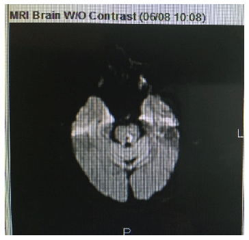
Magnetic resonance imaging (MRI)/magnetic resonance angiography (MRA) revealed left pontine infarction involving the area of the abducens nerve motor nucleus. This result is what spurred the misdiagnosis (Figure 4).

Figure 4: 0.9 × 0.4 cm Area of restricted diffusion involving the left para median, central, and posterior pons/tegmentum consistent with acute infarction involving the area of the abducens nerve motor nucleus (arrow pointing to the lesion).
Management of the case required collaboration with neurology and ophthalmology. Intermittent eye patching was initiated and continued post discharge. Resolution of her ophthalmologic symptoms was confirmed with a telephone follow up 3 months later.
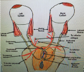
The Oculomotor nerve, controls the levator palpebrae superioris and four of the six extraocular muscles-the superior rectus, inferior rectus, medial rectus and the inferior oblique. It originates from the third nerve nucleus in the midbrain. Oculomotor nerve palsy can result from lesions anywhere along its path between the oculomotor nerve in the midbrain and the extraocular muscles within the orbit (Figure 5). Damage to the axons destined for extraocular muscles can result in medial rectus palsy [3].

Figure 5: Relationship of oculomotor nerve to abducens nerve in the midbrain.
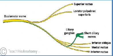
The oculomotor nerve has a superior and inferior nerve branch. The inferior branch supplies the medial and lateral rectus including the inferior oblique and the motor root of the ciliary ganglion. The superior branch supplies the superior recti and Levator palpebrae superior is [4] (Figure 6).

Figure 6: Anatomy of oculomotor nerve (Photo-Credit-TeachMeAnatomy)
Oculomotor nerve palsy, although rare, has several causes (Table 1). It can be bilateral or unilateral, complete or partial. Partial oculomotor nerve palsy can be pupil-sparing or pupil-involving. The pupil-sparing variant oculomotor nerve palsy occurs with ischemic injuries and is the most common type oculomotor nerve palsy. Although, commonly referred to as the diabetic third nerve palsy [5,6], a retrospective study conducted by Lazzaroni et al. [7], found no correlation with uncontrolled diabetes and oculomotor nerve palsy. They concluded, instead, that the atherosclerotic status was a greater contributor. Depending on the etiology and the location of the injury along the nerve tract, oculomotor nerve palsy may be treatable [8,9].
Complete oculomotor nerve palsy will result in esotropia, ptosis, and depression of the affected eye. The lateral rectus innervated by an intact abducens nerve maintains abduction [10]. Complete palsy with pupil sparing is almost never caused by an aneurysm, however a single case has been reported [10,11].
In our case, the presentation of diplopia, absent adduction, and accommodation of the left eye with pupil-sparing raised a suspicion for stroke. With the added atherosclerotic risk factors-uncontrolled diabetes, hypertension, and hyperlipidemia-it was easier to narrow down the exact etiology
Oculomotor nerve palsy usually resolves spontaneously within 3-6 months [7], and may require surgery if symptoms persist more than 6 months. Conservative therapy is directed at alleviating pain and diplopia .Prognosis depends on the etiology.
Oculomotor nerve palsy is rare and can be missed, especially if it is partial, and can resolve with conservative therapy. The close proximity of the oculomotor nerve to the abducens nerve in the midbrain, as seen on the MRI, led to the initial misdiagnosis of abducens palsy. Conducting a thorough neuro-ophthalmologic is paramount in diagnosing and managing patients with acute pontine strokes like the one reported. This case report highlights the role of physical diagnosis, rather than the reliance on imaging to diagnose partial oculomotor nerve palsy.
Download Provisional PDF Here
Article Type: Case Report
Citation: Sikod A, Ahmed A, McMillan-Persaud B, Kelsey-Harris R, Omole F (2016) A Misdiagnosis of Partial Oculomotor Nerve Palsy: A Curious Case of Stroke. J Neurol Neurobiol 2(5): doi http://dx.doi. org/10.16966/2379-7150.132
Copyright: © 2016 Sikod A, et al. This is an open-access article distributed under the terms of the Creative Commons Attribution License, which permits unrestricted use, distribution, and reproduction in any medium, provided the original author and source are credited.
Publication history:
All Sci Forschen Journals are Open Access