Graphene, a single- or few-layered sheet of sp2
- bonded carbon
atoms, whose discovery won the 2010 Nobel Prize in physics. Potential
applications of graphene for nanoelectronics, sensors, and nano
composites have been actively pursued [1]. The biological applications of
graphene oxide (GO, an oxidized form of graphene) has been explored
starting in 2008 and the research in this area is exploded soon after [1,2].
Inspired by its strong near infrared (NIR) absorption, high photo
thermal conversion efficiency, and exceptional large surface area,
graphene has emerged as a new shining star of nanomaterial for biological
applications, especially in the areas of photo thermal therapy including
photo thermal enhanced drug, protein, and gene delivery systems [3-7].
Intriguingly, its intrinsic ability to generate near inferred photoacoustic
signal allows not only to monitor the distribution of multifunctional drug
delivery systems in vivo, to evaluate their post-treatment therapeutic
outcomes in situ, and most importantly, to track the long term fate of
graphene sheets in the human body, but also enables imaging guided
therapy [8,9]. These capabilities could largely facilitate their application in
practical multifunctional nanomedicine regimes, fighting various diseases.
Equally important, although a number of studies have uncovered that
pristine graphene and graphene oxide
could induce toxicity in biological system, recent studies demonstrate that
well pegylated GO or reduced GO (rGO) nanosheets (nanometer in their
lateral dimension) are not toxic in vitro to cells and in vivo to animals,
demonstrating great potential for practical clinical applications.
On the other hand, applications of graphene in biomedical applications,
such as drug delivery require mono dispersed grapheme nanosheets with
lateral dimensions on the order of nanometers (typically <50 nm) [1].
Large effort has been devoted to fabricate nanosized graphene sheets for
variety of applications including the field of nanomedicine. The dominant
approaches typically rely upon Hummer’s or modified Hummer’s
methods which involve rather tedious and time consuming procedures
[10]. In brief, one must first oxidize graphite powder (this step normally
takes hours to a few days depending on the specific chemical recipes);
exfoliate the oxidized product to form GO suspensions. The as-prepared
GO sheets are very poly-disperse, ranging from a few nanometers to
tens of micrometers. To reach a predefined nanometer-sized GO sheet,
extended oxidation and sonication were applied. The final GO products
still have a large distribution of size and shape. To increase the size
uniformity of the GO sheets, extensive size separation steps need to be
performed. Alternatively, size-controlled GO was synthesized using
starting materials which are already small such as graphite nanofibers
or carbon fibers [11,12]. Compared to graphene, GO has very low
absorption capability in the near infrared (NIR) region, various chemical,
electrochemical, or hydrothermal reduction have been explored to
produce rGO nanosheets with some fraction of its NIR absorption, photo
thermal conversion recovered [7,8,13,14]. Furthermore, even though
there were no metal catalyst leftover, the major possible toxicity resources
of carbon nanotube (CNT) based delivery systems, are naturally avoided
in graphene based delivery systems, trace amounts of metal ions and other
chemicals involved in the oxidation and subsequent reduction processes
can participate in unwanted toxic reactions and could be detrimental to
biological applications. However, purification of GO is difficult due to its
gelatin tendency, therefore, extensive purification steps, which requires
large amount of solvents and long time of washing, making the production
of clean GO and rGO very time consuming [15]
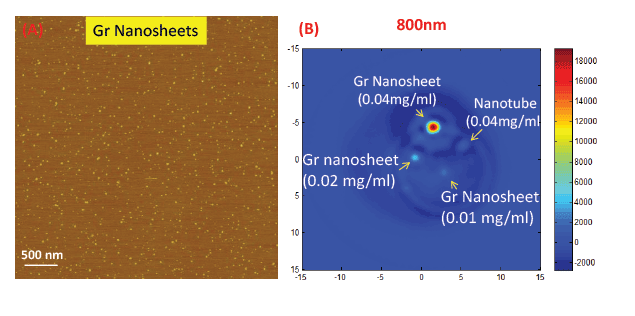
Figure 1: (A) An Atomic Force Microscopy image of the graphene nanosheets fabricated by the microwave enabled direct fabrication approach. (B)
Photo acoustic signal of graphene nanosheets with different concentrations and single walled carbon nanotubes.
Recently, a Ph. D student, Mehulkumar Patel in Prof. Huixin He
research group at Rutgers-Newark has developed an innovative approach,
which was referred to microwave enabled direct fabrication approach,
could solve the above mentioned problems ( Figure 1). This novel approach
allows rapid and direct fabrication of uniform amphiphilic low oxygen
containing (meaning low defects) graphene nanosheets [16], which can
be used as multifunctional drug delivery carrier. This novel fabrication
approach does not involve toxic chemicals and is much easier for cleaning
and surface modification to render them physiological stability and
biocompatibility. Compared to the commonly used GO, or chemically
reduced graphene oxide nanosheets (rGO), the grapheme nanosheets
contain much increased graphene domains, which ensures much higher
drug loading capability, especially for hydrophobic anticancer drugs.
Without the requirement of a post-reduction process, the fabricated
grapheme nanosheets exhibits strong NIR absorption, high opticalthermal
conversion efficiency for external controlled “on demand’ release
capabilities [17]. It could also enhance the photo thermal therapeutic
efficiency [18]. They also show strong NIR photo-acoustic conversion
efficiency, stronger than GO and single walled carbon nanotubes with the
same concentrations (Figure 1B), which provides great potential for the future to build
in vivo imaging capabilities for in-situ evaluation of therapeutic effects
and/or imaging guided therapy.
Download Provisional PDF Here
Article Information
Aritcle Type: Editorial
Citation: He H (2016) Microwave enabled Direct
Fabrication of Graphene Nanosheets for NIR
Photoacoustic Imaging Guided Therapy. Int
J Nanomed Nanosurg 2(2): doi http://dx.doi.
org/10.16966/2470-3206.e105
Copyright: © 2016 He H. This is an open-access
article distributed under the terms of the Creative
Commons Attribution License, which permits
unrestricted use, distribution, and reproduction
in any medium, provided the original author and
source are credited.
Publication history:
Received date: 25 Apr 2016
Accepted date: 26
Apr 2016
Published date: 30 Apr 2016