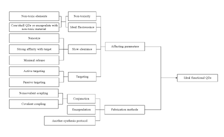
Figure 1: Schematic diagram of an ideal functional QDs used for diagnosis and therapy.
Dongxiu He1,2# Danxia Wang2# Wenjie Quan2 Cui-yun Yu1,2,*
1Hunan Province Cooperative Innovation Center for Molecular Target New Drug Study, Hengyang, 421001, China*Corresponding author: CY Yu, Hunan Province Cooperative Innovation Center for Molecular Target New Drug Study, Hengyang, 421001, China, Tel: +86 734 8282614; fax:+86 734 8282914; E-mail: yucuiyunusc@aliyun.com; yucuiyunusc@hotmail.com
Functional nanoparticles are considered to have the potential as novel molecular probes for both carcinoma imaging and targeting delivery, which plays a critical role in cancer diagnosis and therapy. Functional quantum dots (QDs), conjugated with types of targeting ligands or anticancer drug/gene to simultaneously cancerous image, treat cancer through selective binding to the receptors over-expressed on cancerous cells and tissue surface, were introduced. A series of significant progress in the fields of the fabrication of functional QDs and their applications in cancer diagnosis and therapy were summarized in this review.
Functional quantum dots; Fabrication; Applications; Cancer diagnosis; Cancer therapy
The diagnosis and treatment of cancer remains a key challenge for biomedical technology. The diagnosis and therapy of cancer at the cellular level will be greatly improved with the development of techniques that enable the delivery of molecular probes and therapeutic agents into cells and cellular compartments. Functional nanoparticles, one of the cutting-edge materials of the twenty-first century, are considered to have the potential as novel molecular probes for both imaging and targeting delivery, which plays a critical role in biomedicine. One major merit of using functional nanoparticles is that one can control and tailor properties in a very predictable manner to meet the needs of specific application. Recently, functional nanoparticles have received great interest in biomedical applications [1], especially their future application in the field of carcinoma diagnosis and therapy [2-5].
QDs are semiconductor inorganic nonmaterials ranging from 1-10 nm. QDs, containing elements found in groups II-IV (eg, CdSe, CdTe, CdS, and ZnSe) or III-V (eg, InP and InAs) of the periodic table, have shown great potential interest to medical scientists because of their unique advantages over traditional fluorescent dyes, such as broad excitation spectra, narrow and symmetric photoluminescence bands, large two-photon absorption cross-section, size-tunable absorption and photoluminescence spectra, exceptional photo stability, high quantum yield, and versatility in surface modification [3]. QDs have initially been used for carcinoma cell imaging. Additionally, QDs provide a versatile nanoscale scaffold for designing multifunctional nanoparticles with both imaging and drug delivery vehicles [4]. Herein, QDs, conjugated with types of targeting ligands or anticancer drug/gene to simultaneously cancerous image, cancer treatment through selective binding to the receptors over-expressed on cancerous cells and tissue surface, have the potential to considerably improve the efficiency of fluorescence imaging and target delivery [6-7]. This article provides a brief review on the recent developments of the fabrication of functional QDs and promising applications in cancerous image and cancer treatment.
Although functional QDs as contrast agent or drug delivery vehicle brings about many benefits to us, its fabrication is still a big challenge, because an ideal contrast agent or drug delivery vehicle must meet the need of increasing imaging or curative effect and simultaneously reducing the unintended severely side effects. In addition, to gather more information during specific cancer imaging or target delivery, QDs must be coupled with molecules which have the capabilities of recognizing the target. Surface chemistry modifications on the QDs are necessary for biomedical application. These surface modifications can also reduce the nonspecific binding, help prevent aggregation, and be critical to achieving specific target imaging and delivery. However, they often reduce the fluorescence quantum yield of the QDs.
Several recent studies have already discussed the synthesis and the biomedical imaging and targeting delivery applications of functional QDs [8-10]. There are many parameters that have to be taken into consideration and several fabrication methods that can be chosen in the design of the ideal functional QDs for cancer imaging (diagnosis) and targeting delivery (cancer therapy) (Figure 1). The main parameters are as following: i) the toxicity; ii) the fluorescence properties, including its quantum yield, its absorption cross-section, its lifetime, its resistance; iii) the ability to modify its surface chemistry without losing the QDs fluorescence; and iv) the specific target.
The toxicity of the functional QDs strongly depends on especially QDs’ composition and surface chemistry [11]. The fluorescence properties of the functional QDs rely on the surface chemical modifications and properties of the QDs. The design to obtain low toxicity, ideal fluorescence, slow clearance and targeting as well as the strategies currently available to fabricate them will be firstly introduced.
Both low toxicity and ideal fluorescence are the primary factors which should be taken into account. Many efficient attempts were made to reduce the toxicity and preserve a good fluorescence of the QDs.
One attempt to reduce the toxicity was to choose non-toxic and biodegradable nano-materials as drug/gene carriers and to find the substitutions for heavy mental-based QDs. Up to now, non-toxic elements such as silica, zinc, sulfur and copper were often used in the synthesis of the QDs [5,12-14]. Due to the low toxicity, graphene quantum dots were also pursued as possible alternatives in the field of cancer imaging [15,16]. Nevertheless, low photoluminescence quantum yield was the main concern.
Figure 1: Schematic diagram of an ideal functional QDs used for diagnosis and therapy.
Another attempt to reduce the toxicity and preserve a good fluorescence of the functional QDs was developed to synthesize core/shell QDs or encapsulate QDs by non-toxic materials [11,16-24]. QDs encapsulated in non-toxic materials including cross linked dendrimers [11], silica [11,16- 19], nanogel [20,21], mucleotide [22], and copolymer [23,24] were verified to remarkably reduce the cytotoxicity of QDs. Moreover, shells or nontoxic materials offer platform for conjugating molecule for cell and tissue targeting. QDs became stable once silica shell formed on their surface and excess surfactants were removed [11,25]. PEGlyated phospholipids anchoring onto carbon nanotube surface have shown better targeting efficacy, better entrapment in spleen and liver without any degradation and toxicity [26].
In general, emission in the near infrared (NIR) range (700-1000 nm) is characterized by far more penetration power than emission in the visible spectrum, thus making it possible to visualize the structure of living tissues in vivo [12-14,27]. Once a large particle enters the vasculature, it is likely to be removed through the reticuloendothelial system, however, if the size of the particles is small enough i.e., in nanosize (<100 nm) they would bypass this process. Additionally, there are possible interactions of functional QDs with blood components and stimulation of immune response. Herein, the functional QDs applied for deep tissues infrared imaging or targeting delivery must meet more criteria: near infrared emitting, nanosize, and strong affinity for their target and minimal release back into the blood or lymph circulation. Most of these functional QDs are coated with long chain PEG for minimization of non-specific adsorption and prolonged circulation [11].
The entry of functional QDs into the cell requires transit across the lipid bilayer. This may occur through endocytosis or receptor-mediated uptake, which are also known as passive or active transport. Passive targeting is much slower and less efficient than active targeting. When passive targeting is used, nanocomposites can be easily accumulated at tumor site with high concentration due to the pathophysiological differences between normal tissues and tumor tissues known as enhanced permeability and retention (EPR) effect [28]. While the functional QDs have been successfully active targeted to different tissues, such as blood or lymphatic vessel [12,29], or liver [30], it involves adding molecules such as antibodies, peptides or other ligands to the QDs surface. Because tumor cells often express unique proteins on their membranes that can be identified by the specific ligand, antibody or targeting-peptide binding [31-33].
Recently, QDs conjugated with targeting moieties that include aptamer [6], lectin [23], hyaluronic acid [29], folic acid [34], transferrin [35], antibodies against tumor-specific antigens [36,37], were fabricated. Fabrication methods of the functional QDs for cancerous imaging and targeting delivery can be classified into several categories listed below.
Conjunction: Conjunction, which can be grouped into two general strategies: noncovalent coupling [38] and covalent coupling [39,40], is a common fabrication method. Some functional QDs have been fabricated by noncovalent coupling such as electrostatic interactions [20] or π-π stacking interactions [31]. While more functional QDs have been prepared by noncovalent coupling [6,8,22,34,39,40]. For example, CdSe/ ZnS-QDs coated with a polymer shell and covalently functionalized with streptavidin were attached onto pristine side-walls of CNTs by the surface amino groups of the streptavidin. Based on the strong affinity between biotin and streptavidin, these hydrophilic functional QDs was used as a multivalent intracellular fluorescent imaging in Jurkat T leukemia cells to which biotinylated mouse anti-human CD3 antibody bonded to surface CD3 receptors [41].
To fabricate the functional QDs, other intermediate molecules including 1-ethyl-3-(3-dimethylaminopropyl) carbodiimide (EDC) and/ or N-hydroxysuccinimide (NHS) have been involved [6, 11, 38]. QDs were first covalently coupled with aptamer specific for mutated MUC1 mucin overexpressed in ovarian carcinoma cells, followed by doxorubicin covalently coupled with QDs -MUC1 aptamer conjugates by EDC and NHS [6].
Encapsulation is a physical loading technique. The functional QDs for carcinoma imaging and targeting delivery were encapsulated in various materials such as silica [16-19,42,43], glycopolymer [23], micelle [12, 44], and liposome [45]. Among of them, silica has been applied most to encapsulate both hydrophobic and hydrophilic QDs because silica encapsulation has the following advantages: dramatically reducing nonspecific adsorption on the QDs, reducing the release of harmful ions, and decreasing environmental interferences on the brightness of QDs [19,43,46,47]. Silica encapsulation method studies recently focused on sol– gel process (so-called Stöber method) [46,47] and reverse microemulsion method [43].
Wu’s group reported that the hybrid nanogels were prepared by in-situ immobilization of CdSe QDs in the interior of the pH and temperature dual responsive hydroxypropylcellulose-poly (acrylic acid) (HPC-PAA) semi-interpenetrating polymer networks [21].
Over the past decade, functional QDs have been used in many different aspects of biomedical field. First used for bioimaging [6,22], they later became useful tools for bioanalysis [1], then biolabeling and medical therapy [2-5]. Here, we will focus on the two main applications of functional QDs: cancer diagnosis and cancer therapy (Figure 2).
Early diagnosis plays an important role in cancerous diagnosis. In most cases, detection of stage I cancers is associated with a higher than 90% 5-year survival rate [48] due to availability of curative treatment. Fluorescence labeling/imaging techniques are especially valuable tools in the arsenal of clinical diagnosis. Functional QDs have been wide used for improving fluorescence imaging [6,22,37,38] due to the photochemical stability and high fluorescence intensity of QDs.
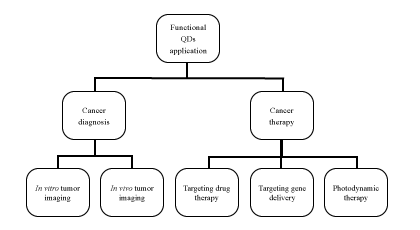
Figure 2: Functional QDs in the applications for cancer diagnosis and cancer therapy.
In vitro tumor imaging: One of the most advancing applications of functional QDs is in vitro imaging of cancer cells. Many research groups applied functional QDs for in vitro fluorescence imaging of human cancer cells derived from ovarian carcinoma [6], melanoma [22], breast cancer [36], pancreatic cancer [37], glioblastoma [38], ovarian epidermoid carcinoma [49], lung adenocarcinoma [50], and hepatocellular carcinoma [24,51].
Zhang and his colleagues [36] found that QDs conjugated with anti-type 1 insulin-like growth factor receptor (IGF1R) is a promising candidate for targeting and imaging in breast cancer cells. The key in this targeting was the detection of up-regulated IGF1R in MCF-7 breast cancer cells by QDanti-IGFR1 conjugate.
Epidermal growth factor receptor (EGFR or HER1) or human epidermal growth factor 2 (HER2) is transmembrane glycoprotein that constitutes one of four members of the erbB family of tyrosine kinase receptors, is over-expressed in many cancers and can bind to epidermal growth factor (EGF) [52]. Based on that, the QD-EGF conjugate can be utilized for various cancer cells fluorescence imaging because EGFR is over-expressed in many cancers. Nakane and coworkers [53] reported most KPL-4 cells (breast cancer cells) were stained by the anti-HER2 antibody conjugated PbS QDs, whereas only very weak fluorescence signals were observed when stained with bovine serum albumin (BSA) conjugated PbS QDs after the incubation of anti-HER2 antibody conjugated PbS QDs and BSA-conjugated PbS QDs. This result clearly shows the specific targeting imaging ability of PbS QDs to HER2 surface receptors in KPL-4 cells.
Kawashima et al. [49] also explored intermolecular interactions involved in the lateral propagation of cell-signaling by EGFR singlemolecules in human ovarian epidermoid carcinoma cells (A431) using nanocomposites loaded CdSe/ZnS QDs. Kawashima found that CHO and A431 cells were efficiently labeled by QD-EGF conjugates due to the specific binding of EGF to EGFR.
In research by Yong [37], they selectively detected human pancreatic cancer cells using QDs conjugated with anti-Claudin-4 antibody and antiprostate stem cell antigen (anti-PSCA). These conjugates were recognized by the membrane proteins Claudin-4 and PSCA which are over-expressed in both primary and metastatic pancreatic cancer cells. This result clearly shows fluorescence images of human pancreatic cancer cells (MiaPaCa) treated with various nanocomposites loaded QDs. From these results, it clearly showed that the nanocomposites loaded InP/ZnS QDs can serve as a potential biocompatible targeted nanoprobe to specifically diagnose human pancreatic cancer cells.
In vivo tumor imaging: The basic principles underlying in vitro targeting of cancer cells can be applied in vivo. However, in vivo applications of functional NIR QDs are more complicated and challenging. One main challenge for in vivo targeting and imaging using the functional QDs is their biodistribution and pharmacokinetics. Additionally, the type and structure of organic/bioorganic shells of QDs determine their biocompatibility and are crucial for their application in imaging in vivo, due to the effects of the shell on the following properties: colloidal stability, solubility in physiological fluids, influence of the basic physiological parameters, and cytotoxicity [11]. Chen et al. have monitored the dynamic distribution of CdHgTe/SiO2 nanocomposites in vivo by near infrared fluorescence imaging system. They also show that CdHgTe/SiO2 nanocomposites acted as a novel fluorescence probe have a maximum fluorescence emission of 785 nm and high photo-stability and the hydrodynamic diameter of CdHgTe/SiO2 nanocomposites could be adjusted to 122.3 nm [54]. Another main challenge for in vivo targeting and imaging is the fluorescence emitting property of the functional QDs. Visible emitting QDs provide poor signal-to background ratio in deep tissue and when imaging targets in small animals [23]. While NIR QDs offer several advantages for the non-invasive visualization of living tissues because of its deeper photon penetration, low absorption and scattering. So, functional NIR QDs are considered to have the potential as novel probe for tumor diagnostics, for example, carcinoma cell labeling or tracking [55-57], tumor markers targeted imaging [58,59], tumor vessel imaging [60,61].
Carcinoma cell labeling or tracking in living organisms was monitored efficiently and sensitively, which may provide tools to locate tumors and metastases or map tumor margins during surgery. This could be achieved by functional QDs, then administrating conjugated QDs for in vivo targeted imaging of various human cancer cells such as oral squamous carcinoma cell [52], brain cancer [55], hepatacarcinoma cell [56], and colon carcinoma cell [57]. Fang et al. reported that in vivo fluorescence imaging of a breast tumor in a mouse were taken through anti-HER2 antibody conjugated PbS QDs injected into the tail vain. Immediately after the injection of QDs, 2nd-NIR fluorescence signals for the QDs were observed over the whole body with time: after about 1 h QDs were detected in the liver and after 48 h the breast tumor was clearly detected [51]. Yu et al. subcutaneously implanted HCCLM6 cancer cells in mice, and intravenously injected the QDs conjugated with an antibody to alpha-fetoprotein (anti-AFP). The QD-anti-AFP conjugate was effectively accumulated in human hepatocarcinoma cells, which have demonstrated that the QD-anti-AFP conjugate was an ideal candidate for in vivo targeted imaging of HCCLM6 human hepatacarcinoma cells [56].
Tumor markers play a critical role in the detection and diagnosis of cancer [58]. Biomarker assays may be useful for screening and diagnosis of cancer if a set of tumor markers can be quantified and statistically differentiated between cancerous cells and normal cells [58].
Angiogenesis, the formation of new blood vessels from preexisting vasculature, is essential for tumor growth and progression. Integrin αv β3 , which binds to RGD-containing components of the interstitial matrix, plays a key role in tumor angiogenesis and metastasis [60]. Functional QDs represent excellent tool for tumor vessel imaging [61] or multimodal molecular imaging of angiogenesis [62].
Targeting drug therapy applications: Recently, targeted therapy has been widely explored. Drug/gene targeting by nanoparticles or nanocapsules offers the following enormous advantages, as examples: reduces dosage, ensures the pharmaceutical effects, and minimizes side-effects, and enhances drug stability. QDs are newer luminescent nanoparticles with rich surface chemistry and unique optical properties that make them useful as visualization probes or carriers for traceable targeted delivery and therapy applications [63]. Thus, it has been developed functional QDs that can perform imaging and drug delivery tasks without the need for external dyes [64]. We will briefly describe developments of functional QDs as probes and carriers in targeting drug delivery applications.
By directly noncovalent coupling or covalent coupling drug molecules to the QD surface, drug-conjugated QDs can be delivered to specific sites and subsequently release drug molecules from the QD surface in response to local biological conditions such as pH or the presence of enzymes. Several research groups have demonstrated the integration of drug molecules with conjugated QDs and targeting moieties for targeting drug delivery in vitro and in vivo. Chakravarthy et al. [65] found that doxorubicin (Dox) can be effectively released from the functional CdSe/CdS/ZnS QDs conjugated with Dox and accumulated in the cell nucleus. They also demonstrated that the functional QDs can provide targeted macrophage-selective therapy for the treatment of pulmonary disease. And, several researches recently suggested that QDs conjugated therapeutic antibody [66] or drug [67] can be used as a suitable nanoparticles for breast cancer treatment. Xu et al. [66] demonstrated for the first time that the functional QDs conjugated with anti-GRP78 scFv could be efficiently internalized by cancer cells, thus up-regulate phophosphate-AKT-ser473 and possess biological antitumor activity by inhibition of breast cancer growth in a xenograft model. Fibrinogen (fib) coated CdTe/ZnTe and paclitaxel (PTX) [PTX-fib-QDs] can target MCF-7 cells and effectively deliver PTX towards breast cancer cells via the α5β1-integrins [67]. Furthermore, Jeyadevia’s study revealed that using TGA-CdTe QDs as nanocarrier of quercetin could enhance the anti-arthritic effect of quercetin even at a lower concentration of the drug in rheumatic complications [68].
All these studies have laid an important foundation in the functional QDs as probes and carriers for target drug delivery research and applications.
Targeting gene delivery applications: Gene therapy has emerged as a powerful strategy for cancer treatment over the past several decades because of the genetic link associated with tumor development and progression. The delivery of nucleic acid therapeutics to down-regulate or replace mutated genes, and to silence unexpected gene expression, is becoming an attractive approach to suppressing tumor cell growth and invasion. There have been intensive efforts to develop safe and efficient gene delivery carriers to provide high transfect ion efficiency at the desired target.
Besides the delivery of small molecule drugs, QDs have also been proved to have prospect for delivery of more intricate genes, such as small interfering RNA (siRNA). The short and double-stranded therapeutic siRNA works by silencing the expression of unwanted, disease-causing genes. Nevertheless, free forms of them owe high negative charge and are easy to degrade in body environment. Thus, in order to achieve optimal function in physiological conditions, they must be delivered via combination with cationic nanocariers. Because of appropriately surface functionalized with cationic moieties, nanocomposites including QDs are good choices of siRNA carriers as they not only render these genetic drugs with physiological stability and target specificity, but also the entire nanocomposites can be optically traced. Therefore, nanocomposites loaded with QDs were specifically designed to overcome barriers in siRNA delivery such as siRNA protection, cellular penetration, endosomal release, carrier unpacking, intracellular transport and gene silencing. Li et al. confirmed that functional QDs could efficiently delivery siRNA into HeLa cells and silence a target gene, and the nanocomposites loaded with QDs could also be used as fluorescence probes, allowing real-time tracking and localization of QDs during delivery and transfection in vivo [69]. More importantly, multifunctional QDs have been demonstrated to deliver an active siRNA to knockdown EGFRvIII receptors in human glioma cells, and subsequently monitor the resulting down-regulated signaling pathway with high efficiency [70], suggesting that functional QDs could be designed to deliver gene to a specified target cell type.
Photodynamic therapy of cancer: Photodynamic therapy (PDT) is still considered to be a new and promising invasive antitumor strategy that can exert a selective cytotoxic activity toward malignant cells. The antitumor strategy involves administration of a photosensitizing (PS) agent followed by irradiation at a wavelength corresponding to an absorbance band of the photosensitizing agent. Clinical studies revealed that PDT can be curative, particularly in early stage tumors because sensitizer may lead to direct tumor cell death, damage to the microvasculature, and induction of a local inflammatory reaction [71]. QDs can be designed to have fluorescence peaks at different wavelengths by adjusting their size. The unique property of the QDs allows QDs to be fabricated for specific functions such as Fluorescence Resonance Energy Transfer (FRET), in which a specific emission wavelength can transfer energy to drug molecules or photosensitizers. Consequently, FRET phenomenon can be potentially used for the development of novel therapeutic approaches for cancer treatment. Additionally, photo stability of QDs is the most promising property for PDT. Recent reports showed that QDs can be used in photodynamic and radiation therapy of cancer [72,73].
Although targeted delivery of QDs in cancer cells and tumor milieu by using QDs conjugated with anticancer antibodies or other bio molecules have became possible recently, compared with conventional PS drugs such as porphyrins and phthalocyanines, the efficiency of QDs to produce ROI under direct photoactivation is low. Herein, preparation of conjugates between QDs and conventional PS drugs, investigation of energy transfer efficiencies from QDs to PS drugs and ROI production by the conjugates are being widely investigated.
Real-time imaging and targeting delivery have been hot topics in cancerous diagnosis and therapy fields. Current investigation of the functional QDs in vitro and in vivo has offered less invasive imaging and targeting drug delivery. However, up to date the clinical applications of the functional QDs have been limited due to side-effects. Various novel nanoparticles such as CNTs have been explored with recent developments in the preparation and characterization techniques of nanoparticles. The emergence of functional QDs, targeting moieties, and other nanoparticles enabled to improve imaging and targeting delivery applications because of their better biocompatibility, lower toxicity and longer circulation time in vivo, which were better applied for high-resolution noninvasive cancer diagnosis and therapy
The authors gratefully acknowledged financial supports from National Natural Science Foundation of China (81471777, 81102409) and financial supports from Natural Science Foundation of Hunan province (13JJ6096).
Download Provisional PDF Here
Aritcle Type: Review Article
Citation: He D, Wang D, Quan W, Yu C (2015) Functional Quantum Dots for Promising Cancer Diagnosis and Therapy. J Nanomed Nanosurg 1(1): doi http://dx.doi.org/10.16966/2470-3206.103
Copyright: © 2015 He D, et al. This is an openaccess article distributed under the terms of the Creative Commons Attribution License, which permits unrestricted use, distribution, and reproduction in any medium, provided the original author and source are credited.
Publication history:
All Sci Forschen Journals are Open Access