Abstract
Objective: To examine the appearance of children complex coronary artery fistula using digital subtraction angiography (DSA) and to conclude
whether DSA provides comprehensive information in interventional treatment.
Materials and Methods: Five children with complex coronary artery fistula were evaluated by DSA and ultrasonography (US) between
2008 and 2013. Preoperative US was performed on five children at 1, 11, 4, 12, and 5 years old following DSA evaluations that demonstrated
complex coronary artery fistula. The preoperative US and DSA findings were compared with the physical examination, then decided to perform
interventional treatment of three cases.
Results: In all five cases, two cases were left coronary artery to right ventricle fistulas, two cases were left coronary artery to right atrium
fistulas, the other one case was right coronary artery to right atrium fistula, three cases of successful interventional treatment.
Conclusions: DSA is effective in the assessment of children complex coronary artery fistula, and may Guide interventional procedures. US
may provide a useful adjunction in assessing children complex coronary artery fistula.
Keywords
Digital subtraction angiography; Ultrasonography; Children; Coronary artery fistula
Introduction
Coronary artery fistula (CAF) is a rare congenital cardiovascular
malformations, the rate was 0.25% to 0.40% in congenital heart disease [1].
CAF is the presence of abnormal blood flow path between coronary artery
and cardiac chambers or thirds, or other blood vessels, children are more
common than adults. CAF was firstly reported by Krause in 1865, 60%
to 78% of independent malformations, and other associated with atrial
septal defect (ASD), patent ductus arteriosus (PDA), ventricular septal
defect (VSD), etc.. US is the primary imaging modality for evaluating CAF
[2]. However, since US provides limited anatomical information, DSA has
provided a useful complementary tool to assess anomalies in children in
recent years, coronary angiography is the “gold standard” for the diagnosis
of coronary artery fistula [3].
Here, we report the findings from examinations of six children with
complex coronary artery fistulas. Two children had left coronary artery to
right ventricle fistula, one children had left coronary artery to right atrium
fistula, the other two had right coronary artery to right atrium fistulas. Both
DSA and US were used to demonstrate the CAF. This report demonstrates
the importance of DSA for successful diagnosis and treatment.
Materials and Methods
This study presents five consecutive cases diagnosed for coronary
artery fistula, who were referred for DSA and US evaluations between July
2008 and August 2012. Before the examination, all the patients provided
their informed consent. In all the cases, the diagnosis was confirmed
by combined findings of postoperative physical examinations, serial
ultrasound examinations, or pathology.
Coronary angiography was performed with a 850 mA digital
subtraction angiography (Neusoft, NSX6000). General anesthesia, the
right femoral artery puncture, indwelling arterial sheath, routine cardiac
catheterization, angiography and interventional cardiovascular treatment.
Aortic root angiography and selective left and right coronary angiography,
observing coronary anatomy, coronary artery fistula origin, course and
drainage site; presence or absence of coronary artery aneurysm, aneurysm
formation. After repeated coronary angiography, plugging effect was
observed, while observing the patient’s symptoms, signs, and ECG
(Echocardiography) changes. When determining the CAF was completely
blocked off, the patient no discomfort, no ischemic ECG changes and other
signs, then the occluder is completely released. The cardiac ultrasounds
were performed with a GE Vivid 7 ultrasound system with a 3–5 MHz
curved-array transducer.
All of the DSA images were interpreted by two authors (Lv HT and
Yang FB). The US results were known to the DSA imaging radiologist at
the time of acquisition and at the time the DSA images were interpreted.
Results
All five children showed different and complex DSA and US findings.
In each case, the DSA detected abnormal traffic vessels between coronary
artery and heart chamber or large blood vessels. Echocardiography could
only find coronal artery fistula abnormal blood flow but poor specificity
for small fistula or shunt. In one of the cases, we detected a complex
left coronary artery to right ventricle fistula, and branches of the left
coronary artery tortuosity and expansion, the drain port is small (3.8
mm in diameter). The second case was also left coronary artery to right
ventricle fistula, the performance of DSA was similar to the first case,
and the drain port is small (3.2 mm in diameter). In all five cases, two
cases were left coronary artery to right atrium fistulas, the other one case
was right coronary artery to right atrium fistula, three cases of successful
interventional treatment.
Case 1
A 1-year-old boy, aortic root DSA revealed a left coronary artery to
right ventricle fistula with significant expansion of the left coronary
artery, running tortuous( Figure 1a), distal stenosis (3.8 mm diameter).
Cardiac ultrasound showed dilatation of coronary section, Doppler
US imaging demonstrated the direction of blood flow (Figure 1b). By
X-ray fluoroscopy-guided interventional treatment of coronary artery
fistula was implemented, firstly established channels by the aorta, the
left coronary artery and right ventricle fistula, then placed the guide
sheath. An amplatzer occluder was placed on the lower end of the
narrowest part of the left coronary artery, first opening an umbrella at
the distal of the stenosis (Figure 1c), another umbrella at the proximal
stenosis, fistula was blocked. Postoperative aortic root DSA showed no
obvious contrast agent shunt, amplatzer umbrella being normal position
(Figure 1d).
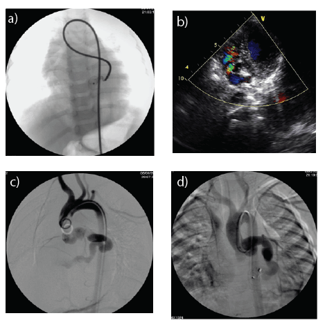
Figure 1: A 1-year-old boy with a left coronary artery to right ventricle fistula. (a) Aortic root DSA revealed expansion of the left coronary artery, running
tortuous (arrows). (b) Cardiac US showed dilatation of coronary section, Doppler US demonstrated the direction of blood flow. (c) The first umbrella
of amplatzer occluder was opened at the distal of the stenosis. (d) Postoperative aortic root DSA showed no obvious contrast agent shunt, amplatzer
umbrella being normal position.
Case 2
An 11-year-old boy, aortic root DSA revealed a left coronary artery
anterior descending branch to right ventricle fistula (Figure 2a). Selective
left coronary artery anterior descending branch angiography showed
obvious expansion of the left coronary artery, running tortuous, a small
right ventricle fistula (Figure 2b). Cardiac ultrasound showed dilatation
of left coronary artery anterior descending branch, a small right ventricle
fistula (3.2 mm diameter) (Figure 2c). Under X-ray fluoroscopy a
amplatzer occluder was tried to place on the right ventricle fistula (Figure
2d), finally, no success.
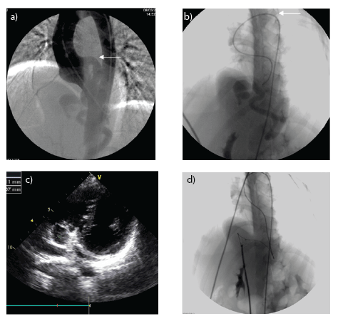
Figure 2: An 11-year-old boy with a left coronary artery to right ventricle fistula. (a) Aortic root DSA revealed expansion of the left coronary artery, running
tortuous (arrows). (b) Selective left coronary artery anterior descending branch angiography showed a small right ventricle fistula. (c) Cardiac ultrasound
showed dilatation of left coronary artery, a small right ventricle fistula (3.2 mm diameter). (d) Under X-ray fluoroscopy an amplatzer occluder was tried
to place on the right ventricle fistula.
Case 3
A 4-year-old girl, aortic root DSA revealed a left circumflex
coronary artery to right atrium fistula with significant expansion
of the left circumflex coronary artery, running tortuous (Figure
3a), distal right atrium fistula stenosis. Cardiac ultrasound showed
dilatation of left circumflex coronary artery section, Doppler US
imaging demonstrated the direction of blood flow (Figure 3b). This
case had no interventional treatment.
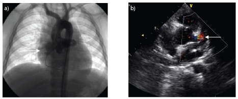
Figure 3: A 4-year-old girl with a left circumflex coronary artery to right atrium fistula. (a) Aortic root DSA revealed expansion of the left circumflex
coronary artery, a right atrium fistula (arrows). (b) Cardiac US showed dilatation of coronary section, Doppler US demonstrated the direction of blood
flow (arrows).
Case 4
A 12-year-old boy, aortic root DSA revealed a left circumflex coronary
artery to right atrium fistula with significant expansion of the left
coronary artery (Figure 4a). Selective left coronary artery angiography
showed obvious expansion of the left coronary artery, running tortuous,
a small right atrium fistula (Figure 4b). By X-ray fluoroscopy-guided
interventional treatment of coronary artery fistula was implemented.
An amplatzer occluder was placed on the fistula, an umbrella at the right
atrium, another umbrella at the expansion of left coronary artery, fistula
was blocked (Figure 4c). Postoperative aortic root DSA showed no obvious
contrast agent shunt, amplatzer umbrella being normal position (Figure 4d).
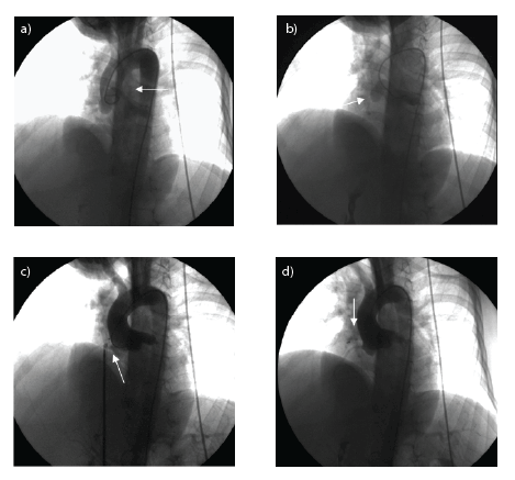
Figure 4: A 12-year-old boy with a left coronary artery to right atrium fistula. (a) Aortic root DSA revealed expansion of the left circumflex coronary artery,
because of the overlap with the aorta, indicating poor (arrows). (b) Selective left circumflex coronary artery angiography showed an expansion section
and right atrium fistula (arrows). (c) The umbrella of amplatzer occluder was opened at the right atrium fistula. (d) Postoperative aortic root DSA showed
no obvious contrast agent shunt, Amplatzer umbrella being normal position.
Case 5
A 5-year-old girl, aortic root DSA revealed a right coronary artery to
right atrium fistula with significant expansion of the right coronary artery.
Selective right coronary artery angiography showed obvious expansion of
the right coronary artery, a small right atrium fistula (Figure 5a), fistula
stenosis (3.6 mm diameter). By X-ray fluoroscopy-guided interventional
treatment of coronary artery fistula was implemented, an amplatzer
occluder was placed on the fistula, an umbrella at the right atrium, another
umbrella at the expansion of right coronary artery, fistula was blocked.
Postoperative aortic root DSA showed no obvious contrast agent shunt,
amplatzer umbrella being normal position (Figure 5b).
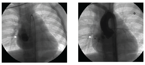
Figure 5: A 5-year-old girl with a right coronary artery to right atrium fistula. (a) Selective right coronary artery angiography showed an expansion section
and right atrium fistula (arrows). (b) Postoperative aortic root DSA showed no obvious contrast agent shunt, Amplatzer umbrella being normal position
(arrows).
Discussion
Congenital coronary artery fistula is a rare congenital coronary artery
disease, the main reason is due to occur during embryonic development,
trabecular cardiac muscle and coronary sinus-shaped gap communicates
with the cardiac development, sinus-shaped gap is compressed and
gradually back into the capillaries, causing the formation of abnormal
blood vessels [4]. CAF can be originated from the right coronary artery,
the left coronary artery or bilateral. Clinically, coronary artery with the
right atrium, right ventricle, left atrium, left ventricle or pulmonary form
CAF, mainly with right heart [5]. In all five cases in this group, four cases
were originated from the left coronary artery, one case was originated
from the right coronary artery, all five cases were right heart fistula, and
this is consistent with the literature.
CAF imaging techniques, including US, Multi-slice CT and DSA,
provide critical information to diagnose fistula. Obtaining accurate
imaging diagnoses of CAF remains challenging [6]. US is the primary
imaging modality to evaluate CAF. Color Doppler can observe the
degree of expansion of the initial segment of the right coronary artery
and application flow imaging of the heart chamber and abnormal blood
flow within the pulmonary artery. However, due to the expansion of the
artery cannot display the full image, especially drainage to the pulmonary
artery, a small fistula, shunt less prone coronary artery fistula and easy to
find, easy to cause misdiagnosis. Now, with the continuous development
of improved technology for the rapid development of MSCT scanning and
post-processing functions, MSCT can be used as a relatively safe, fast and
accurate means of checking on the diagnosis for CAF [7]. Nevertheless,
US and MSCT has great limitations in the interventional treatment.
Cardiovascular DSA has provided a useful complementary tool to
diagnose and treat CAF. So far, coronary angiography is considered for
the “gold standard” for the diagnosis of CAF [8]. Cardiovascular DSA
can display the entire coronary artery branches, clear fistula size, number,
location, fistula diameter, and then guide treatment choices, especially
in the presence of fistula blood vessels that supply the heart or less thick
muscle has an advantage. Relative to the surgery, interventional treatment
of coronary artery fistula catheter shorter hospital stay, lower costs, less
invasive, but must be strictly controlled the indications, standardized
operation, avoid surgical complications [9]. Amplatzer Duct-Occluder
and coil are common occluder devices. Amplatzer Duct-Occluder
are applicable in large coronary artery fistula usually, coil for a smaller
coronary fistula. In this group, Amplatzer Duct-Occluder were used
for the first, fourth, fifth cases. Amplatzer Duct-Occluder and coil were
tried to place on the fistula respectively, but no success. Not all cases are
suitable for interventional therapy, in conjunction with imaging findings,
the patient’s electrocardiogram and other clinical situations, we had the
final decision on whether intervention or surgery.
Conclusions
Cardiovascular DSA is effective in the assessment of children
complex coronary artery fistula, and may Guide interventional
procedures. US can add useful informations in assessing children
complex coronary artery fistula. With the development of intervention
techniques, cardiovascular DSA are increasingly used in the treatment
of children complex CAF.
Foundation Content
- Early warning technology in 2013 social development projects in the
province of coronary artery lesions of Kawasaki disease (BE2013632).
- Suzhou Municipal Science and Technology Bureau: Children’s Heart
and Vascular Diseases Laboratory (SZS201411) (key projects) 2014.
- Suzhou City Planning Commission: Key Discipline Children
Cardiology (Szxk201507) (key projects) 2016.
Article Information
Article Type: Research Article
Citation: Yang F, Lv H, Sheng M, Sun L, Huang J,
et al. (2016) Use of DSA in Diagnosing and Treating
Children Complex Coronary Artery Fistula: A Report
of Five Cases. J Hear Health 2(3): doi http://dx.doi.
org/10.16966/2379-769X.124
Copyright: © 2016 Yang F, et al. This is an
open-access article distributed under the terms
of the Creative Commons Attribution License,
which permits unrestricted use, distribution, and
reproduction in any medium, provided the original
author and source are credited.
Publication history:
Received date: 02 Dec 2015
Accepted date: 02
May 2016
Published date: 06 May 2016


