Introduction
Coronary artery disease (CAD) leads to high mortality among men and
women, especially in developed countries [1]. Various imaging modalities,
pharmacological treatment, and invasive interventions are used to
accurately diagnose and treat CAD [2]. Invasive coronary angiography
(ICA) is the gold standard investigation in the assessment of CAD for both
diagnosis and therapeutic purposes [3]. However, with rapid technological
development of computed tomography (CT), a non-invasive alternative
has been introduced to reduce the risk of invasive interventions, namely
coronary computed tomography angiography (CCTA). The potential
clinical application of CCTA includes CAD detection, heart function
assessment, and anatomical structure of coronary arteries analysis [4-6].
The mean sensitivity and specificity ranges from 76% and 93% in a 4-slice
CCTA to 99% and 96% in a 64-slice CCTA [7,8].
A low and consistent heart rate is a key factor to provide a good image
quality with high diagnostic accuracy in CCTA. An increment in the
patient’s heart rate will cause motion artefacts, which leads to poor image
quality and affects the accuracy of CAD detection [9]. A 320-slice CT
scans the entire heart within a single heartbeat; this eliminates stair-step
artefacts due to involuntary cardiac movement and reduces risk of motion
artefact due to chest movement during breath-hold. Moreover, with the
introduction of dual source CT, the temporal resolution is increased even
at elevated heart rate, thereby reducing the problem of motion artefacts
[10,11]. However, such techniques increase the level of radiation exposure
to the patients. Several approaches have been identified to overcome this
problem. For instance, applying tube current modulation mode using
high pitch, low tube voltage and implementing adaptive statistical iterative
reconstruction (ASIR) will reduce the radiation dose [12]. Moreover, step-
and-shoot scanning mode (prospective ECG-triggering CCTA) in CCTA
reduces radiation exposure by 83% as compared to the conventional
CCTA technique (retrospective ECG-gating) [13,14]. However, this
technique is limited to patients who have a consistent heart rate below 65
beats per minute [13]. Thus, pre-medication such as beta-blockers (most
commonly used is metoprololtartarate), calcium antagonists and nitrates
are introduced to reduce the heart rate and stabilize heart rhythms.
The beta-blocker is not widely used as most patients with CAD
would have been on long-term beta blocker therapy [15]. Beta-blockers
are contraindicated in asthmatic patients as it may increase
bronchoconstriction [16]. Calcium antagonist can be used as an
alternate and is of limited value in patients with a history of heart failure
or impaired left ventricular function [17]. Ivabradine is used in the
treatment of chronic angina pectoris and chronic heart failure for decades
[18]. Ivabradine blocks the funny channel (If
) in the sinoatrial node [19]
resulting in reduced heart rate without adversely affecting blood pressure
and cardiac contractility [20,21]. In late 2009, ivabradine was introduced
as a pre-medication heart rate controller for CCTA examination and has
been adopted by institutions at present. The effectiveness in terms of
accuracy is still inconclusive. Therefore, a systematic review was done to
provide information on the effectiveness of ivabradine compared to betablocker
(metoprololtartarate) as a pre-medication in CCTA.
Methods
Literature search
We searched four databases, including PubMed, Medline, Highwire
Press, and Science Direct to identify studies using ivabradine as a premedication
in CCTA examination and published between January 2010
and February 2014. The terms used for identification of the relevant
articles in this study were ‘heart rate control in CCTA’, ‘heart rate control
premedication in CCTA’ and ‘Ivabradine used to control heart rate in
CCTA’. The search was conducted on English literature and selected based
on the following criteria: (a) CCTA was only performed using ivabradine
as premedication; and (b) ivabradine studies provided with complete
heart rate information. Studies with other cardiac drug therapy, case
reports, phantom studies, animal studies, and other non-original articles
were excluded from this review.
Data extraction and analysis
Authors’ names, year of publication and patient numbers were noted in
each article. Variables such as premedication dosage, admission heart rate,
heart rate prior to CCTA, heart rate during CCTA and mean heart rate
reduction were recorded in order to analyze the effectiveness of ivabradine
as premedication prior to CCTA examination. Technical parameters such
as number of detector collimation, gantry rotation time, exposure factors
(mA and kVp), pitch, contrast injection method, and scanning mode were
also recorded.
All images produced from these studies were of diagnostic quality. Each
study was evaluated distinctively either using quantitative or qualitative
method. The qualitative image quality evaluation was performed
referring to American Heart Association classification with a minimum
of two independent reviewers interpreting the CCTA images using 4-
and 5-point grading scales depending on overall image performance
(‘excellent’, ‘good’, ‘moderate’ and ‘not-interpretable’) and presentation of
artifacts (‘no motion artefacts’, ‘minor artifacts, ‘moderate artifacts, ‘severe
artifacts and ‘unreadable’) [22]. However, the image quality of each study
was not discussed comprehensively in this review.
Statistical analysis
All data obtained were analyzed using the Statistical Package for the
Social Sciences (SPSS) version 20.0. One-way ANOVA was used for
comparison of mean total heart rate reduction in between single dose
ivabradine, multiple doses ivabradine, beta-blocker, and combination of
both ivabradine and beta-blockers as heart rate control agents in CCTA
examination.
Results
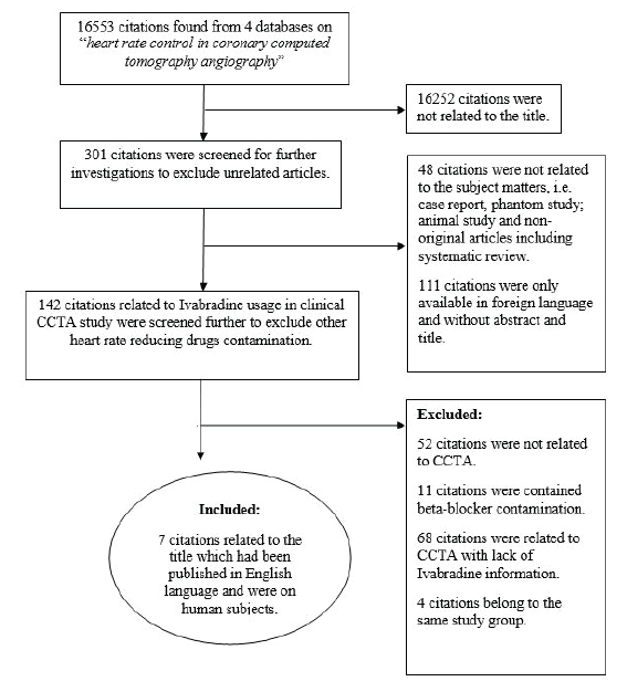
Seven articles out of 16,553 citations found in four databases met the
inclusion criteria and were included in the analysis. The flow chart of study
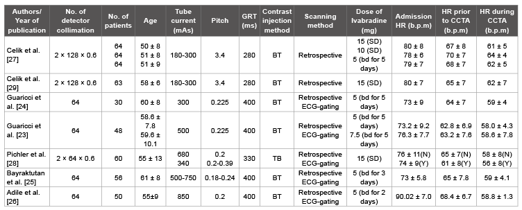
attributions is shown in figure 1. Data on 903 patients from 7 ivabradine
studies [23-29], 6 beta-blocker studies [23-26,28,29] and 2 combination of
ivabradine and beta-blocker studies [23,24] were tabulated in tables 1-3
respectively. The information contains heart rates monitored in several
stages including on arrival (HR admission), right before CCTA scanning
(HR prior to CCTA) and during CCTA examination (HR during CCTA).

Table 1: Ivabradine used as a heart rate controlling agent in CCTA examination.
GRT: Gantry rotation time; HR: Heart rate; NS: Not stated; BT: Bolus tracking; TB: Test bolus; bd: Twice a day; SD: Single dose; N: Patients who without
received long-term beta-blocker therapy; Y: Patients who received long-term beta-blocker therapy

Table 2: Beta-blocker used as a heart rate controlling agent in CCTA examination.
GRT: Gantry rotation time; HR: Heart rate; NS: Not stated; BT: Bolus tracking; TB: Test bolus; bd: Twice a day; SD: Single dose; N: Patients who without
received long-term beta-blocker therapy; Y: Patients who received long-term beta-blocker therapy

Table 3: Combination of ivabradine and beta-blockerused as heart rate controlling agents in CCTA examination.
GRT: Gantry rotation time; HR: Heart rate; NS: Not stated; BT: Bolus tracking; bd: Twice a day; SD: Single dose; N: Patients who without received long-term
beta-blocker therapy; Y: Patients who received long-term beta-blocker therapy.

Figure 1: The flow chart shows the study attribution of eligible references.
Analysis of heart rate reduction in CCTA examination
In ivabradine studies, the dosage used as premedication was
administered either as a single dose (10 mg or 15 mg) or multiple doses (5
or 7.5 mg twice daily between 2 and 5 days prior to CCTA). The detailed
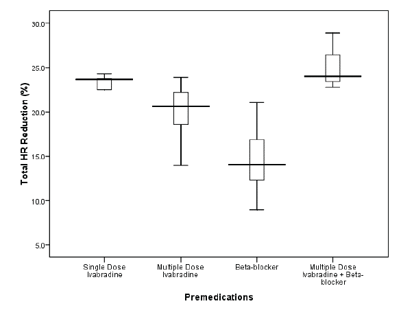
comparison of total heart rate reduction for ivabradine, beta-blocker and
combination of both studies were presented in box plot (Figure 2).

Figure 2: The box plot shows the comparison of total heart rate
reduction CCTA in between single dose Ivabradine, multiple doses
Ivabradine, beta-blocker and combination of multiple doses Ivabradine
and beta-blocker studies. The boxes indicate the first to third quartiles;
each midline indicates the median (second quartile) and the whispers
represent the maximum and minimum percentage of respective
premedication used. The tremendous heart rate reduction was achieved
during CCTA procedure when multiple doses of Ivabradine and betablocker
were applied.
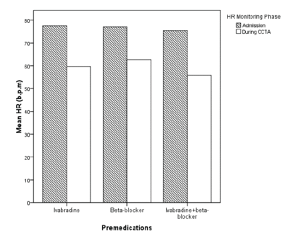
A comparison of HR admission and HR during CCTA among those
three premedication techniques were analyzed and shown in bar chart
(Figure 3). The highest heart rate reduction recorded among those 3
premedication techniques was the combination of ivabradine and beta-
blocker studies with 19.57 b.p.m (25.2%) followed by ivabradine studies
with 17.83 b.p.m (22.4%) and beta-blocker studies with 14.45 b.p.m
(14.6%), respectively.

Figure 3: The bar chart shows the comparison between different premedications
status (Ivabradine study, beta-blocker and combination of
Ivabradine and beta-blocker studies) on mean heart rate. The heart rate
on admission and during CCTA scanning were monitored and recorded.
All 3 premedication conditions showed significant HR reductions in both
HR phases.
Discussion
This systematic review provides a comparative analysis of the mean HR
at the time of admission, HR prior to CCTA, HR during CCTA and total
heart rate reduction between ivabradine, beta-blocker, and combination
of ivabradine and beta-blocker as premedication in CCTA examination.
Our analysis highlights major finding that ivabradine is effective as a
heart, reducing agent prior to CCTA examination.
Although several strategies are widely available in clinical setup to
reduce heart rate by premedication prior to CCTA examination, betablocking
medication such as metoprololtartarate is the most prominent
and commonly used. Many investigators have recommended the
administration of a single dose of oral [30,31] or intravenous [32] betablocker
to decrease the heart rate for CCTA examination preparation.
However, the use of beta-blocker is contraindicated in many diseases.
A study conducted by Shapiro et al. [15] stated that approximately 24%
of the subjects failed to receive beta-blocker due to contraindications.
Replacing beta-blocker with calcium channel blockers (diltiazem and
verapamil) too is not an effective strategy since the heart rate reduction
effect of these drugs is not sufficient for CCTA examination. Further,
they are contraindicated in heart failure and significantly impaired left
ventricle function. In atrial fibrillation cases, drugs commonly used are
beta-blockers, non-dihydropyridine calcium channel antagonists, and
digoxin but their use might be limited by hypotension or other side effects.
Ivabradine, was found to be safe and effective in reducing heart rate in
patients with stable angina. Ivabradine selectively inhibits funny channel
(If
) and has minimal cardiovascular effects and is found to be safe in
patients who were under long-term calcium channel blockers and effective
to overcome limitations of other beta-blockers or calcium channel blockers
side effects [29]. Indeed, the safety of ivabradine has been demonstrated
in large clinical trials with minor side effects such as mild-to-moderate
visual symptoms [27,33,34]. Our analysis reveals that premedication
with ivabradine provides a better reduction in heart rate as opposed to
beta-blocker premedication prior to CCTA examination. Although the
side effects of ivabradine are uncommon, blurred vision is the most likely
symptom which only occurred if doses are taken excessively more than 15
mg BD and thus, unlikely to be observed with the short time use in CCTA.
With the advantages of minimal side effects, lower hemodynamic effects
and better tolerance, ivabradine is a better alternative to beta-blocker [35].
In our analysis of ivabradine studies, both single high dose regimen
and the multiple low dose regimes were found to be safe and effective in
reducing the heart rate prior to CCTA scans. This was supported by Celik
et al. [27] who reported that 15 mg of single-dose oral ivabradine given
150 seconds prior to scan was as effective as 5 mg of oral ivabradine given
twice daily for 5 consecutive days. The single dose ivabradine regime may
be a better option than a 5-day regime because of ease of administration
and feasibility. In terms of drug safety, several studies supports that
ivabradine as a single dose or as multiple doses are effective and safe to
be used in clinical settings regardless of any medical procedures [34,36].
Moreover, in CCTA study, Bax et al. [37] found that a single IV bolus of
ivabradine achieved a safe, rapid, and effective heart rate reducing effect in
facilitating the performance of a successful CCTA examination.
Only two studies have used ivabradine and beta-blocker combination
as a premedication technique prior to CCTA so far and they have shown
that the highest heart rate reduction was successfully achieved compared
to other studies. Guaricci et al. [24] suggested that a combination of 50
mg of beta-blocker (metoprolol) and 5 mg of ivabradine administered
orally, twice a day for five consecutive days prior to CCTA yields the
highest heart rate reduction compared to a single dose of beta-blocker
or ivabradine alone. Furthermore, in their later study, Guaricci et al. [23]
reported that with a slight increment in ivabradine dose by 2.5 mg, further
heart rate reduction was achieved as compared to their previous findings
(5 mg of ivabradine+50 mg of metoprolol).
The studies with ivabradine premedication resulted in superior CCTA
image quality and diagnostic accuracy in the detection of CAD compared
to beta-blocker premedication studies. Bayraktutan et al. [25] reported that
95.5% of coronary segments were diagnostically accepted with ivabradine
studies compared to 89.9% in beta-blocker studies. Moreover, Celik et al.
[27] reported that only 4.5% of the images were of unacceptable quality in
ivabradine studies compared to 10.2% in beta-blocker studies. The most
common cause of CCTA image degradation is a stair-step artifact due to
involuntary cardiac motion. This phenomenon, however, can be avoided
by lowering the heart rate and maintaining the cardiac pace [25]. With a
slow heart rhythm, each image can be registered completely during scan
acquisition resulting in improved image quality [38]. Several literatures
agreed that most CCTA protocols recommend that the most optimal
visibility of vessels with high diagnostic value can be achieved at heart rate
not more than 65 b.p.m. [13,39,40].
CCTA examination has high radiation exposure compared to other
diagnostic radiological investigations. Several approaches had been
introduced to reduce the radiation dose to patient without affecting
image quality [41]. Prospective ECG-triggered CCTA is one of them that
reduces radiation dose by 83% compared to retrospective ECG-gated
technique [13]. However, a low and stable heart rate is essential to perform
this low radiation dose protocol effectively. Therefore, heart rate reduction
remains as an important factor not only to optimize CCTA image quality,
but also to limit radiation exposure. To date, no studies have specifically
addressed the effectiveness of ivabradine as a heart rate reducing agent
in prospective ECG-gated CCTA examination which may have high
possibility to minimize radiation dose.
Only a small number of studies met our inclusion criteria for analysis.
Most literature was excluded from this review due to lack of information
provided such as patient’s heart rate monitoring especially in ivabradine
studies. In fact, almost all of ivabradine studies were described in the
management of stable angina pectoris. Second, only few studies provide the
details of image quality analysis. The total heart rate reduction provided in
the literature was not in the same SI unit. Therefore, a conversion process
to obtain similar unit (SI) is adopted in our data analysis.
Conclusions
The systematic review reveals that Ivabradine is an effective heart rate
reducing agent in CCTA examination. Ivabradine is safe, and effective
in reducing heart rate to generate image of diagnostically acceptable
quality in almost all coronary segments in comparison to beta-blocker.
In addition, further heart rate reduction can be achieved prior to CCTA
examination with combination of ivabradine and beta-blocker regardless
of prospective ECG-triggering or retrospective ECG-gating protocols.
Acknowledgement
We would like to thank Young Researcher Incentive Grant Scheme
(GGPM-2013-099), Universiti Kebangsaan Malaysia, for financial support
of the study review.
Article Information
Article Type: Research Article
Citation: Sabarudin A, Chan AL, Nasir NM (2016)
Ivabradine as an Effective Heart Rate Controlling
Agent in Coronary Computed Tomography
Angiography: A Systematic Review. J Hear Health
2(2): doi http://dx.doi.org/10.16966/2379-769X.123
Copyright: © 2016 Sabarudin A, et al. This is an
open-access article distributed under the terms
of the Creative Commons Attribution License,
which permits unrestricted use, distribution, and
reproduction in any medium, provided the original
author and source are credited.
Publication history:
Received date: 12 Mar 2016
Accepted date: 24
Mar 2016
Published date: 29 Mar 2016


