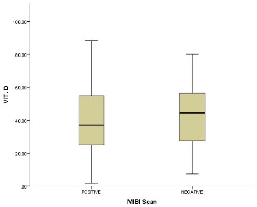
Figure 1: The median of vitamin D levels in relation to positive and negative Tc99 MIBI scans. The median of vitamin D for positive scans is 37.9, while the negative scans have a higher vitamin D median of 45.


Nadia Batawil* Albaraa Algithmi
1Radiology department, Nuclear medicine section, King Abdulaziz University Hospital, P.O. Box 80215, 21589 Jeddah, Saudi Arabia*Corresponding authors: Nadia Batawil, Radiology department, Nuclear medicine section, King Abdulaziz University Hospital, P.O. Box 80215, 21589 Jeddah, Saudi Arabia, E-mail: nbatawil@kau.edu.sa
Background: Patients with low vitamin D levels presented more advanced forms of primary hyperparathyroidism (PHPT) .We assess the effects of Vitamin D level on MIBI scan in PHPT.
Methods: 115 patients with PHPT who were referred for Parathyroid MIBI scan were classified according to their Vitamin D levels (low/ normal). The results of surgery, MIBI scans and parathyroid hormone (PTH) were analyzed to assess the true positive MIBI result.
Results: Out of 115 patients, sixty-six patients (61.7%) had low vitamin D levels. 65 patients had undergone surgery for parathyroidectomy, where 50 (77%) of them had low vitamin D. The overall sensitivity of the MIBI scans was 68%, specificity 80% which is not significantly different from the patients with low Vitamin D (68.9%, 81% respectively). The specificity of MIBI scan in low Vitamin D patients improved to 94% when combined with high PTH levels.
Conclusions: In PHPT the result of MIBI scan is not affected by the level of vitamin D. The specificity of the MIBI scans in low Vitamin D cases increased when the MIBI results is combined to high PTH levels.
Hyperparathyroidism; MIBI scan; Vitamin D; PTH; Diagnosis
Minimal invasive directed par thyroidectomy has become the operation of choice for primary hyperparathyroidism (PHPT). This surgical approach is aided by preoperative imaging and intra-operative parathyroid hormone (PTH) assays. Ultrasonogoraphy and Tc99m-methoxy isobutyl isonitrile or Tc99m sestamibi (MIBI) scans are a well-known techniques for evaluating primary hyperparathyroidism for parathyroid adenoma localization and maximize operative cure .The overall sensetivity of combined ultrasound and MIBI scan in PHPT is 97% [1]. 4D-CT scan has a high diagnostic accurecy >90% for single and multigland disease [2]. With the introduction of single photon computed tomography (SPECT), the reported sensitivity of MIBI sacn increased to 90% [3]. Several factors influence MIBI uptake by parathyroid cells, such as adenoma size, nodular thyroid disease, preoperative parathyroid hormone level, and plasma calcium level [4-7].
Limited studies have examined the correlation between levels of vitamin D (25 hydroxyl cholecalciferol) and Tc99 MIBI scan specificity. In a large study that included 421 patients, patients with low vitamin D levels presented more advanced forms of primary hyperparathyroidism, larger gland sizes, more positive MIBI scans, and more significant decreases in serum calcium at 1 and 6 months post-operation [8]. In the current study, the effects of vitamin D level on the results of Tc99m MIBI scans in patients with PHPT were assessed. The level of high PTH was combined to the MIBI scan results to see if this can help in predicting true positive MIBI scans in relation to the surgical outcome.
Cross sectional cohort study for 122 patients who were referred to the Nuclear Medicine Department at King Abdulaziz University Hospital between 2006-2012 for parathyroid Tc99 MIBI scans with a clinical diagnosis of primary hyperparathyroidism was conducted. Data from these cases were reviewed and included age, sex and preoperative and follow-up laboratory values for serum calcium (Ca; normal values 2.12- 2.52 mmol/L), PTH (normal values 1.18-8.2 Pmol/L), and serum vitamin D (normal values 50-80 nmol/L). The patients were classified according to their Vitamin D levels (low Vitamin D group/normal Vitamin D group). The results of surgery and MIBI scans were analyzed in each group. The results of high Parathyroid hormone level was combined to the results of MIBI scan in low Vitamin D group to determine if this combination will help in predicting the true MIBI result in relation to surgical outcome. Patients with thyroid adenoma [4] and patients for whom laboratory follow-ups could not be found [3] where excluded from the study measurements.
After intravenous injection of 925 MBq (25 mCi) of Tc99 m MIBI, planar images were obtained from patients in supine positions with a low energy, high resolution collimator. For five minutes, dual-phase planar images were obtained using a 256 × 256 matrix both immediately and 120 minutes after MIBI injection. SPECT images were obtained 120 minutes after MIBI injection from the patients’ necks and upper thorax using a dual-head gamma camera for SPECT acquisition, 128 × 128 matrix, and a low power dual-head CT scanner (Siemens, Symbia T6, Siemen).
Data analysis was performed with statistical software (IBM SPSS Statistics 24 IBM Corp, Armonk, NY and MedCalc v.17). We calculated sensitivity, specificity and accuracy of MIBI-SPECT in relation to one fold, two folds and three folds of PTH by using Youden,s index that captures the performance of a diagnostic test and estimate the probability of an informed decision ( J=sensitivity + specificity-1). Average values were reported as mean ± SD, and numeric data were reported as number (%). Statistical significance was defined by P<0.5.
A retrospective statistical analysis was conducted using the data from 115 patients, including 81 women and 34 men , mean age 49.6 +/- 15, mean PTH value 68.7 +/- 88 Pmol/L, mean serum vitamin D level 42.3 +/- 20 nmol/L , and mean serum Ca 2.43 +/- 0.35 mmol/L). Sixty-six patients had low serum vitamin D levels (less than 50 nmol/L), which accounted for 61.7% of the sample group. Tables 1 and 2 present the demographic data for all patients. There was no significant difference in the median for vitamin D in relation to the Tc99 MIBI scan results (i.e., 37.9 nmol/L for negative scans and 45 nmol/L for positive scans; P value, 0.109; Figure 1). The mean vitamin D levels for positive and negative Tc99 MIBI scans were 42.8 and 45.3, respectively. From the same data, 65 patients had undergone surgery for parthyroidectomy, 50 (77%) patients had low vitamin D levels (less than 50 nmol/L), and 15 patients had normal vitamin D levels (more than 50 nmol/L). The overall sensitivity of the MIBI scans for parathyroid adenoma localization was 68%, with specificity being 80% Table 3. The sensitivity, specificity or accuracy of MIBI scan for parathyroid adenoma localization in low Vitamin D group was not significantly different from the patients with normal vitamin D level (68.9, 81,72.5 Vs 63,75 and 65% respectively) Table 3. Out of 65 patients who had parathyroidectomy, 50 had low serum Vitamin D. The performance of Tc99 MIBI scans in this group of patients were analyzed in relation to one-fold, two-fold, and three-fold the normal PTH level by using Youden’s index. A PTH level of 16 Pmol/L (two-fold the normal level) is the best indicator for a true positive Tc99 MIBI scan with the highest test performance (Youden’s index 8.4%) Table 4. Overall Tc99 MIBI specificity in patients with low serum vitamin D level improved to 94% when combined with PTH levels that is two-fold the normal value, Table 4.

Figure 1: The median of vitamin D levels in relation to positive and negative Tc99 MIBI scans. The median of vitamin D for positive scans is 37.9, while the negative scans have a higher vitamin D median of 45.
Variable |
Mean ± SD |
Levels | ||
| Low | Normal | High | ||
| Age PTH | 49.62 ± 15.13 68.79 ± 88.61 Pmol/L |
- | 9 (8.2%) |
101 (91.8%) |
| Ca | 2.43 ± 0.35 mmol/L | 21 (19.4%) |
44 (40.7%) |
43 (39.8%) |
| Vit. D | 42.37 ± 20.66 nmol/L | 66 (61.6%) |
37 (34.5%) |
4 (3.4%) |
| Thyroxine (T4) | 13.48 ± 3.2 Pmol/L | |||
| Thyroid Stimulating Hormon (TSH) | 3.95 ± 9.79 uIU/L | |||
Table 1: Clinical and laboratory results of 118 patients with primary hyperparathyroidism
| Number | Percent | ||
| Total | 118 | 100.0 | |
| Gender | Male | 30 | 25.4 |
| Female | 88 | 74.6 | |
| MIBI Scan | Positive | 54 | 46.6 |
| Negative | 62 | 53.4 | |
| Pathology | Positive | 50 | 76.9 |
| Negative | 15 | 23.1 | |
Table 2: Demographic data for 118 patients with primary hyperparathyroidism
Table 3: The Sensitivity, Specificity,and Accuracy of MIBI scan in low and normal Vitamin D in 65 patients with PHPT and surgery
| PTH Pmol/L | Tc99 MIBI Scan | Sensitivity | Specificity | Accuracy | Test Performance Positive Negative (Youden’s Index) | ||
| Positive | Negative | ||||||
| PTH | < 8.2 | 1 | 0 | 0.029 | 1.000 | 34.0% | 2.94% |
| > 8.2 | 33 | 16 | |||||
| 2 × PTH | ≤ 16 | 5 | 1 | 0.147 | 0.937 | 40.0% | 8.40% |
| > 16 | 29 | 15 | |||||
| 3 × PTH | ≤ 24 | 7 | 3 | 0.206 | 0.812 | 40.0% | 1.84% |
| > 24 | 27 | 13 | |||||
Table 4: The performance of Tc99 MIBI scans in relation to multiple values of PTH (one-, two-, and three-fold the normal level) in 50 patients with parathyroid adenoma at surgery and low Vitamin D
Low vitamin D is not limited to elderly patients and has been observed in adult patients of all ages. In our region ,Vitamin D deficiency is common in healthy adults ,more pronounced in females and young age group (more than 50%) likely related to deliberate avoidance of the sun, lack of physical activity, wearing traditional clothing and inadequate dietary intake [9]. Genetic susceptibility and skin pigmentation can play role in vitamin D metabolism. [10,11]. In the current study, 77% of the patients who had Para thyroidectomy demonstrated low vitamin D levels. The sensitivity, specificity or accuracy of MIBI scan for parathyroid adenoma localization in low Vitamin D group was not significantly different from the patients with normal vitamin D level. However the specificity of theTc99 MIBI scans in patients with low serum Vitamin D increased from 80% to 93% when combined with high PTH levels that were two-fold the normal value ( >16 Pmol/L). A PTH level of 16 Pmol/L (two-fold the normal level) is the best indicator for a true positive Tc99 MIBI scan in low Vitamin D population with the highest test performance (Youden’s index 8.4%) Table 4. Patients with low vitamin D levels presented more advanced forms of primary hyperparathyroidism and positive scans [8]. Silverberge et al. reported that patients with low vitamin D had higher PTH and plasma alkaline phosphatase levels and rapid bone turnover [12]. Rao et al. [13] proposed that vitamin D deficiency can stimulate PTH production and have a substantial impact on biochemical indices indicative of mild PHPT. Reduced Vitamin D receptors (VDR) polymorphisms likely to impairethe 1, 25 (OH)D2 mediated control of parathyroid function and be an important in the pathogenesis of PHPT [14].
The reported sensitivity of Tc99 MIBI scans for parathyroid adenoma varied from 39% to 90% in a large meta-analysis of 52 studies [15]. Several factors can affect the scan’s accuracy for parathyroid adenoma localization in patients with PHPT, such as small adenoma size, parathyroid hyperplasia, PTH levels, and calcium channel blockers [2, 3,16]. The positive predictive value of high levels of PTH is more than 88% with a linear correlation to the size of a parathyroid adenoma. The combination of adenoma size, high PTH level, and scans can improve preoperative localization of parathyroid adenoma.
In patients with primary hyperparathyroidism the sensitivity, specificity or accuracy of MIBI scan for parathyroid adenoma localization is not affected by the level of serum vitamin D. However the specificity of theTc99 MIBI scans increased from 80% to 93% in patients with low Vitamin D level when MIBI scan results is combined with high PTH levels that is two-fold the normal value ( >16 Pmol/L). Limitation of the study include the retrospective design and small number of patients with normal Vitamin D level (out of 65 patients who had surgery, only 15 with normal Vitamin D level), which may affect the statistical power of the study. Prospective study that compares equal number of PHPT patients with normal Vitamin D level and abnormal Vitamin D level is recommended.
The authors express their gratitude to Mr. Salah Barnawi for his outstanding support.
Download Provisional PDF Here
Article Type: Research Article
Citation: Batawil N, Algithmi A (2017) The Effects of Vitamin D Level on the Results of Tc99 Sestamibi Scans for Parathyroid Adenoma Localization in Patients with Primary Hyperparathyroidism. Int J Endocrinol Metab Disord 3(2): doi http://dx.doi.org/10.16966/2380-548X.136
Copyright: © 2017 Batawil N et al. This is an open-access article distributed under the terms of the Creative Commons Attribution License, which permits unrestricted use, distribution, and reproduction in any medium, provided the original author and source are credited.
Publication history:
All Sci Forschen Journals are Open Access