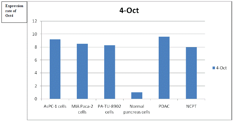
Chart 1: Expression rate of Oct4.

Prof. Dr. Seyed Saeid Zamanieh Shahri, MD* Prof. Dr. Sonia Sayyedalhosseini, MD*
Faculty Member in California Northstate University, CNSU-University Professor in Losrios Community College District, USA*Corresponding author: Prof. Dr. Seyed Saeid Zamanieh Shahri, MD, Faculty Member in California Northstate University, CNSU-University Professor in Losrios Community College District, USA, Tel: (916)724-9485; E-mail: saeid.zamanieh@cnsu.edu, zamanis@flc.losrios.edu
Prof. Dr. Sonia Sayyedalhosseini, MD, Faculty Member in California Northstate University, CNSU-University Professor in Losrios Community College District, USA, E-mail: sonia.sayyedalhosseini@cnsu.edu, sayyeds@flc.losrios.edu
Introduction: Oct4 gene is one of the important genes involved in stem cell self-renewal control. Recently, this gene has been proposed as a novel molecular marker for cancer detection. Therefore, in this study we examined the expression of Oct4 gene in MIA Paca-2, PA-TU-8902 and AsPC-1 cell lines as well as pancreatic cancer.
Methods: In this experimental study, MIA Paca-2, PA-TU-8902 and AsPC-1 cell lines were cultured in DMEM [1] (GibcoDulbecco’s Modified Eagle Medium is a widely used basal medium for supporting the growth of many different mammalian cells) and RPMI [2] - 1640 (RPMI, also known as RPMI 1640 or Roswell Park Memorial Institute Medium, was first described by Moore, 1967, in successfully culturing and expanding human lymphoid cells) medium containing 10% Fetal Bovine Serum (FBS) in a 37 degree centigrade, incubator containing 5% CO2 and 90% moisture. Pancreatic tumor and non-tumor specimens were purchased from Tumor Bank of Iran. RNA extraction and cDNA synthesis were then performed. Oct4 expression levels were determined using Real-time PCR. Protein expression levels of target genes in cell lines were evaluated using flow cytometry and immunocytochemistry.
Results: The expression level of Oct4 was higher in cancer cell lines than in control (normal tissue) samples. The protein expression levels of the target genes in the goal cell lines were confirmed by flow cytometry and immunocytochemistry.
Conclusion/Implications: Oct4 could be considered as a molecular marker of cancer in pancreatic cells. This gene may play an important role in the uncontrolled proliferation of cancer cells.
Pancreatic Ductal Adenocarcinoma (PDAC) is the deadliest solid cancer and it is currently the fourth most common cause of cancer death in the United States. PDAC has a late diagnosis due to the absence of symptoms in the early stages of cancer, extensive metastasis at the time of diagnosis and high resistance to chemotherapy and radiation therapy. Despite the expansion of research into pancreatic cancer over the past decades, little therapeutic progress has been made in this area [3].
Indeed, despite the declining trend for most cancers, the incidence and mortality of PDACs continue to rise, with pancreatic cancer predicted to be the second most common cause of cancer death in Western societies by 2030 [4]. The incidence of pancreatic cancer (85% of which is adenocarcinoma) is reported to be between 1 and 10 per 100,000 people worldwide and it is more prevalent among men than women [5]. This cancer is rarely diagnosed in people under 40 years old and the average age of diagnosis is 71. This disease is the eighth leading cause of cancer mortality in men and the ninth leading cause of cancer mortality in women [6].
In Iran, the prevalence of this cancer was lower than in Europe and the United States. Most people have metastases at the time of diagnosis, so surgery or chemotherapy interventions are least effective in these patients. One of the underlying barriers to clinical challenges in the treatment of pancreatic cancer is limited understanding of the molecular mechanisms of PDAC progression. In fact, the occurrence of defects in epigenetic pathways is one of the key mechanisms involved in the development of this type of cancer. Studies have shown that in pancreatic cancer, like many cancers, defects in the epigenetic regulation of cell cycle-regulating genes, DNA repair, cell connections, and cellular signaling pathways lead to tumor development, progression, and resistance to medication therapy [7].
Epigenetic disorders in the early stages of pancreatic cancer overcome the genetic disorders. In general, cancer is believed to be caused by the accumulation of multiple genetic mutations, but there is growing evidence that cancer cells also undergo epigenetic abnormalities during cancer formation, maintenance, and progression [8].
In another word, tissue neoplasms contain a heterogeneous population of cells that are biologically distinct and they have the potential for self-regeneration [9].
According to the clonal evolution model, cancer tumor cells are caused by the accumulation of genetic changes in the cells and the gradual selection of existing clones. Most conventional therapies for the destruction of cancer cells are based on this theory [10].
The limitations of these methods, including the poor prognosis for patients with advanced cancer, suggest that tumor cells contain a population of cells that are responsible for initiating tumor growth and expansion, as well as the ability to metastasize and recur. These cells were named Cancer Stem Cells (CSCs) because of the similarities in multiplication between these cancer cells and the fundamental stem cells. This model estimates the following characteristics of CSCs:
1) Self-rehabilitation;
2) Heterogeneity, such as multidimensional differentiation potential;
3) Resistance to apoptosis.
These traits appear to lead to a reduction in the effect of conventional therapies, which do function more on differentiated or under differentiation tumor cells. The undifferentiated stem cell population forms a small fraction of the tumor mass. CSC theory estimates that these cells are descended from progenitor stem cells that are somewhat differentiated and they have limited proliferative potential [11].
Recent studies indicate that specific transcription factors of embryonic stem cells such as Oct4 and Nanog play a very important role in the stability of CSC function and they are very important targets for inhibiting the proliferation of these cells, and producing medications to suppress growing of cancer cells [12,13].
In fact, The Oct4 protein is a transcription factor that encoded by the Pou5f1 gene, which Pou5f1 gene is a gene from the family of DNAbinding proteins located on chromosome 6 in human genome. Oct4, whose expression is associated with pluripotent properties of stem cells, is an essential factor for controlling early stages of mammalian embryogenesis [14].
To a certain degree, this factor is vital for maintaining the selfrenewal property of embryonic stem cells, and any increase or decrease in its expression, changes the fate of the cell. So it is involved in the process of cancer formation. To date, there has been no report of Oct4 expression in differentiated cells [15].
Many recent studies have shown the expression of Oct4 gene at protein levels in bladder, breast and osteosarcoma cancers. Besides that; Tai et al. confirmed the presence of Oct4 in several cancer cell lines, including liver, kidney, gastric, cervical and bone cancers. However, this gene is not expressed in normal cells and this, presents it as a very suitable candidate to be considered as a molecular marker of cancer cells [16,17].
Therefore, in this study, we examined the expression of Oct4 in pancreatic cancer cells compared to normal pancreatic cells.
The main purpose of this study is to demonstrate that Oct4 is a specific tumor promoter that is expressed in a wide range of tumors, particularly in this study in pancreatic cancer, but it is not expressed in normal tissues. In the present study, for the first time, the expression of Oct4 gene promoter as a specific tumor promoter in MIA Paca-2, PATU-8902 and AsPC-1 cell lines was compared with healthy pancreatic tissue cells.
PDAC and Non-cancer Pancreatic Tumor (NCPT) specimens, AsPC-1, MIA Paca-2 and PA-TU-8902 cell lines were purchased from Pasteur Institute of Tehran Iran. After morphological examination and ensuring cell health, 105 cells per flask were cultured in a culture flask after several passages, and the genes were analyzed to determine the expression pattern.
The samples were placed under direct supervision and as biopsy tissue in sterile and RNase-free micro tubes and they were transferred to the laboratory by American MGM nitrogen tank and kept at -80°C until RNA extraction. After culturing and reaching a density of 70%, the cell lines were isolated from the culture medium (by trypsinization) and after washing twice with PBS buffer, total cell RNA was isolated. The culture medium which is used for the PDAC, NCPT specimens, MIA Paca-2 and PA-TU-8902 cell line is DMEM with high glucose level. The specific culture medium for the AsPC-1 cell line is RPMI 1640+10% Fetal Bovine Serum. Culture media was supplemented with 1% penicillin/streptomycin and cells were cultured at 37°C and 5% CO2 at 90% humidity. The doubling time of the cells is 25-40 hours. Each biopsy specimen was divided into cancer and healthy tissue under the supervision of a pathologist and RNA purification was performed separately. After ensuring the quality of RNA extracted by electrophoresis and light absorption by spectrophotometer, all mRNA molecules were converted to cDNA and they were transferred to a freezer at -20°C, with the help of Oligo (dT) 12-18 Primer.
The sequence of Forward and Reverse primers of Oct4 gene was designed using Primer design software and then BLAST [18] (Basic Local Alignment Search Tool) on NCBI [18] (National Center for Biotechnology Information) to ensure its accuracy (Table 1).
| Target gene | designed Oligo | Primer’s order | Length |
| OCT4 | F | CGCAAGCCCTCATTTCAC | 111 |
| R | CATCACCTCCACCACCTG |
Table 1: Oligo (dT) 12-18 Primer
In order to amplify the desired gene, first primers were prepared and then real-time PCR was used to amplify the mRNA (cDNA) of the target genes. 4 microliters of proprietary Forward and Reverse primer, 3 microliters of cDNA, 10 microliters of MastermixSyber green and 3 microliters of DNase free water (with a final volume of 20 microliters) added to each PCR, then the plate was covered with a special adhesive to prevent evaporation. The plate was placed in a Real-Time PCR machine with the recommended program (one cycle of 95°C for 30 seconds and 45 cycles with conditions of; 95°C for 10 seconds, 52- 58°C for 15 seconds and 72°C for 20 seconds). All mentioned tests were repeated at least 3 times.
The human samples used in this study included six biopsy specimens of pancreatic tissue. The AsPC-1 cell sample from standard laboratory kit derived from a 62-year-old female Caucasian patient with pancreatic adenocarcinoma. The MIA Paca-2 cell sample from standard laboratory kit took from a 65-year-old patient with pancreatic carcinoma in the body and tail of pancreas. The PATU-8902 cell sample from standard laboratory kit took from a 44-yearold woman with pancreatic adenocarcinoma. The Pancreatic Ductal Adenocarcinoma (PDAC) sample took from a 55-year-old man and NCPT specimen took from a 50-year-old male Persian and normal pancreas tissue from a 45-year-old male Persian, all were purchased at Tumor Bank of Tehran Iran.
The expression of Oct4 gene in biopsy specimens of the mentioned tumor cell lines as well as healthy tissue of pancreatic biopsy specimens were examined by RT PCR method. The studied genes showed statistically higher expression of Oct4, 8-9 times more, in MIA Paca2, PA-TU-8902, AsPC-1, PDAC and NCPT compared to the control group (normal pancreas tissue).
Expression rate of the target gene (Oct4) in PDAC and NCPT, MIA Paca-2, PA-TU-8902, AsPC-1cell lines, compared to the control group (normal) showed in table 2, chart 1.
Chart 1: Expression rate of Oct4.
| Cell line sample | AsPC-1 Cells |
MIA Paca- cells | PA-TU-8902 cells |
Normal pancreas cells | PDAC | NCPT |
| Target gene: Oct4 | 9.2 | 8.5 | 8.3 | 1 | 9.6 | 8 |
Table 2: Expression rate of Oct4.
By recognizing and presenting the theory of CSCs and the similarities between the behavior of these cells and embryonic stem cells, the researchers found that Oct4gene, which is one of the critical embryonic stem cell genes, is a major factor in pluripotency and selfregeneration in these cells and it is also involved in cancer cells [19].
In this study, simultaneous expression of Oct4 gene in three pancreatic tumor-derived cell lines (AsPC-1, MIA Paca-2, and PATU-8902), healthy cells around pancreatic tumor tissue, PDAC and NCPT were evaluated. The results confirm that the expression of this gene is increased in pancreatic cancer cell lines but not in normal cells adjacent to cancerous tissue. This result confirms that the cells in the mass of cancer tissue have the ability to regenerate themselves with uncontrolled divisions that would lead to formation of cancer tissue.
The results of this study show that pancreatic cancer cells express major genes that control self-regeneration, and since the expression of the Oct4 gene has been shown to be a sign of stem cell fundamental, it can therefore be stated that these cells are actually CSCs.
Download Provisional PDF Here
Article Type: RESEARCH ARTICLE
Citation: Shahri SSZ, Sayyedalhosseini S (2020) The Evaluation of Gene Oct4 Expression as a New Tumor Marker in Pancreatic Tumor and Non-Tumor Cell Lines. J Diab Res Ther 6(2): dx.doi.org/10.16966/2380-5544.154
Copyright: © 2020 Shahri SSZ, et al. This is an open-access article distributed under the terms of the Creative Commons Attribution License, which permits unrestricted use, distribution, and reproduction in any medium, provided the original author and source are credited.
Publication history:
All Sci Forschen Journals are Open Access