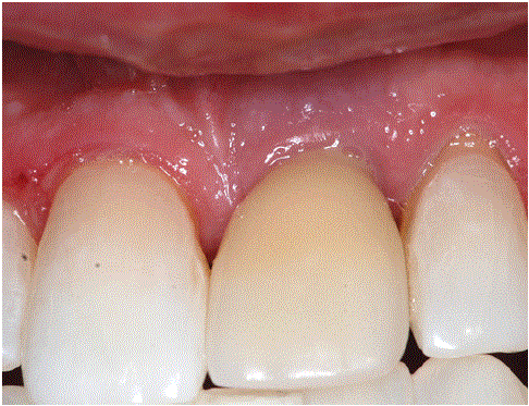
Figure 1: Facial view of tooth #9 (porcelain fuse to metal implant supported crown) with signs of inflammation.

Dumitru Gogarnoiu1-3*
1Private Practice, Lancaster Avenue, Wynnewood, PA, USA*Corresponding author: Dumitru Gogarnoiu, Private practice: 300 E. Lancaster Avenue, Wynnewood, PA, USA, E-mail: [email protected]
This case report details the diagnosis and management of peri-implantitis in the esthetic zone of a 68-year-old male patient. The patient presented with clinical signs of inflammation and radiographic evidence of bone loss around a 30-year-old implant-supported crown. The treatment plan was executed in three phases: initial removal of the pathogenic biofilm and cemented crown, surgical correction of hard tissue deformities using guided bone regeneration (GBR), and the fabrication of a new screw-retained zirconium crown. The comprehensive approach, incorporating pharmacological, surgical, and prosthetic interventions, resulted in the successful restoration of the implant site with optimal esthetic and functional outcomes. This report highlights the importance of a perio-prosthetic management approach for peri-implantitis.
As the prevalence of implants increases, so does that of periimplantitis [1-3]. A standardized definition of peri-implantitis has been established: it is defined as radiographic bone level loss of 3+ mm from the top of the implant, combined with profuse bleeding and/ or suppuration, and probing depths of 6+ mm [4]. Managing periimplantitis in the esthetic zone adds another degree of challenge. As an inflammatory disorder, peri-implantitis leads to tissue breakdown and bone loss around dental implants. The treatment alone can result in remodeling of the hard and soft tissue that may not be esthetically acceptable.
Tailored treatment approaches for specific clinical scenarios have been published and classified based on the presence or absence of bone loss and soft tissue loss [5]:
• Soft Tissue Deformity without Bone Loss
• Bone Loss without Soft Tissue Deformity
• Combined Hard and Soft Tissue Loss
Key factors influencing the esthetic outcome have also been published and organized into [6]:
• Patient-related factors: Willingness to undergo treatment, smile line, and esthetic awareness.
• Implant-related factors: Implant position relative to the bony housing and adjacent teeth.
• Site-related factors: Defect morphology and severity, presence of keratinized mucosa, and mucosal thickness.
• Prosthetic-related factors: Type of prosthesis and its emergence profile.
A comprehensive preoperative assessment should include a correct diagnosis [7,8], the ability to predict the esthetic outcome, and the ability to manage patient expectations. The goal of this case report is to illustrate how this clinical case fits into the new landscape of diagnosis, treatment planning, and treatment endeavors from surgical, prosthetic, and technological perspectives when it comes to peri-implantitis in the esthetic zone.
A 68-year-old male was referred for clinical and radiographic evaluation of the implant and the implant-supported crown of tooth #9 (Figures 1-3). Based on past dental history, the patient received the implant and the crown 30 years ago with the original external hex Branemark implant and a cemented porcelain-fused-to-metal implant-supported crown [9,10].
Figure 1: Facial view of tooth #9 (porcelain fuse to metal implant supported crown) with signs of inflammation. Figure 2: Palatal view showing deficient mesial and distal papillae and Inflammatory exudate at the gumline. Figure 3: Bone loss visible radiographically and unsated crown over the abutment.
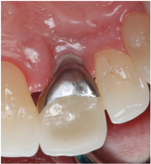
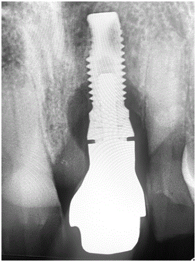
Clinically, the soft tissue showed signs of inflammation: redness, discomfort to palpation, and evidence of inflammatory exudate (Figure 3). No major soft tissue deformities were present. Probing depths were beyond 7 mm with bleeding and inflammatory exudate present. Radiographically, there was evidence of bone loss and signs of incomplete seating of the crown associated with the cementation process.
Based on clinical and radiographic findings, the diagnosis was periimplantitis characterized by probing depths beyond 5 mm with the presence of bleeding, inflammatory exudate, and radiographic evidence of bone loss. The peri-implantitis was considered a prosthetic-related factor [6] presenting bone loss without soft tissue deformities [5].
The treatment plan was designed in phases [7,8]:
• Phase one therapy addressed the immediate etiologic factors, such as removing the pathogenic biofilm and the cemented implant supported crown that showed evidence of excess cement [11] and replacing it with a screw-retained implant-supported provisional crown.
• Phase two, the surgical phase, aimed to correct hard tissue deformities with the goal of restoring the lost volume and shape of the bony structure with localized guided bone regeneration (GBR).
• Phase three, the restorative phase, involved the fabrication of a new screw-retained implant-supported zirconium crown, followed by addressing the possibility of protecting the crown and the implant from occlusal parafunctional activity with an occlusal appliance.
First visit: Under local anesthesia, deep irrigation with chlorhexidine 0.12% and a 3% solutionof hydrogen peroxide [12,13] was performed using a plastic syringe with a closed-ended double side vented irrigation needle. The goal was to change the acute nature of the peri-implantitis.
Second visit: The old crown and the prosthetic abutment associated with the crown were removed (Figure 4), followed by chairside fabrication of a new provisional restoration. The provisional restoration was block carved from acrylic PMMA [7,8] and used a titanium temporary cylinder as anchorage to the implant (Figures 5,6). The emergence profile was developed based on the critical and subcritical contour principle [14,15] to optimize gingival health, esthetics, and maintenance (Figures 7,8). Supplemental deep irrigation with chlorhexidine and hydrogen peroxidewas repeated [12,13].
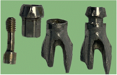
Figure 4: Cera One abutment was specifically lunched for cementationbased restoration in the esthetic zone. The retentive part of the abutment was hexagonal in shape with long parallel walls made the cementation difficult specially with crown abutment interface placed too far sub gingivally. The gold screw slightly bends as proof of past occlusal demand on this crown.
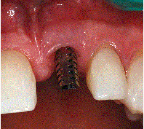
Figure 5: Externa hex engaging titanium temporary cylinder.
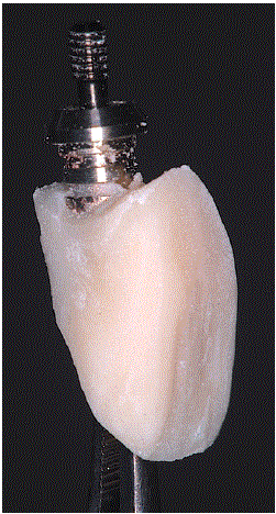
Figure 6: Temporary crown without the emergence profile established yet.
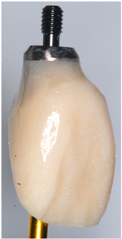
Figure 7: Temporary crown with new emergence profile.
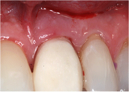
Figure 8: Soft tissue support with papillary contour recovered as a result of new established therapeutic emergence profile.
A new X-ray was taken to evaluate the fit of the new provisional crown and evidence of removal of the previous etiologic factors (Figure 8). Occlusion was checked for proper occlusal interaction statically and dynamically for evidence of interferences and hyper occlusion on the implant and teeth (Figure 9).
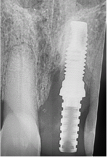
Figure 9: Despite of still presence of bone loss radiographically a better adaptation of the provisional crown represents an improvement over previous porcelain fuse to metal crown.
At two weeks, reevaluation of tissue response to biofilm removal and the emergence profile established in the provisional crown was very good (Figure 10). At three weeks, the case was considered ready for phase II of therapy, and the patient was scheduled for surgery.
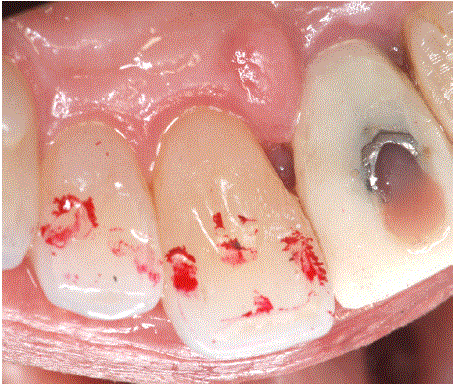
Figure 10: Occlusion was checked for proper loading of teeth and implant supported provisional crown.
Surgery: Under local anesthesia, a sulcular incision around teeth 8, 9, and 10 with a full mucoperiosteal envelope flap was raised, followed by degranulation of the bony defect and decontamination of the implant surface by applying cotton pellets soaked in chlorhexidine and peroxide (Figure 11). A collagen membrane was tailored to cover the regenerative space, with a punched hole added to accommodate the provisional crown (Figure 12), which played the role of membrane stabilization and prosthetic seal of the regenerative space [16,17]. Xenograft bone graft was placed in the infrabony defect [18] and molded to a positive architecture after the provisional crown and the membrane were secured in place (Figure 13).
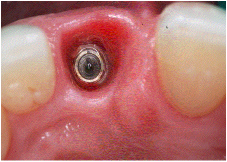
Figure 11: Sot tissue response to phase one therapy is adequate and acute signs of inflammation they are no longer present.
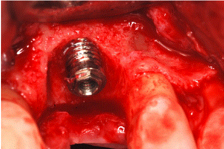
Figure 12: The original Branemark implant with external hex connection and machine surface made the decontamination apparently achievable versus rougher surfaces of today implants.
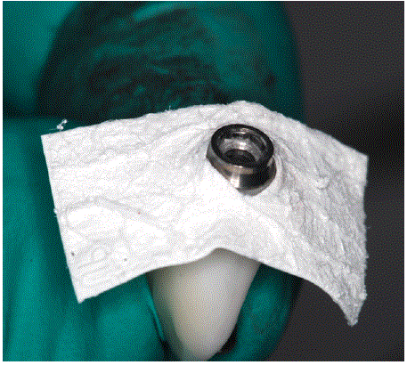
Figure 13: Collagen membrane received a punch cut allowing the provisional crown to play the role in its fixation and stabilization.
The flaps were secured with interrupted glycolon suture 06 (Figure 14). A new radiographic image was taken to visualize the bony fill and its relationship with the provisional restoration (Figure 15). Postop instructions, pain medication (Advil 600 mg every 4-6 hours as needed for pain), antibiotic coverage (Amoxicillin 500 mg three times a day for a week), along with a prescription for chlorhexidine mouth rinse were given.
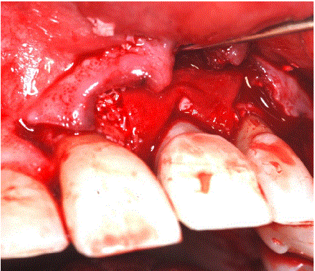
Figure 14: The provisional restoration plays a very important role in closing the surgical phase by providing adequate emergence profile for soft tissue closure and protection of the regenerative space.
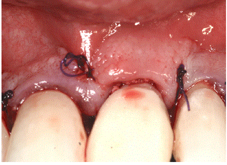
Figure 15: Sutures and prosthetic seal of the surgical site.
At four months, a new reevaluation was done, revealing excellent tissue healing and architecture based on the emergence profile of the provisional restoration (Figure 16). A digital impression was taken with a scan body, and an analog impression with the provisional crown was also taken (Figures 17-19). The analog impression was digitized and merged with the previous digital one.
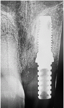
Figure 16: Radiographic evidence of proper sitting of the provisional crown and the bone graft distribution.
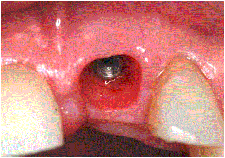
Figure 17: Excellent tissue healing and maturation based on the emergence profile provided by provisional crown.
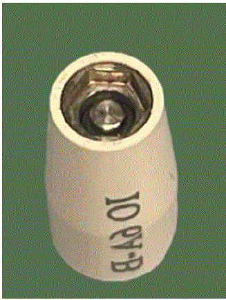
Figure 18: Scan body for Branemark external hex implant.
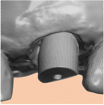
Figure 19: Digital impression with scan body.
A perfect replica of the emergence profile and crown contour was designed using CAD software (Figure 20).
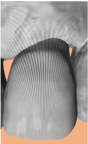
Figure 20: Analog impression that was digitized and merge with the intra oral digital impression.
The STL file was downloaded to a milling machine for nesting and milling out of a zirconium block (Figures 21,22), followed by coloring (Figure 23), sintering, cut back for porcelain veneering, porcelain application to the cut back (Figures 24-26) and finally glazing and polishing (Figures 27,28). The critical and subcritical portions of the crown were highly polished with rubber wheels and diamond paste [19].
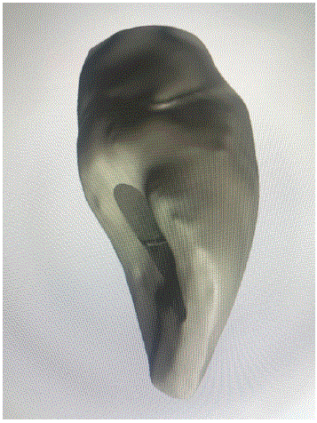
Figure 21: The new digital crown that is a copy of the analog version of the provisional crown.
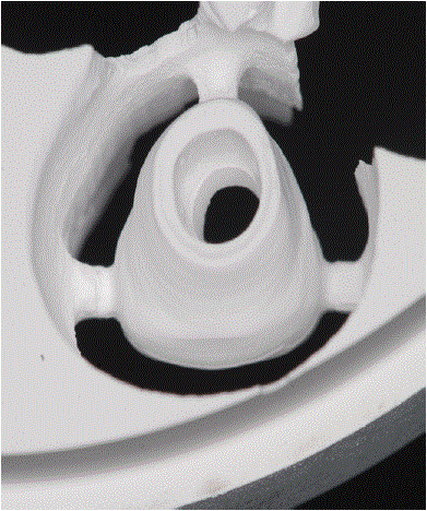
Figure 22: Post milling of the crown out of a zirconium disk.
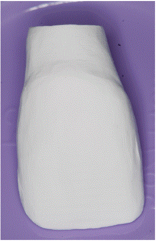
Figure 23: Full contour unsintered crown.
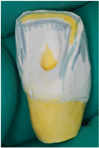
Figure 24: Color shading before sintering.
The color is very easily absorbed internally helping the natural and artistic look off the crown. Chromatic accents added to cervical area.
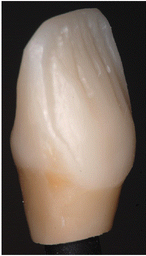
Figure 25: Zirconia still lacks translucency compare with felspathic porcelain or lithium disilicate. Some veneering facially is necessary to add the translucency that is lacking.
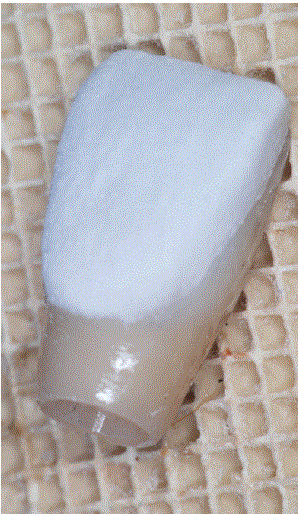
Figure 26: Transparent blue felspathic porcelain was added and sintered.
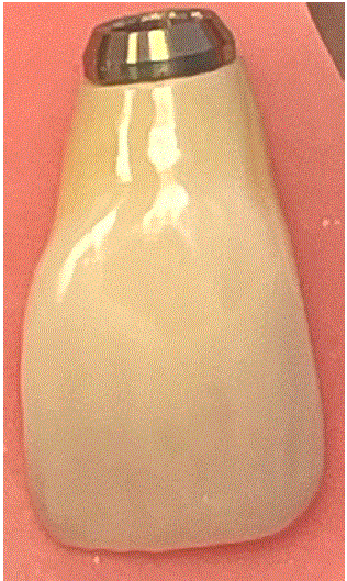
Figure 27: Facial view of the finished crown.
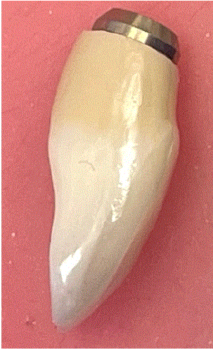
Figure 28: Proximal view of the final crown.
The crown was cemented to the titanium base with resin cement. The crown was inserted into the patient's mouth and checked for contact points and occlusal relationship (Figure 29). An X-ray was taken to ensure proper seating of the crown and the final outcome of the GBR therapy (Figure 30).
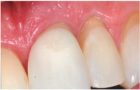
Figure 29: Tissue is very healthy the crown blends very well with the adjacent teeth in shape and color.
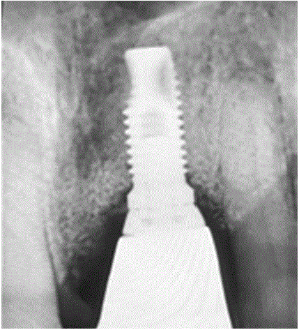
Figure 30: Final result shows the bone architecture recaptured covering all the treads that were previously exposed radiographically.
The implant was torqued according to manufacturer recommendations and covered with cotton pellets and composite. An impression for an occlusal appliance [7,8] was taken, and a Hawley bite plane appliance was delivered two weeks later (Figure 31). The patient was scheduled for recall maintenance.
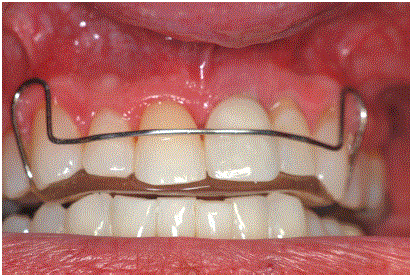
Figure 31: The occlusal appliance will provide protection to the implant and the crown from parafunctional habits like excessive clenching and grinding.
The management of peri-implantitis, especially in the esthetic zone, presents a unique set of challenges that require a multifaceted approach. This case report highlights the significance of properly diagnosing the condition and implementing a structured treatment plan that addresses both biological and esthetic concerns. The successful intervention developed in three phases reveals the importance of comprehensive management strategies, particularly the integration of pharmacological, surgical, and prosthetic solutions.
In this case, the initial phase focused on immediate etiologic factors, including the removal of pathogenic biofilm and the old cemented crown, which were pivotal in controlling the inflammatory processes. The importance of biofilm management is underscored by its role in the progression of peri-implantitis. By switching to a screwretained provisional crown, a more accessible restorative approach was established for future maintenance and monitoring.
The surgical phase employed guided bone regeneration (GBR) to restore lost hard tissue volume. This is particularly critical in the esthetic zone, where both hard and soft tissue architecture influences the final outcome. The integration of GBR techniques alongside the provisional restoration demonstrates the need for a synergistic approach that not only addresses the immediate anatomical defects but also optimizes aesthetics and functional outcomes.
Post-surgical assessments at four months yielded promising results, with excellent tissue healing and successful integration of the GBR graft. This emphasizes the importance of adequate healing time and followup evaluations in ensuring the long-term success of implant therapies. The use of advanced technology, such as digital impressions and CAD/ CAM workflows in the prosthetic phase, further refined the esthetic outcome. The precise fit and contour of the final zirconium crown contributed to both aesthetic appeal and functional effectiveness.
Overall, this case reinforces the critical nature of thorough preoperative assessments and the classification of peri-implantitis. As the prevalence of dental implants increases, so does the incidence of peri-implantitis, necessitating ongoing research into effective management strategies.
In conclusion, this case report highlights the effective management of peri-implantitis in the esthetic zone through a structured, threephase treatment approach. It emphasizes the importance of integrating pharmacological, surgical, and prosthetic techniques tailored to individual patient needs and site-specific factors. This comprehensive strategy not only addresses the underlying causes of peri-implantitis but also optimizes both aesthetic and functional outcomes.
Screw-retained restorations offer clear advantages [20] in the esthetic zone by eliminating residual cement, enhancing retrievability for maintenance, and reducing peri-implantitis risk. Mastering the emergence profile [21] is essential both in an analog way (provisional shaping to sculpt soft tissue) and digitally (CAD/CAM contour and Scan-to-Mill workflows) to achieve stable papillae and optimal esthetics. Therapy progresses in three phases: Phase I-biofilm/cement removal and placement of a provisional screw-retained crown; Phase II-localized GBR and soft-tissue optimization with a provisional to guide healing; Phase III-fabrication and placement of the final screw retained zirconia crown with occlusal stabilization, all within an integrated digital-analog prosthetic workflow.
The author has no conflict of interest and all the surgical, prosthetic and laboratory work was done by the author in his private practice.
Download Provisional PDF Here
Article Type: CASE REPORT
Citation: Gogarnoiu D (2026) Surgical and Prosthetic Management of Peri-Implantitis in a Single Tooth Implant-Supported Crown in the Esthetic Zone. Int J Dent Oral Health 12(1): dx.doi.org/10.16966/2378-7090.439
Copyright: © 2026 Gogarnoiu D. This is an open-access article distributed under the terms of the Creative Commons Attribution License, which permits unrestricted use, distribution, and reproduction in any medium, provided the original author and source are credited.
Publication history:
All Sci Forschen Journals are Open Access