
Figure 1: Six Anterior maxillary teeth were sculpted by the author out of wax using carving instruments and using measurement caliper to accurately reproduced form and dimension according with dental anatomy textbook’s.

Dumitru Gogarnoiu1,2*
1Private Practice, Lancaster Avenue, Wynnewood, PA, USA*Corresponding author: Dumitru Gogarnoiu, Private practice: 300 E. Lancaster Avenue, Wynnewood, PA, USA, E-mail: gogarnoiu@aol.com
The cemento-enamel junction (CEJ) is essential to the morphology of the dental alveolar process as the tooth emerges into the oral environment. The aesthetic outcomes of implant-supported restorations (ISRs) in the anterior maxilla are closely linked to this bone morphology. Furthermore, the soft tissue gingiva that covers the bone is influenced by the dental-alveolar architecture, particularly in terms of preserving papillary structures. Existing literature has noted challenges associated with papilla loss and the formation of "black triangles" when implants are placed, especially during the remodeling phase of the alveolar process, which must adapt to accommodate a dental implant designed quite differently from the natural tooth it replaces.
This article investigates the role of the cemento-enamel junction (CEJ) in the dental-alveolar architecture of maxillary anterior teeth and its influence on the aesthetic outcomes of implant-supported restorations (ISRs). The study aims to provide insights that can enhance decision-making in implant placement and restorative outcomes.
Cemento enamel junction; Biologic width; Dental implants; Periodontics; Dental papilla; Black triangle
Tooth replacement in the anterior zone with implant-supported restorations (ISR) is quite demanding, but it can lead to excellent aesthetic outcomes [1,2].
However, the way an implant is positioned in the bony site of an extracted tooth in the anterior maxilla can result in a deficiency of the papillae, which is aesthetically unacceptable. For example, an excessively deep placement of the restorative platform can cause significant saucerization and extensive peri-implant bone remodeling, as well as an undesirable proximity to neighboring teeth [3].
Belser evaluated early-placed maxillary anterior single-tooth implants using objective aesthetic criteria [4] known as the Pink Esthetic Score (PES) [5].
The scores for the mesial and distal papilla surrounding ISRs placed in maxillary incisor sites were 1.84 and 1.30, respectively, with two being the highest (most aesthetic) possible score.
While these scores indicate that the results are not perfect, they may be considered good enough under certain circumstances.
These clinical realities cannot be fully understood without knowledge of the dental-alveolar structure of the anterior maxilla. It is known that the vertical distance between the bone crest and the contact point is 5 mm when the papilla is present [6].
Additionally, the vertical distance between the bone crest and the contact point between a tooth and an adjacent implant-supported crown largely dictates the papillary response [7].
Oscar showed that the position of the mesial cemento-enamel junction (CEJ) of maxillary lateral incisors is located slightly apical to the distal CEJ of the central incisors [8].
Despite the scattered information available, the structure of the dental alveolar morphology in the maxillary anterior zone remains an unresolved puzzle. It is understood that, in a healthy state, the cementoenamel junctions play a crucial role in determining the profile of the alveolar crests [9].
The CEJ is located in the cervical region of teeth, where the enamel meets the cementum. Following tooth eruption, the biological width complex, as described by Gargiulo AW, et al. [10], will establish around the CEJ and play a major role in protecting the periodontium, as described by Lisgarten MA and Schroeder HE [11].
By utilizing the data, we have regarding tooth anatomy and the CEJ it may be possible to accurately reconstruct the dental alveolar morphology of the six maxillary anterior teeth.
The article adheres to the STROBE observational study that is part of EQUATOR reporting guidelines. Ethics approval was not required for this study since it was based solely on wax, carving tools, and the author's expertise in dental anatomy.
The six maxillary incisor teeth were carved from wax [12] (Figure 1). According to the dimensions outlined in dental anatomy textbook [13] (Table1).
Figure 1: Six Anterior maxillary teeth were sculpted by the author out of wax using carving instruments and using measurement caliper to accurately reproduced form and dimension according with dental anatomy textbook’s.
| Maxillary Central Incisor | Maxillary Lateral Incisor | Maxillary Canine | |
| Tooth length | 23.5 mm | 22 mm | 27 mm |
| Crown length | 10.5 mm | 9 mm | 10 mm |
| Root Length | 13 mm | 13 mm | 17 mm |
| Angulation M-D | 20 | 70 | 170 |
| Angulation F-P | 280 | 260 | 160 |
| CEJ M | 3.5 mm | 3 mm | 2.5 mm |
| CEJ D | 2.5 mm | 2 mm | 1.5 mm |
| At High of Contour F-P | 7 mm | 6 mm | 8 mm |
| At High of Contour M-D | 8.5 mm | 6.5 mm | 7.5 mm |
| At Cervix F-P | 4 mm | 4 mm | 5.5 mm |
| At Cervix M-D | 7 mm | 5 mm | 5.5 mm |
Table 1: Tooth dimensions used in this study.
Mesial=M, Distal=D, Facial=F, Palatal=P CEJ=Cemento Enamel Junction
Digital photography was conducted to capture the mesial, distal, facial, and palatal aspects of each carved tooth. The analysis of the images was performed with the teeth oriented parallel to their long axes (Figures 2a, 2b).
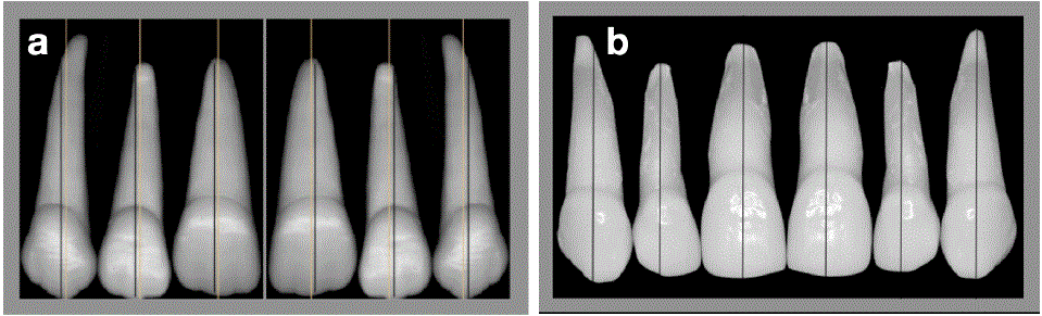
Figure 2a: Wax models of idealized maxillary anterior teeth were created.
Figure 2b: Natural dentition of maxillary anterior teeth as evidence of similarity with idealized wax model.
The coronal portion of each carved tooth was removed following the curvature of the cement-enamel junction (CEJ).
New digital photographs were taken from the mesial, distal, facial, and palatal views. The analysis of these photographs revealed a parabolic shape of the CEJ on both the buccal and palatal surfaces, with an elevation of the CEJ mesially and distally, showing greater height mesially than distally (Figure 3a).

Figure 3a: Facial view reveals a parabolic shape of the CEJ, starting higher on the mesial and finishing lower on the distal side. When these points are connected, a descending diagonal from mesial to distal becomes noticeable.
Figure 3b: The proximal view shows that the facial and palatal levels of the CEJ share the same position when analyzed from a perspective parallel to the vertical axis.
Figure 3c: The angulation between 28° and 16° with the vertical axis will change the relationship between the facial and palatal positions of the CEJ, resulting in a lower facial position and a higher palatal position.
By removing the coronal section according to the CEJ outline, the root itself exhibited a top profile that sloped diagonally from mesial to distal (Figure 3a and figure 4a).
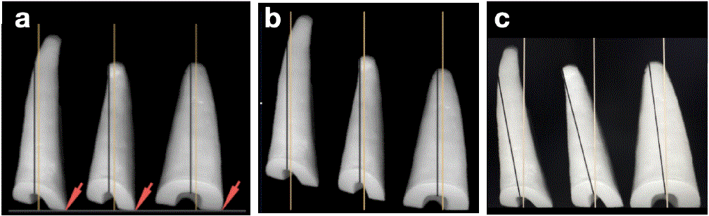
Figure 4a: Along the long axis, the roots exhibit a slanted profile at the cemento-enamel junction (CEJ).
Figure 4b: When the cemento-enamel junction (CEJ) is aligned along the long axis, a diagonal formation becomes evident.
Figure 4c: When the cemento-enamel junction (CEJ) is aligned horizontally, the roots will exhibit a mesiodistally angled inclination.
Interproximally, the buccal and palatal aspects were at the same level (Figure 3b). When the root is positioned facial-palatally angled, the facial side is lower than the palatal aspect (Figure 3c).
Based on these observations, the following conclusions can be drawn: In a mesiodistal direction, the CEJ is aligned as it typically is, showing a diagonal orientation (Figure 4b) that anatomically suggests increased bone height from distal to mesial. For a horizontal arrangement of the CEJ, the tooth must have a more pronounced angulation in a mesiodistal orientation, which would correlate with flatter bone architecture (Figure 4c).
Knowing that the CEJ is parallel to the bone level and positioned 1mm to 3mm supracrestally, a bony architecture can be visualized. From this perspective, the following conclusions can be made: the CEJ is highest mesially and lowest facially with the distal aspect second and the palatal aspect third (Figure 5 and figure 6).
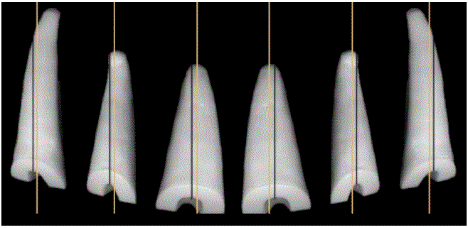
Figure 5: Together, they form a diagonal pattern that converges in the middle.
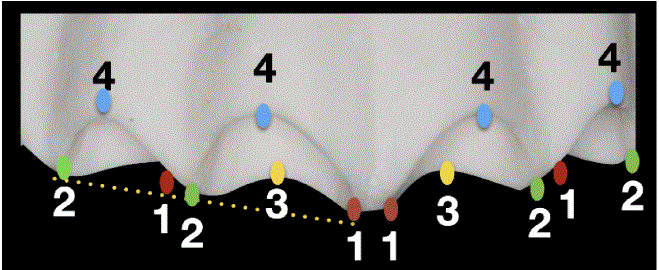
Figure 6: Representation of numerical data, as well as the position and elevation of the crestal bone associated with the maxillary incisors.
When restoring the bony architecture based on the spatial orientation of the CEJ, the bone exhibits a diagonal orientation that extends from the mesial of the central incisors to the distal of the canine (Figure 6).
Only at the midline is the bone equally positioned, displaying a horizontal profile between the two central incisors; thereafter, it exhibits a step-like pattern towards the distal of the canine (Figure 6, figures 7a, 7b).
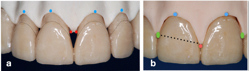
Figure 7a: Teeth and alveolar bone together as the CEJ is followed in a gradual step like fashion from middle to distal. This arrangement is called positive architecture.
Figure 7b: Soft tissue and the interproximal papillae mirroring the bone morphology.
To validate this model, 3D models derived from cone beam computed tomography (CBCT) scans were analyzed for similarity (Figure 8a), along with bone and tooth image segmentation models that were compared to the proposed model (Figure 8b).

Figure 8a: CBCT 3D model is enhanced with graphical representations of the biological width complex as it pertains to dental alveolar architecture.
Figure 8b: Image segmentation of central incisors 8 and 9 reveals a similar pattern of bone morphology surrounding the maxillary central incisors with the model proposed. Also, the relationship with the CEJ and the bone is also visible.
Additionally, a radiographic evaluation was conducted on anterior teeth, particularly the central incisors, which are the most frequently replaced with dental implants. Assessments were made before tooth replacement and after the completion of the implant placement and restorative phases (Figures 9a, 9c).
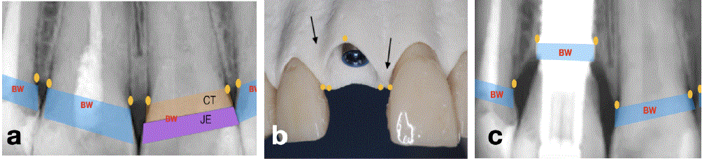
Figure 9a: The X-ray indicates that the central incisors share the same bone level at the highest point in the midline, with a diagonal run toward the distal where the distal aspect of the central incisor is higher than the mesial aspect of the lateral incisor. The biological width (BW), which encompasses both the connective tissue attachment (CT) and the junctional epithelium attachment (JE), follows a similar diagonal pattern.
Figure 9b: Implant placed using the buccal bone as point of reference. That position will induce remodeling mesially and distally.
Figure 9c: When the implant is positioned using the facial bone as a reference point, the interproximal bone will undergo remodeling. The biological width (BW) is then repositioned subcrestally, becoming circumferentially symmetric, flat, and smaller in size compared to its previous configuration around the natural tooth. The mesial papilla will maintain its position due to the neighboring central incisor remaining intact. However, the distal papilla will lose its previous position because the mesial aspect of the lateral incisor is now positioned lower than before.
Clinical cases were also examined to reveal the interaction between implant placement and the dynamics of bone remodeling in the replacement of maxillary anterior teeth with dental implants (Figures 10a, 10c).
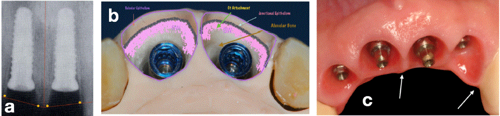
Figure 10a: Two adjacent implants are being used to replace the central incisors. It's important to note that the distance from the implant platform to the crestal bone is greatest in the center. This difference will lead to bone remodeling, which will cause the middle papilla to collapse significantly, resulting in an aesthetic challenge.
Figure 10b: The biologic width complex will have to reorganize to adapt to the implant shape that is circumferentially smaller and also flat.
Figure 10c: Despite achieving excellent tissue health, the positive architecture of the hard and soft tissues that teeth previously maintained before implant placement is becoming flatter. While the midline papilla used to be the highest point, it is now losing its position to the mesial papilla of the canine.
The use of wax models of idealized teeth (Figure 2a) is a subject of debate, and an alternative that may seem more effective is the use of real teeth (Figure 2b). However, obtaining six periodontally compromised anterior teeth from multiple patients is likely infeasible due to the limited availability of such cases. Additionally, modifying natural teeth to meet the study's objectives would be challenging because of the hardness of enamel and dentin, as well as the specialized burs and cooling systems required for the process.
Wax models of six maxillary incisors were created based on the average dimensions outlined in dental anatomy textbooks, ensuring representation of diversity. These dimensions are supported by more recent studies, such as one conducted by Gallucci GO [14], who evaluated the dimensions and characteristics of the cementoenamel junction (CEJ) of maxillary anterior teeth. He used standardized digital images of 137 extracted human teeth (45 central incisors, 46 lateral incisors, and 46 canines) to measure CEJ curvature. The study found that the curvature value (parabolic value) was consistently greater at the mesial aspect compared to the distal aspect, with measurements of 3.46 mm versus 3.13 mm for central incisors, 2.97 mm versus 2.38 mm for lateral incisors, and 2.55 mm versus 2.60 mm for canines. He also described the shape of the CEJ curvature, which extends facially and palatally in a coronal direction while running mesially and distally in an apical direction.
Thus, we can say that bone architecture is determined by the presence of teeth. The shape that a tooth assumes as it emerges in the oral cavity is influenced by the geometry of the cementoenamel junction (CEJ), and this architecture is referred to as positive architecture (Figure 6, figures 7a, 7b).
Reverse engineering the architecture of dental alveolar morphology based on the cemento-enamel junction (CEJ) of the teeth has been a labor-intensive process. However, utilizing Cone Beam Computed Tomography (CBCT) has proven to be the most rewarding aspect of this effort. The model provided is undeniably valid, both as a 3D rendering of the maxilla (Figure 8a) and as an image segmentation of the teeth and supporting alveolar bone (Figure 8b).
While CBCT currently offers only the bone topography, the soft tissue (interproximal papillae) can be anticipated as a mirror of the underlying bone; in a healthy state, they share the same characteristics of positive architecture (Figures 7a, 7b). Any alteration in bone architecture would also be reflected in the soft tissue.
In contrast, dental implants have an entirely different design, characterized by a circumferential flat structure. During the immediate extraction followed by the placement of the implant, the facial bone serves as a reference point. Typically, the implant is positioned 3 mm below the free gingival margin in a mesiodistal orientation. The central incisor is the tooth most frequently replaced with an immediate dental implant after extraction.
Considering the previously described dental alveolar structure, it is evident that the facial reference point is the lowest, resulting in more remodeling occurring on the mesial and distal sides.
Despite of that remodeling does not affect the presence of the papilla, resulting in an aesthetically pleasing outcome.
As comparing figure 9a versus figure 9c, there is noticeable bone remodeling and a reorientation of the biological width (BW).
The X-ray (Figure 9a) reveals that the central incisors have the same bone level at the highest point in the midline, with a diagonal slope toward the distal aspect-where the distal surface of the central incisor is higher than the mesial surface of the lateral incisor. The biological width, which includes both connective tissue attachment (CT) and junctional epithelium attachment (JE), follows a similar diagonal pattern.
In the figure 9b and X-ray (Figure 9c), the implant is positioned using the facial bone as a reference point, displaying interproximal bone remodeling. The biological width is then repositioned subcrestally, becoming circumferentially symmetric, flat, and smaller than its previous configuration around the natural tooth (Figure 9c).
The presence of two adjacent teeth helps maintain the papilla, despite the bone remodeling that occurs. However, even with this positive outcome, there is a noticeable difference in the quality of papilla presence between the mesial and distal sides. The mesial papilla fills in perfectly, while the distal papilla is less prominent and may need to be addressed through modifications to the crown contour [8].
This difference can be attributed to the morphology of the bone, as illustrated earlier in figure 6 and figure 7. The mesial papilla retains its position because it is adjacent to the neighboring central incisor, which shares a similar height. In contrast, the distal papilla loses its original position because the mesial aspect of the lateral incisor is now lower than the distal part of the replaced central incisor [8].
The situation changes when the central incisors are replaced by two adjacent implants (Figures 10a, 10b). In this case, the mesial papilla loses its highest position in the midline, leading to the formation of a "black triangle" that needs to be addressed restoratively. As shown in figure 10a, the distance from the implant platform to the crestal bone is greatest in the center, as illustrated in figure 10a. This significant remodeling will cause the collapse of the middle papilla. Therefore, placing two adjacent dental implants can result in excessive mesiodistal remodeling, which is undesirable [15].
If a distance of more than 3 mm is maintained between adjacent implants, it is possible to position implants adjacent to each other. However, the natural architecture of the bone and soft tissue is compromised, leading to a maximum papilla height of no more than 4 mm, resulting in the loss of the prominence of the central papilla as the highest point [16] (Figure 10c).
In other words, replacing all four incisors with dental implants that are adjacent to one another will change the topography of the bone and soft tissue from a well-defined, positive architecture to a flat architecture.
A proposed model outlining the dental alveolar architecture of maxillary anterior teeth has been presented. This model enhances our understanding of the esthetic challenges we face and current implant design, which features a flat, rotation-symmetric shoulder. However, further investigation is needed, especially with the advent of new tools such as imaging technology and artificial intelligence, which can provide deeper insights into implant therapy in the anterior zone.
The author declares that there is no conflict of interest. The study was conducted in a private office and was supported by the author through time, expertise, and necessary materials. All carving, photography, and analysis were performed by the author. Additionally, the literature review and the writing of the article were solely completed by the author.
Download Provisional PDF Here
Article Type: REVIEW ARTICLE
Citation: Gogarnoiu D (2025) The Role of Cemento Enamel Junction in Maxillary Anterior Teeth. Int J Dent Oral Health 11(3): dx.doi.org/10.16966/2378- 7090.436
Copyright: © 2025 Gogarnoiu D. This is an open-access article distributed under the terms of the Creative Commons Attribution License, which permits unrestricted use, distribution, and reproduction in any medium, provided the original author and source are credited.
Publication history:
All Sci Forschen Journals are Open Access