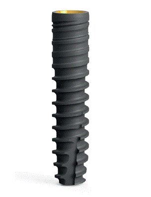
Figure 1: BioniQ implant - straight 2,9 x 14 mm (bone level).

Starosta M1 Maďar R2
1Department of Dentistry, Faculty of Medicine, University of Ostrava, Ostrava, Czech Republic*Corresponding author: Starosta M, Department of Dentistry, Faculty of Medicine, University of Ostrava, 70300 Ostrava, Czech Republic, E-mail: martin.starosta@osu.cz; mstarosta@seznam.cz
The type of implant and the method of implantation are believed to have a significant influence on primary failure. The aim of this retrospective study was to determine the reliability of osseointegration of BioniQ implants (LASAK s.r.o., Czech Republic) based on the rate of primary failure and to analyze the causes of failure in detail. The primary failure rate of BioniQ implants was 2.35% of all implants and 0.6% when augmentation procedures are excluded. This represents a result, which is in agreement with the data found in current published literature.
Dental implants; Early failure; Local and systemic factors
Dental implants are now considered a reliable replacement for lost teeth and are often chosen as the primary treatment option. However, despite the high success rate, early or late failure can occur [1-5]. When implant failure occurs, it is always a question of what caused the problem. In general, factors affecting implant durability can be divided into those related to the type of implant, iatrogenic factors, and patient-related factors [6]. The type of implant includes its macrostructure (shape, diameter, length, arrangement and shape of threads, connection to the abutment), microstructure (implant surface), and material (type of titanium alloy and its purity, materials other than titanium alloy). Iatrogenic factors depend mainly on the expertise of the attending physician. The patient then determines the conditions in which the implant is inserted (general health, medication, habits, hygiene level, bone quality, location). From this list, it is clear that implant failure can be considered multifactorial, and identifying the main cause of failure is challenging. However, the question is not so unclear given the more than 50-year history of dental implant use in clinical practice [7-12]. We can assume that the type of implant and the method of implantation have a significant influence on primary failure. The question of other more patient-related influences remains unclear. The aim of this retrospective study was to determine the reliability of osseointegration of BioniQ implants (LASAK s.r.o., Czech Republic) based on the rate of primary failure and to analyze the causes in detail.
This retrospective study analyzed patients treated by a single implantologist with over 20 years of experience. The study period encompassed treatments performed during 2023, involving a total of 312 patients aged 18 to 84 years, with an average age of 51 years. The cohort comprised 152 men (age range: 18-77 years, mean age: 52 years) and 160 women (age range: 19-84 years, mean age: 53 years). All patients received BioniQ implants (LASAK s.r.o., Czech Republic) with varying diameters and lengths based on clinical requirements. These implants shared common features, including titanium grade 4, BIO-surface treatment (hydrophilic, nanostructured surface), and similar thread styles. They were available in both straight and tapered shapes. Most implants were designed for bone-level insertion, with the exception of type BioniQ Plus which featured a 1.7mm smooth collar (marked C1.7) for tissue-level insertion. The appearance of the implants is illustrated in figures 1-7.
Figure 1: BioniQ implant - straight 2,9 x 14 mm (bone level).
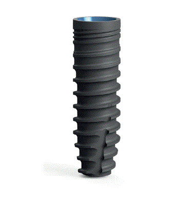
Figure 2: BioniQ implant - straight 3,5 x 12 mm (bone level).

Figure 3: BioniQ implant - straight 4 x 12 mm (bone level).

Figure 4: BioniQ implant - tapered 4 x 12 mm (bone level).

Figure 5: BioniQ implant - straight 5 x 12 mm (bone level).
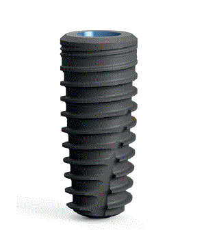
Figure 6: BioniQ implant - tapered 5 x 12 mm (bone level).

Figure 7: BioniQ implant - Straight Plus, C1.7, 4 x 12 mm (tissue level).
The study excluded patients with absolute contraindications to oral surgery, such as acute microbial or viral infections, acute leukemia, agranulocytosis, uncompensated hemorrhagic conditions, uncompensated diabetes mellitus, recent tumor ablative therapy of the orofacial region, tumor ablative therapy with bisphosphonates, and scleroderma. Patients with untreated periodontitis or those requiring immediate implant placement following tooth extraction were also excluded. Inclusion criteria encompassed healthy individuals or those with well-managed systemic diseases (e.g., diabetes mellitus, heart disease, hypertension, osteoporosis), smokers consuming up to 20 cigarettes per day, and cooperative patients demonstrating good oral hygiene. For patients with a history of treated periodontitis, the absence of periodontal pockets deeper than 5mm at the time of implantation and maintenance of good oral hygiene (Papilla Bleeding Index not exceeding 2 locally) were required.
In cases of local bone deficiency, appropriate implant types or bone augmentation techniques were employed. The study focused on primary implant failures observed during 2023, defined as spontaneous implant loss, inflammatory complications necessitating premature implant removal, or failure to achieve osseointegration (detected by implant rotation during healing cap removal). All cases of failure occurred prior to prosthetic loading.
All implants were placed using a standardized protocol under local anesthesia (Supracain, Zentiva®) with antibiotic prophylaxis (Augmentin 1g, Glaxo Smith Kline® or Dalacin C 300 mg, Pfizer®). The antibiotic regimen varied based on the procedure: a single dose was administered for simple implantations, while a week-long course was prescribed for cases involving augmentation procedures. The open healing method was consistently employed, wherein a healing cap was placed post-implantation, and the gingiva was sutured to ensure tight contact with the cap's surface.
The duration of the healing period adhered to the manufacturer's recommendations, with consideration given to the implant location and any additional procedures performed. Upper jaw implants were allowed a minimum of 3 months for healing, while lower jaw implants required at least 2 months. In cases involving alveolar augmentation, the healing period was extended to 6-9 months, tailored to individual circumstances.
Following the prescribed healing period, prosthetic treatment commenced. This phase typically began with the removal of the healing cap and the subsequent application of an impression pin or scanning body. If implant mobility was detected at this stage, it was classified as a primary failure.
Statistical analyses were conducted using Statistica 12 software. A chi-square test was employed to evaluate the potential dependency between implant failure and implant diameter, with the significance level set at p < 0.05.
Between February 29, 2022, and November 2, 2023, a total of 468 BioniQ implants were placed. These included various diameters and lengths to accommodate different anatomical locations: 20 implants of 2.9 mm diameter (14 mm length) for upper lateral incisors and lower incisors; 42 implants of 3.5 mm diameter (12 and 14 mm lengths) for lower lateral incisors and premolars; 224 implants of 4 mm diameter (10, 12, and 14 mm lengths) for upper middle incisors, canines, and premolars; and 182 implants of 5 mm diameter (8, 10, and 12 mm lengths) for molar regions. Table 1 provides a detailed breakdown of implant types, characteristics, and primary failure rates.
| Specification | Shape | Diameter | Length | No. Of Implants |
No. of failed implants |
| S2.9 x 14 mm | S | 2.9 | 14 | 20 | 0 |
| S3.5 x 12 mm | S | 3.5 | 12 | 21 | 5 |
| S3.5 x 14 mm | S | 3.5 | 14 | 21 | 0 |
| T4 x 10 mm | T | 4 | 10 | 11 | 0 |
| T4 x 12 mm | T | 4 | 12 | 30 | 0 |
| T4 x 14 mm | T | 4 | 14 | 28 | 0 |
| S4 x 10 mm | S | 4 | 10 | 29 | 0 |
| S4 x 12 mm | S | 4 | 12 | 46 | 1 |
| S4 x 14 mm | S | 4 | 14 | 47 | 1 |
| T5 x 8 mm | T | 5 | 8 | 2 | 0 |
| T5 x 10 mm | T | 5 | 10 | 51 | 0 |
| T5 x 12 mm | T | 5 | 12 | 27 | 0 |
| S5 x 8 mm | S | 5 | 8 | 9 | 1 |
| S5 x 10 mm | S | 5 | 10 | 48 | 2 |
| S5 x 12 mm | S | 5 | 12 | 45 | 1 |
| C4 x10 mm | S | 4 | 10 | 15 | 0 |
| C4 x 12 mm | S | 4 | 12 | 18 | 0 |
Table 1: The number and type of inserted implants and number of primary failure.
Out of 468 implants, 11 experienced primary failure, resulting in a failure rate of 2.35%. The failures were distributed across implant diameters as follows: five 3.5 mm implants, two 4 mm implants, and four 5 mm implants. No failures were observed in the 2.9 mm diameter group. The dependence of implant diameter on primary non-healing was further statistically processed. Specifically, the implant diameters were 2.9 mm, 3.5 mm, 4 mm, and 5 mm. The characteristics are shown in table 2.
| Diameter | Successes | Failures | Total |
| 2.90 | 20 | 0 | 20 |
| 3.50 | 37 | 5 | 42 |
| 4.00 | 222 | 2 | 224 |
| 5.00 | 178 | 4 | 182 |
Table 2: The number of failures and successes for implant diameter.
Statistical analysis using the Chi-square test for independence yielded a Chi-Square statistic of 17.09 with a p-value of 0.000679, which is statistically significant at the 0.05 level.
Null Hypothesis (H0): The failure rate is independent of the implant diameter.
Alternative Hypothesis (Ha): The failure rate is dependent on the implant diameter.
In table 3 shows dates for the calculation of observed frequencies.
| Diameter | Successes | Failures | Failure rate % | Success rate % |
| 2.90 | 20 | 0 | 0.00 | 100.00 |
| 3.50 | 37 | 5 | 11.90 | 88.10 |
| 4.00 | 222 | 2 | 0.89 | 99.11 |
| 5.00 | 178 | 4 | 2.20 | 97.80 |
Table 3: The failure and success rate for implant diameter.
Chi-Square statistic
Χ2 =17.09
p=0.000679*
*Statistically significant at a 0.05 level
This result led to the rejection of the null hypothesis, indicating a significant association between implant diameter and failure rate. Further pairwise comparisons using Fisher's Exact Test (Table 4) revealed that the failure rate of 3.5 mm implants was significantly higher than that of 4.0 mm and 5.0 mm implants, while other comparisons showed no significant differences.
| Implant diameter | 2.9 | 3.5 | 4.00 | 5.00 |
| 2.90 | - | 0.165 | 1.000 | 1.000 |
| 3.50 | - | - | 0.0013* | 0.013* |
| 4.00 | - | - | - | 0.415 |
| 5.00 | - | - | - | - |
Table 4: Fisher’s Exact Test.
*Statistically significant at a 0.05 level.
The 11 patients who experienced primary implant failure comprised 8 men and 3 women, with an age range of 30 to 70 years (mean age: 56 years). Within this group, one patient had non-insulin dependent diabetes, three were occasional smokers, one had coronary artery disease, and two had hypertension. None were undergoing bisphosphonate treatment. Analysis of local conditions at implant sites revealed a clinical correlation with both quantitative and qualitative alveolar bone status. Four patients required bone augmentation procedures, and four presented with marginal alveolar bone supply for implant placement. Only three patients had no issues with local alveolar bone supply.
This comprehensive analysis of both implant and patient-related factors provides valuable insights into the multifactorial nature of primary implant failure, highlighting the importance of considering both technical and biological aspects in implant dentistry.
This retrospective study evaluated the success rate of BioniQ implants in primary osseointegration. All procedures were performed by an experienced surgeon, which likely contributed to a lower failure rate compared to less experienced practitioners. Research suggests that surgeons with less than 5 years of implant experience may have failure rates up to five times higher [11]. Despite the expertise involved, our failure rate still exceeded 2 percent, which is consistent with findings from other studies. Comparing our results to existing literature, we find our failure rate to be within expected ranges. Krisam reported primary failure in 9 of 186 implants (4.8%) [13], while Staedt observed failure in 293 of 9080 implants (3.2%) [14]. These studies demonstrate higher failure rates than ours, though it's important to note the considerable difference in sample sizes. In contrast, Lin's large-scale study of over 30,000 implants reported a remarkably low primary failure rate of 0.6% [15], highlighting the potential variability in outcomes across different clinical settings and sample sizes.
Regarding the effect of implant shape on primary failure, many studies suggest that implants with smaller diameters and lengths are more prone to primary non-healing [14,16,17]. Interestingly, our study found no primary failures in 2.9 mm diameter implants, which were used only at 14 mm length. However, we observed primary failure in five 3.5 mm diameter implants with a 12 mm length, making this type the most represented in the primary failure group. Due to the limited sample size of implants with this diameter, definitive conclusions cannot be drawn, emphasizing the need for larger-scale studies to validate these observations.
This discussion highlights the complex interplay of factors influencing implant success, from implant characteristics and surgical technique to sample size and study design. The variability in failure rates across different studies underscores the importance of considering multiple factors when evaluating implant success. Further research with larger sample sizes and more detailed analysis of implant characteristics is needed to draw more definitive conclusions about the factors contributing to primary implant failure, particularly regarding the impact of implant diameter and length on osseointegration success.
Sufficient qualitative and quantitative bone supply is fundamental for successful implant healing. In our study, we observed some interesting patterns related to implant diameter selection and alveolar bone conditions. Notably, 3.5 mm diameter implants were used in three cases where the alveolus was actually not sufficient for a 4-mm diameter implant without requiring horizontal alveolar augmentation. This decision to use a smaller diameter implant without augmentation could be considered risky and may have contributed to failure, potentially reflecting an error in clinical judgment.
In five other patients, augmentation techniques were employed to achieve sufficient alveolar volume. The choice of augmentation method typically depends on the specific site characteristics and the surgeon's experience with various procedures. However, it's important to note that several studies, including those by Olmedo-Gaia, Lin, and Chang [18,15,19], have reported negative effects of augmentation techniques on primary implant failure. While some researchers advocate for the quality of augmentation techniques, we cannot ignore the potential complications associated with bone restoration. These challenges may arise from the materials used, the operator's experience, and the individual patient's biological response.
If we were to exclude the eight unhealed implants associated with borderline alveolar volume or augmentation cases in our study, our failure rate would drop dramatically to 0.6%. However, this adjustment would be misleading and potentially inaccurate, as it would ignore important clinical scenarios that are common in practice. We must consider all cases, including those with borderline alveolar volume or requiring augmentation, to gain a comprehensive understanding of implant success rates.
It's worth noting that in only three cases where primary failure occurred, the adequacy of alveolar quality and quantity was not in question. This observation underscores the complexity of factors influencing implant success and highlights the need for careful consideration of local bone conditions in treatment planning.
These findings emphasize the critical importance of thorough preoperative assessment of alveolar bone quality and quantity, as well as judicious decision-making regarding implant size selection and the need for augmentation procedures. Future studies with larger sample sizes and more detailed analysis of local bone conditions could provide further insights into optimizing implant success rates in various clinical scenarios.
Our study found a primary failure rate of 2.35% for BioniQ implants, which is comparable to the current state of the published literature, supporting their reliability. BioniQ implants show a reliable performeance in terms of osseointegration succes rate. When implants affected by bone augmentation procedure are excluded, the success rate of osseointegration reaches 99,4% which represents top osseointegration performace. From a patient perspective, our findings suggest that the volume and quality of the alveolus at the implant site likely play a more significant role in implant success than overall health status. This emphasizes the critical importance of thorough preoperative assessment of local bone conditions and careful treatment planning.
Download Provisional PDF Here
Article Type: RESEARCH ARTICLE
Citation: Starosta M, Maďar R (2024) Retrospective Analysis of Primary Failure of Dental Implants (Set of 468 BioniQ Implants). Int J Dent Oral Health 10(3): dx.doi.org/10.16966/2378-7090.424
Copyright: © 2024 Starosta M, et al. This is an open-access article distributed under the terms of the Creative Commons Attribution License, which permits unrestricted use, distribution, and reproduction in any medium, provided the original author and source are credited.
Publication history:
All Sci Forschen Journals are Open Access