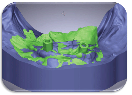
Figure 1: Patient IO scan superimposed on a patient model to locate the position of the implants concerning the physical model.

Mostafa Helmy Mostafa Ahmed1* Amr Hosney Elkhadem2
1Associate Professor of Prosthodontics, Faculty of Oral & Dental medicine, Cairo University, Cairo, Egypt*Corresponding author: Mostafa Helmy Mostafa Ahmed, Associate Professor of Prosthodontics, Faculty of Oral & Dental medicine, Cairo University, Cairo, Egypt, Tel: 00201001817812; E-mail: mostafa.helmy@dentistry.cu.edu.eg
Objectives: This study was performed to compare implant locators versus customized mandibular implant-retained overdentures considering patient satisfaction and prosthesis maintenance.
Materials and methods: 12 completely edentulous patients from the prosthodontic department-Cairo university outpatient clinic were selected according to specific inclusion criteria. The patients were then allocated randomly into two groups using closed envelopes.
For both groups, each patient received dental implants in the planned position. The patients were left for 3 months for implant healing. For group (A) the patients received two locator attachments. The attachment collar height was selected based on the soft tissue thickness keeping the retentive part 1 mm above the soft tissue level. The denture was relieved opposite the locator housing and the attachment was picked up using cold-cured resin.
For group B, the implants were exposed, and healing collars were placed. After 2 weeks, the implants were scanned using an intraoral scanner. The denture was readapted using a light body closed mouth impression. The relined denture was scanned from both the fitting and the occlusal side. The scanned fitting surface was used to create an inverted virtual model and preceded till creating custom locator attachments. The generated custom attachment abutment was milled from grade V titanium blanks using a 5-axis milling machine. The abutments were anodized into gold color using an electric anodizer. The attachments were then sterilized and placed in the patient mouth as set before in the design. The pick-up of the housing was done in the same manner as group A.
For both groups, the patients were given instructions for using and maintaining the overdenture. Initial denture corrections were made in the first 2 weeks if any complaint existed. The patients were recalled periodically at 3 months, 6 months, 9 months & 12 months respectively.
Results: Likert scale out of 5 was followed for patient denture satisfaction along 3, 6, 9, and 12 months for groups A & B. Applying an Independent t-test for significance evaluation between group A and group B, precluded the higher significance of group B than group A for all follow up as P-value <0.05.For prosthetic maintenance along twelve months follow-up, group B revealed a lower insignificant different incidence of prosthetic complications than group A using the Chi-square test for significance testing between proportions as P-value >0.05.
Conclusions: Customized locator attachments are highly recommended to retain mandibular overdentures, as they exhibited better maintenance values and more wear resistance with minimal loss of retention. Moreover, can be utilized by any presented implant system in the market, in addition, Patients were completely satisfied with their customized locators retaining their implant-supported overdentures.
Overdenture; Patient satisfaction; Locators; Dental implants; Attachments
Edentulous patients repeatedly experienced problems with their mandibular complete dentures; a treatment modality of employing two implants to support a mandibular overdenture has been advised to improve the retention & stability of the mandibular denture. Besides, maintaining healthy residual alveolar bone [1].
Numerous worldwide research studies examined the influence of implant-assisted overdentures on satisfaction and patient life quality and revealed that; individuals with implant-aided overdentures exhibited higher values of satisfaction and developed improved oral health quality than others with conventional dentures. Additional advancements involve a better chewing ability with the implantretained overdenture. Likewise, it has been recommended that implant placement to support an overdenture will maintain the remaining alveolar ridge [2-4].
Implant-assisted overdentures showed a simpler, cheaper, and more successful prosthetic solution in comparison to fixed restorations during the rehabilitation of edentulous mandibles.
Likewise, they provide enhanced masticatory function, and greater patient satisfaction, than traditional complete dentures [3].
The Preferability of any attachment system relies on the amount of retention needed, arch form, patient expectation, cost, and stress distribution over the implants and related surrounding tissue [4].
Systematic reviews evaluating implant-supported overdentures retained by utilizing a wide range of attachment mechanisms were recently published [5].
Rehabilitation of any edentulous arch necessitates a sufficient vertical space between the opposite arches to guarantee sample restorative material thickness, space for the retentive elements, and cleansability. The needed inter-arch space for an implant-assisted overdenture was determined from the implant shoulder towards the incisal edge and revealed nearly 12-14mm [6]. Moreover, 2- 3 millimeters of soft tissue thickness is generally present above the implant. In various height mucosal thicknesses, locator attachments dispersed the load better in comparison to the ball attachments of the implant and its surrounding structures [7].
Deficient inter-arch space was observed to be one of the key factors of the acrylic denture base mass fracture.
Additionally, it might precede unacceptable positioning of the denture teeth with consequent esthetic and phonetic hazards [8].
The implant-assisted treatment option could be manifested as splinted implants (e.g., bar-retained overdentures), or un-splinted implants (as in the case of the ball, locator, or magnetic implant attachments). Due to the smaller space requirements, cleansability, higher economical achievement, and lower technique sensitivity; un-splinted attachments have been preferred more than splinted attachments [9].
The most popular maintenance necessity of every overdenture attachment is noticed to be the renewal or replacement of its retentive element. Additionally, attachment systems show signs of wear during function, with consequent decrease and even loss of retention [10].
Outcomes recommend that; depending on the attachment system employed, the degree of patient satisfaction is directly influenced by the amount of retention and stability of the overlying implantsupported overdenture [11].
Locators presented the lowest profile of the presently available stud attachments. Besides, they propose simplicity and reasonable space requirements. Also, they provide double-retention (gained through inner and outer contact surfaces between their male and female parts) and after all, easily manipulated by the patient with less costeffectiveness [12]. Furthermore, locator attachments are presented in the market with numerous vertical heights, they are resilient, retentive, durable, and have some built-in angulation compensation. Additionally, repair and replacement are faster and easier than others. It is also possible to integrate the present denture into the new prosthesis [13].
Nowadays, the need for any customized implant-prosthetic component to be utilized by any presented implant system in the market has been widely increased, in addition, Patients will be completely satisfied when finding very simple, affordable customized solutions for retaining their implant-supported overdentures [14].
12 completely edentulous patients from the prosthodontic department-Cairo university outpatient clinic were selected. The inclusion criteria included patients with sufficient inter-foraminal bone volume and class I skeletal relationship. Patients with uncontrolled diabetes (HbA1c >7) were excluded. The patients were then allocated randomly into two groups using closed envelopes.
All patients were knowledgeable about the treatment plan and asked for approval on it with written consent forms according to the ethical principles stated in human studies approved by the ethical committee department-Cairo university & signed by the patient himself.
For both groups, after complete denture construction, a CBCT was taken, and the implants were planned to be placed in the lateral incisor/canine region bilaterally. Each patient received two 3.7 × 12 mm implants (S-clean, Dentis, South Korea) in the planned position. The patients were left for 3 months for implant healing (Figure 1).
Figure 1: Patient IO scan superimposed on a patient model to locate the position of the implants concerning the physical model.
For group A the patients received two locator attachments. The attachment collar height was selected based on the soft tissue thickness keeping the retentive part 1 mm above the soft tissue level (Figure 2). The denture was relieved opposite the locator housing and the attachment was picked up using cold-cured resin.
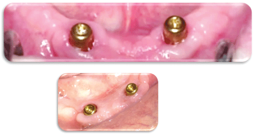
Figure 2: Patients of the group (A) received two locator attachments.
For group B, the implants were exposed, and healing collars were placed. After 2 weeks, the implants were scanned using an intraoral scanner (Medit i700, Medit Corp, South Korea). The denture was readapted using a light body closed mouth impression. The relined denture was scanned from both the fitting and the occlusal side. The scanned fitting surface was used to create an inverted virtual model. The scan of the implant scan bodies was aligned over the virtual model using best surface matching (Figure 3).
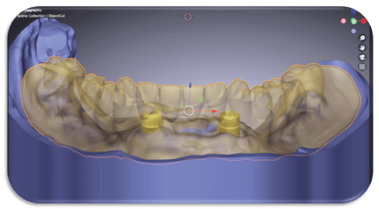
Figure 3: Patient wax up superimposed to the model in a transparent model to estimate the position of the attachment.
The scan bodies were used to place virtual analogues and connections in the recorded implant position (Figure 4).
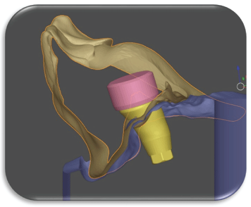
Figure 4: Cross-sectional view to adjust the custom attachment and housing position in the denture body.
Using a blender for dental software a custom low-profile attachment was designed. The overall height is 3.5 mm having a 0.25 mm circumferential undercut on its top (Figure 5).
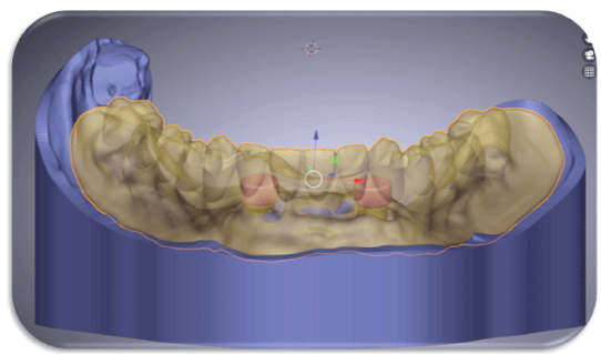
Figure 5: Assessment of the attachment and housing related to the denture in transparent mode.
A retentive cap was also designed with 70 microns tolerance (Figure 6). The attachments were joined to the implant connection using the software crown implant module (Figure 7).
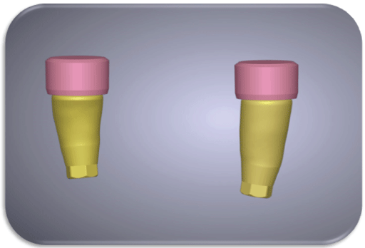
Figure 6: The custom attachment and housing final design.
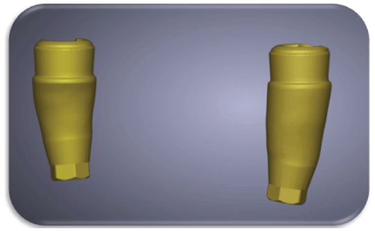
Figure 7: The custom attachments joined to the implant connection to achieve perfect parallelism in the same vertical position.
The attachments were placed virtually in such a way that both were parallel and had the same vertical level (Figure 8).
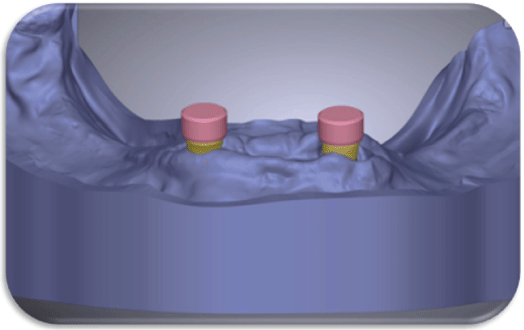
Figure 8: The custom attachments are located at the same vertical position with the housing placed at least 1 mm above the soft tissue level.
The screw access for the abutment screw was then opened using the screw whole function of the software (Figure 9). Afterward, the generated custom attachment abutment was milled using a 5-axis milling machine (Emar 5X, Egypt). The abutments were milled from grade V titanium blanks (Dentaurum, Germany). The abutments were anodized into gold color using an electric anodizer. The attachments were then sterilized and placed in the patient mouth as set before in the design (Figures 10 and 11).
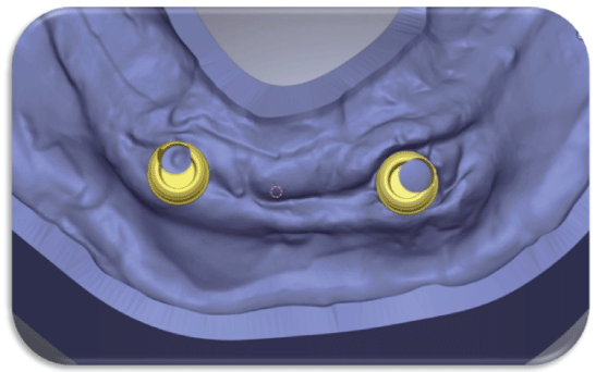
Figure 9: The position of the screw access hole in the attachment design will vary based on the implant position.
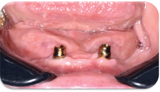
Figure 10: The milled custom attachment (front view).
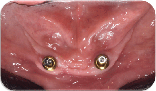
Figure 11: The milled custom attachment (occlusal view).
The pickup of the housing was done in the same manner as group A (Figures 12-14).
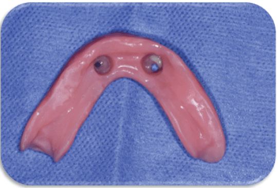
Figure 12: 3D printed denture with sockets for the housing pickup.
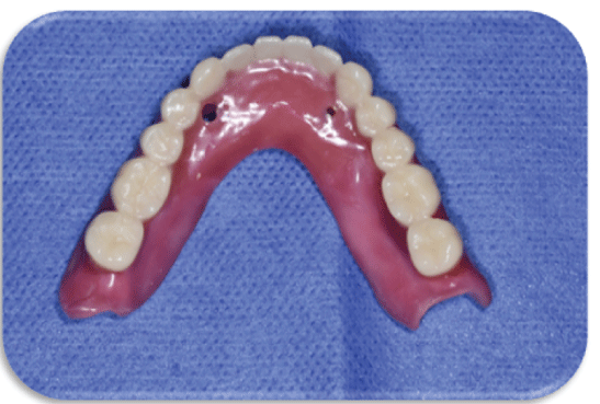
Figure 13: 3D printed denture with lingual vents ready for pickup.
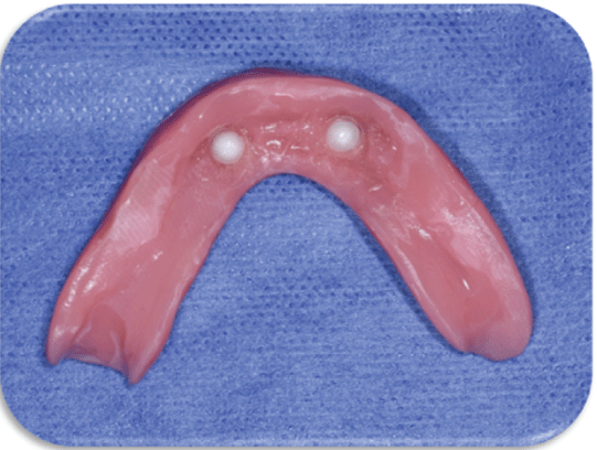
Figure 14: Overdenture after pickup of the custom housing.
For both groups, the patients were given instructions for using and maintaining the overdenture. Initial denture corrections were made in the first 2 weeks if any complaint existed. The patient was recalled periodically at 3 months, 6 months, 9 months & 12 months respectively.
A continuous response variable from separate control and experimental individuals was designed for the study, with one control subject for every experimental subject. In a previous study [15], the responses within each subject group had a standard deviation of 0.08 and were normally distributed. To be able to reject the null hypothesis that the population means of the experimental and control groups are equal with probability (power) 0.8, we need to analyze 6 experimental participants and 6 controls, assuming that the real difference between the experimental and control means is 0.15. The likelihood of a Type I error in this test of the null hypothesis is 0.05 [15].
SPSS 20®, GraphPad Prism®, and Microsoft Excel 2016 were used for the statistical analysis. The means and standard deviations (SD) of the data.
Likert scale out of 5 was followed for patient denture satisfaction along three, six, nine, and twelve months for group A and group B. Mean ± standard deviation of fast obtaining of the satisfactory prosthesis after three months for group A and group B were (2.45 ± 0.25) and (4.28 ± 0.19) respectively. After six months, the mean ± standard deviation for group A and group B were (2.26 ± 0.36) and (4.57 ± 0.74) respectively. While after nine months, the mean ± standard deviation for group A and group B were (2.84 ± 0.94) and (4.39 ± 0.66) respectively. Finally, after twelve months, the mean ± standard deviation for group A and group B were (2.67 ± 0.72) and (4.92 ± 0.28) respectively, all listed the table 1. Using an Independent t-test for significance evaluation between group A and group B revealed the higher significance of group B than group A for follow-up as P-value <0.05, listed in the table (1) (Graphs 1-3).

Graph 1: Bar chart showing patient Denture Satisfaction via Likert Scale at different intervals.
Graph 2: Bar chart showing different Wear Events along Different Overdenture Groups.
Graph 3: Bar chart showing the distribution of Prosthetic Maintenance for Different Attachment Systems.
| Group A | Group B | P-value | ||||
| M | SD | M | SD | |||
| Fast Obtaining of Satisfactory Prosthesis | Three Months | 2.54 | 0.25 | 4.28 | 0.19 | <0.0001** |
| Six Months | 2.26 | 0.36 | 4.57 | 0.74 | <0.0001** | |
| Nine Months | 2.84 | 0.94 | 4.39 | 0.66 | 0.0079* | |
| Twelve Months | 2.67 | 0.72 | 4.92 | 0.28 | <0.0001** | |
| Simplicity of Technique | Three Months | 1.78 | 0.16 | 3.87 | 0.53 | <0.0001** |
| Six Months | 1.96 | 0.24 | 3.52 | 0.29 | <0.0001** | |
| Nine Months | 1.58 | 0.49 | 3.94 | 0.64 | <0.0001** | |
| Twelve Months | 1.73 | 0.76 | 3.26 | 0.59 | 0.003* | |
| Longevity of the Restoration | Three Months | 1.25 | 0.36 | 4.45 | 0.18 | <0.0001** |
| Six Months | 1.66 | 0.27 | 4.83 | 0.93 | <0.0001** | |
| Nine Months | 1.18 | 0.51 | 4.29 | 0.56 | <0.0001** | |
| Twelve Months | 1.63 | 0.73 | 4.97 | 0.39 | <0.0001** | |
Table 1: The Mean ± Standard Deviation of Patient Denture Satisfaction
via Likert Scale.
M: Mean; SD: Standard Deviation; P: Probability Level
*Significant Difference P<0.05 using Independent T-test
**Highly Significant Difference P<0.0001 using Independent T-test
For simplicity technique, after three months for group A and group B were (1.78 ± 0.16) and (3.87 ± 0.53) respectively. After six months, the mean ± standard deviation for group A and group B were (1.96 ± 0.24) and (3.52 ± 0.29) respectively. While after nine months, the mean ± standard deviation for group A and group B were (1.58 ± 0.49) and (3.94 ± 0.64) respectively. Finally, after twelve months, the mean ± standard deviation for group A and group B were (1.73 ± 0.76) and (3.26 ± 0.59) respectively, all subscribed in the table 1. Using the Independent t-test for significance evaluation between group A and group B revealed the higher significance of group B than group A for all follow up as P-value < 0.05, listed in table 1.
For longevity of the restoration, after three months for group A and group B were (1.25 ± 0.36) and (4.45 ± 0.18) respectively. After six months, the mean ± standard deviation for group A and group B were (1.66 ± 0.27) and (4.83 ± 0.93) respectively. While after nine months, the mean ± standard deviation for group A and group B were (1.18 ± 0.51) and (4.29 ± 0.56) respectively. Finally, after twelve months, the mean ± standard deviation for group A and group B were (1.63 ± 0.73) and (4.97 ± 0.39) respectively, all listed the table 1. Using an Independent t-test for significance evaluation between group A and group B revealed the higher significance of group B than group A for follow-up as P-value < 0.05, listed in the table 1.
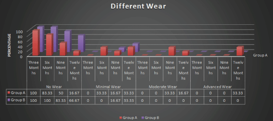
Different wear events were scored as “no wear”, minimal wear”, “moderate wear” and “advanced wear” along three, six, nine, and twelve months for group A and group B. Counts and percentages of “no wear” were 6 (100%) for both groups after three months. After six months, they were revealed 5 (83.33%) and 6 (100%) respectively. While after nine months, counts and percentages of “no wear” were 3 (50%) and 5 (83.33%) respectively. Finally, after twelve months, they were revealed 1 (16.67%) and 4 (66.67%) respectively. Using the Chisquare test for significance evaluation between proportions, it was revealed a higher insignificant difference of group B than group A for all follow up of “no wear” as P-value >0.05. In contrast to other scores of wear events, group A revealed a higher insignificant difference than group B as P-value >0.05, listed in the table 2.
| Group A | Group B | P-value | ||||
| N | % | N | % | |||
| No Wear | Three Months | 6 | 100.00 | 6 | 100.00 | --------- |
| Six Months | 5 | 83.33 | 6 | 100.00 | 0.3173 NS | |
| Nine Months | 3 | 50.00 | 5 | 83.33 | 0.241 NS | |
| Twelve Months | 1 | 16.67 | 4 | 66.67 | 0.0926 NS | |
| Minimal Wear | Three Months | 0 | 0.00 | 0 | 0.00 | --------- |
| Six Months | 2 | 33.33 | 0 | 0.00 | 0.1380 NS | |
| Nine Months | 1 | 16.67 | 1 | 16.67 | --------- | |
| Twelve Months | 2 | 33.33 | 2 | 33.33 | --------- | |
| Moderate Wear | Three Months | 0 | 0.00 | 0 | 0.00 | --------- |
| Six Months | 0 | 0.00 | 0 | 0.00 | --------- | |
| Nine Months | 2 | 33.33 | 0 | 0.00 | 0.1380 NS | |
| Twelve Months | 1 | 16.67 | 0 | 0.00 | 0.3173 NS | |
| Advanced Wear | Three Months | 0 | 0.00 | 0 | 0.00 | --------- |
| Six Months | 0 | 0.00 | 0 | 0.00 | --------- | |
| Nine Months | 0 | 0.00 | 0 | 0.00 | --------- | |
| Twelve Months | 2 | 33.33 | 0 | 0.00 | 0.1380 NS | |
Table 2: Count and Percentages of Different Wear Events along Different
Overdenture Groups.
N: Count; %: Percentage; P: Probability Level
NS: Insignificant Difference P>0.05 using Chi-Square test
*Significant Difference P<0.05 using Chi-Square test
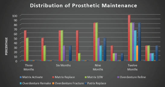
For prosthetic maintenance along twelve months follow-up, group B revealed a lower insignificant different incidence of prosthetic complications than group A using the Chi-square test for significance testing between proportions as P-value >0.05 except after nine months which were significant differences for wear and dislodgement and after twelve months for matrix replacement and overdenture relining which were significantly different as P-value <0.05, listed in the table 3.
| N (%) | Three Months | P-value | Six Months | P-value | Nine Months | P-value | Twelve Months | P-value | |||||
| Group A | Group B | Group A | Group B | Group A | Group B | Group A | Group B | ||||||
| Matrix | Activate | 0 (0%) | 0 (0%) | ---- | 0 (0%) | 0 (0%) | ---- | 0 (0%) | 0 (0%) | ---- | 0 (0%) | 0 (0%) | ---- |
| Replace | 4 (66.67%) | 3 (50%) | 0.5750 NS | 4 (66.67%) | 4 (66.67%) | ---- | 5 (83.33%) | 2 (33.33%) | 0.0926 NS | 6 (100%) | 2 (33.33%) | 0.0190* | |
| D/W | 3 (50%) | 2 (33.33%) | 0.5750 NS | 4 (66.67%) | 1 (16.67%) | 0.0926 NS | 5 (83.33%) | 1 (16.67%) | 0.027* | 5 (83.33%) | 2 (33.33%) | 0.0926 NS | |
| Over Denture | Reline | 0 (0%) | 0 (0%) | ---- | 2 (33.33%) | 0 (0%) | 0.1380 NS | 3 (50%) | 1 (16.67%) | 0.2410 NS | 5 (83.33%) | 1 (16.67%) | 0.027* |
| Remake | 0 (0%) | 0 (0%) | ---- | 0 (0%) | 0 (0%) | ---- | 2 (33.33%) | 0 (0%) | 0.1380 NS | 4 (66.67%) | 1 (16.67%) | 0.0926 NS | |
| Fracture | 0 (0%) | 0 (0%) | ---- | 0 (0%) | 0 (0%) | ---- | 0 (0%) | 0 (0%) | ---- | 2 (33.33%) | 0 (0%) | 0.1380 NS | |
| Patrix | Replace | 0 (0%) | 0 (0%) | ---- | 2 | 0 (0%) | 0.1380 NS | 3 (50%) | 1 (16.67%) | 0.2410 NS | 5 (83.33%) | 2 (33.33%) | 0.0926 NS |
Table 3: No. and Distribution of Prosthetic Maintenance for Different Attachment systems.
N: Count; %: Percentage; P: Probability Level; D/W: Dislodged and Worn
NS: Insignificant Difference P>0.05 using Chi-Square test
*Significant Difference P<0.05 using Chi-Square test
In the current study, all factors that might influence the osseointegration of implants were meticulously considered during patient selection and later after restoration. Those factors could be biological or mechanical or even both; The biological factors could be related to the patient's selection, the procedures of implant instalments, and the degree of oral hygiene measures afterward [16].
Twelve fully edentulous, healthy patients of age ranging from 45-60 years old were included in this study to keep away from any fluctuation in bone changes that might affect the resultant outcomes [17].
Maintenance of good oral hygiene has a great influence on the success of this study to the extent that it has a great impact on the osseointegration process. The oral hygiene of each patient was, therefore, evaluated at the beginning of the study and then throughout the whole investigation period [18].
Patients with superior general health were only selected, to avoid the reflection of any systemic disorder on the bone condition, and hence, osseointegration [19].
Merely cases exhibiting normal maxilla-mandibular relation were included in the study to avoid the effect of transmission of unfavorable loads onto the implants [20].
Heavy smokers were omitted as smoking is considered an essential factor in early implant failure due to anoxia of the oral cavity altogether with a considerable increase in plaque accumulation and calculus deposits, as recommended by numerous authors [21].
Uncooperative patients were eliminated from the investigation, whereas only cooperative patients were incorporated into the study to guarantee their dedication to oral hygiene measures and the regular follow-up visit [22].
Bone quality, as well as quantity, was assessed radiographically to make sure the primary stability of the implant at the time of placement. Furthermore, patients with appropriate bucco-lingual width at sites of implant placement were only chosen to ensure at least one mm. thickness of bone buccally & lingually surrounding the implant after its placement [23].
Provisional jaw relations have been made for the patients to emphasize adequate inter-arch space. Additionally, it helped in the determination of ridge relationship where patients only with Angle class Ị were incorporated in the study to facilitate implant insertion and preclude any possible implant overloading [24].
Regarding infection control throughout implant installation, preand post-surgical antibiotics and chlorohexidine mouthwash were imposed [25].
All used implants were threaded, self-tapping, root-form implants, 12 mm in length and 3.7 mm in width. This implant design was employed to ensure primary stability during the initial healing period, along with increasing the contact area between the implant and the surrounding bony structures for enhanced osseointegration [26].
Appropriate control of the frictional heat generation during the preparation of the implant bed was gently considered to avoid any possible necrosis of the surrounding bony cells which represents a major cause for osseointegration failure [1].
The covering screws were placed onto the implants to prevent the possible entrance of contaminants or food accumulation between the visits.
The cases were followed up for one year to ensure appropriate prosthesis maintenance as well as patient satisfaction throughout a suitable period.
The use of an intraoral scanner was a trial to facilitate the customization of locator attachment components in a simple, feasible & accurate method [27].
Oral rehabilitation with implant-assisted overdentures in totally edentulous mandibles presents a wide-ranging treatment modality not only based on the variable number of implants utilized but also relied on the assortment of different retentive options supplied [28].
the use of implant-assisted overdentures applying the Locator systems appears to be one of the ideal treatments in cases of edentulousim accompanied by retention problems while using a traditional removable prosthesis, especially in the mandible [29].
Treatment of a mandibular edentulism using two osseointegrated implants to support a mandibular overdenture with locator attachments is a recognizable treatment modality [30].
Throughout the recall periods of all patients, there were no complaints from the installed implant and all the patients followed the prescribed oral hygiene measures to overcome any hazardous effects which might influence the results of this study [31].
Several studies informed that the patient's quality of life expressed improvement by the increased retention and stability of their implantassisted overdentures [32].
Attachment adjustment was found to be the most frequent complication in implant overdenture [33]. The consequence of the angulation of the implant in overdenture retention or any possible deterioration appeared in the components of these systems by attachment and dis-attachment of the overdenture. These variables under review revealed the nature of complications most frequently associated [34]. With the utilization of customized locator attachments, the above-mentioned problem could be avoided and obtaining satisfactory implant parallelism. This in turn, enhanced the prosthetic maintenance of the whole prosthesis [35].
Both groups exhibited reduced retention values over the whole study period, which might be attributed to the wear of the retentive nylon inserts. But it was found that; group A revealed a higher insignificant difference than group B represented as a P-value >0.05 [36].
For prosthetic maintenance along twelve months follow-up, group B revealed a lower insignificant different incidence of prosthetic complications than group A, which might be attributed to better parallelism of customized locator attachments and less wear occurring.
Customized locator attachments are highly recommended to retain mandibular overdentures, as they exhibited better maintenance values and more wear resistance with minimal loss of retention. Moreover, can be utilized by any presented implant system in the market, in addition, Patients were completely satisfied with their customized locators retaining their implant-supported overdentures.
This clinical study was self-funded by the authors, with no conflict of interest.
Download Provisional PDF Here
Article Type: RESEARCH ARTICLE
Citation: Ahmed MHM, Elkhadem AH (2022) Patient Satisfaction and Maintenance Requirements of Classical Implant Locators versus Customized Attachments in Mandibular Implant-Retained Overdentures. Int J Dent Oral Health 8(4): dx.doi.org/10.16966/2378-7090.401
Copyright: © 2022 Ahmed MHM, et al. This is an open-access article distributed under the terms of the Creative Commons Attribution License, which permits unrestricted use, distribution, and reproduction in any medium, provided the original author and source are credited.
Publication history:
All Sci Forschen Journals are Open Access