
Figure 1: Non Carious Cervical Lesions (NCCL) different losses of dental structure with predominance of different factors: a-abrasion; b-erosion; c-abfraction-d; stress-corrosion.


Nelida Cunibert1 Guillermo Rossi2*
1Former Chair Associate Professor of Operative Dentistry II, Universidad del Salvador, Asociación Odontológica Argentina Buenos Aires, Argentina*Corresponding author: Guillermo Rossi, Chair Professor of Periodontics III, Universidad del Salvador, Asociación Odontológica Argentina, Buenos Aires, Argentina, Tel: 00-54-11-4-961-5619; E-mail: guinellrossi@fibertel.com.ar
The aims of this study is to demonstrate that not all the lost of dental structure is caused by acid neither the abrasivity of tooth brushing nor the toothpaste.
Abfraction is called the wedge-shaped lesion in the Cement-enamel Junction (CEJ) caused by eccentric occlusal forces that lead to the tooth bending.
An injury strictly of the cervical enamel that rebounds in dentine and cementum that causes the dental flexion in where periodontium plays an important role.
Although the processes of lost of dental structure are due to multifactorial causes, the action of the para funcional forces is acting in one or several pieces of the same sector.
During the development of the present study, different causes are mentioned and the reasons why not all the aetiology of non carious cervical lesions, are the response to the action of acids.
When the harmful force that generates the loss in wedge shape is present, the structure of the enamel is open giving rise to acid elements that become risk factors to trigger a greater loss of dental structure.
Non carious cervical lesions; Abfraction; Erosion; Abrasion; Stress corrosion; Wedge shaped lesions; Abfraction and biological width; Periodontal surgery; Abfraction and steps; Gingival fluid and erosion
The most prevalent mouth diseases are caries and periodontal diseases.
Science has focused on their prevention, thus the percentage of mouth diseases has decreased. Even though they have been controlled, nowadays the percentages of non-carious cervical lesions and loss of dental structure are increasing.
So are we paying attention to the loss of tooth structure in the same way that is made with carious lesions?
Food elaboration, ingestion habits and high stress rates to which men have been subject in the last decades has been contributing to the increase of range and type of loss of dental structures.
Represent the loss of dental structures detected in Cementumenamel Junctions (CEJ) appearing in various ways, with or without sensitivity, risking dental pulp. They are not caused by bacterial etiology
It is important to take into account that these lesions are being seen frequently, although most of them may remained unnoticed by professionals, and even more by patients.
Therefore, professionals should focus on their diagnosis, prevention and treatment.
Abrasion, erosion or corrosion, abfraction and its numerous combinations (Figures 1a-d, and 2a-b).

Figure 1: Non Carious Cervical Lesions (NCCL) different losses of dental structure with predominance of different factors: a-abrasion; b-erosion; c-abfraction-d; stress-corrosion.

Figure 2: Dental pieces prepared for MEB with different losses of dental structure.
a-abrasion
b-erosion
c-abfraction.
Even though erosion and abrasion can appear in the different tooth faces, through this investigation we will refer to those lesions that only appear in cervical thirds.
Erosion: is a chemical dissolution of hard tissues which is not related to bacterial plaque [1-4], but to intrinsically or extrinsicallyoriginated acid or chelating agents.
In this group of dental structure loss (NCCL) a lesion enunciated and defined by Grippo JO, et al. [5] called abfraction stands out. The term abfraction derives from the Latin root ab: division; fraction: division of fractions. (Figures 3a-b).
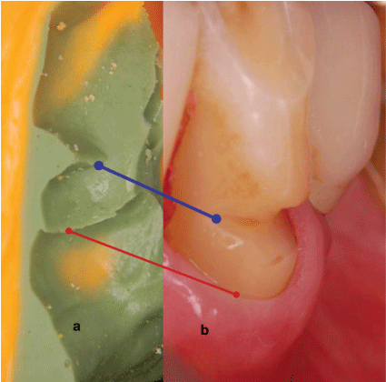
Figure 3: a-Impression of bottom of one abfraction.
b-Characteristic wedge shaped cervical lesion. The angled bottom is observed.
In 1984, Lee WC and Eakle WS [6] stated the dental flexion hypothesis.
Abfraction: “Abfraction is a wedge-shaped lesion appearing in the CEJ and caused by eccentrically applied occlusal forces that lead to dental flexion” Grippo JO, et al. [5].
Lee WC and Eakle WS [7] confirmed this hypothesis ten years later arguing that it comprised the breakage of crystal structures, enamel, cementum and dentin.
Grippo JO, et al. [8] and his team confirm his hypothesis about abfraction in 2012. Abfraction is most related to failure of cervical restorations and to hypersensitivity in treatment-refractory tooth necks when lesions show up.
When said lesions combine with non-bacterial acids, we are in the presence of stress corrosion.
We refer to chemical abrasion when abrasion and acid are combined, as in the case of patients with eating disorders -bulimia-who brush their teeth immediately after vomit.
Corrosion: Corrosion known as acid erosion implies losing teeth structure surfaces due to chemical action caused by demineralizing agents, specially chelating agents and non-bacterial acids [9-12].
Erosion shows a soft, slightly rough-like and opaque faulty surface.
As for corrosion and acid erosion etiology, intrinsic and extrinsic factors may be involved:
Extrinsic factors can be caused by:
Diet is a very important factor, as acid diets contribute to dental structure dissolution.
Intrinsic factors are divided in two big groups: somatic or involuntary intrinsic factors and psychosomatic or voluntary
Dentists play a leading role as they are almost the first ones to diagnose said symptoms due to enamel appearance (showing etched surface) or to evaluation of margins and surfaces of restorations.
Diluted enamel prisms may carry away more easily if teeth are brushed, turning into a combined lesion known as chemical abrasion.
Acid dissolves hydroxyapapite crystals due to the combination of acid hydrogen and enamel calcium. Thus, the brushing movement breaks prisms weakened by acid, making the situation even worst.
Previous investigations comprehend non-carious lesions in cervical thirds [13,14].
In the last conferences, expert’s statements have specially made reference to acid erosion.
Nowadays, almost all toothpaste advertisements focus on erosion as if it were the only responsible for hypersensitivity and loss of dental structure.
This statement demonstrates that acid is not the only responsible for wedge-shaped cervical lesions and that they can be caused by occlusal forces, among other factors.
It is true that the loss of dental structure caused by chelating agents and acid substances is related to in vivo or in vitro research projects which are experimentally and clinically proved.
That is why, abfraction, that is to say loss of dental structure in cervical thirds caused by non-axial forces leading to dental flexion, has not been as diffused as erosion has.
This is promoted due to the lack of in vivo and in vitro reproducibility of periodontal membrane. Moreover, the way patients apply their occlusal forces regarding direction, frequency and intensity may contribute to this deficiency.
Up-to-date published investigations aiming to clarify abfraction etiology include hinged models, force measurers, tension analysis, replication, computerized studies, 3D asymmetric finite elements, micro endoscopy, scanning electron microscopy and photo elasticity studies [15-18].
Through in this communication efforts are made to demonstrate clinically and reasonably that in non-carious cervical lesions, probably caused by erosion, loss of dental structure is not always caused by acid or chelating agents.
In the oral cavity, the loss of tooth structure is not exclusive of a single causal etiologic agent, they respond to a multifactorial etiology
It shall be exposed the reasons why authors argue that wedgeshaped cervical lesions do not react to brushing or acids or chelating agents and by contrast, dental flexion caused by lateral occlusal load may play an important role.
If brushing causes abfraction as mentioned in several investigations why does not the gingiva show any erosion if enamel and gingiva are different in hardness? In an improper brushing, the gingiva is eroded or ulcerated, but the tooth remains undamaged. Therefore the concept which the hard bristle brushing causes the cervical lesion should be replaced by the action of the abrasive toothpaste filled in soft bristles flexible may contribute to cervical lesion.
Consequently, NCCL may not be caused by abfraction but abrasion. Thus, this case confirms that brushing does not intervene in abfraction etiology.
As regards abrasion, loss of dental structure is not only found in one tooth. Lesions may also be evidenced in adjacent teeth, unless we refer to a tooth which can be reached by the toothbrush and is not aligned with the neighboring teeth.
It is important to remember that the appearance of an abrasion looks like a broad wear, with a shiny and polished surface.
It should be noted that in the case of subgingival abfractions the gingiva shall protect the dental structure from acid deletions and also from brushing abrasion, lesions which appear outside said area.
Brush bristles do not enter the pocket therefore subgingival steps that are often found in wedge shaped lesions are not a product of the brush.
The literature mentions the hardness of bristles; today dental practitioners already do not indicate hard bristle brushes.
Patients do not place bristles in the same tooth every time they brush their teeth although they may do so in the same area of the mouth. They neither brush their teeth with the same intensity, frequency, quantity and dilution of saliva in toothpaste.
To date investigations demonstrate that abfraction is originated in the enamel.
Investigations from Kuroe T, et al. [17] to those from Peck C, et al. [19] evidence that lesions move apically contributing to the loss of periodontal support, being less harmful.
Para function variations regarding position, direction and frequency of occlusal forces denote stress-marked changes in the cervical area (Figure 4) [20,21].
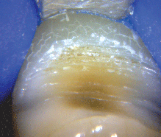
Figure 4: Modifications of dental tissues due to stress in the cervical zone. It is observed the fissure enamel and the bottom of the injury with steps and presence of calculus.
Thus, it is correct to state that abfraction changes from active to inactive, depending on parafunctional forces as bone crests reshape when trying to reestablish the biologic width (Figure 5).
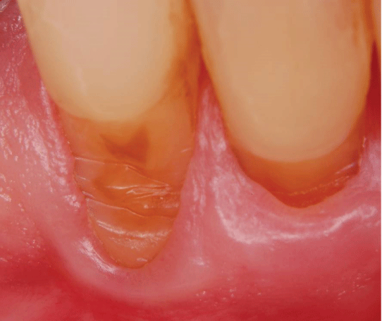
Figure 5: The lost of structure was discovered after a gingivectomy (intervention that is necessary to reveal the apical edge of the injury to be able to recover it). Is possible to observe the different moments of activity simultaneously and how periodontium migrated apically in its attempt to maintain the biologic width.
Periodontium and bone insertion level play an important role in the distribution of forces.
Due to the progressively loss of periodontal support, the lesion migrates apically showing supragingival steps as the gingiva retracts (Figure 6).
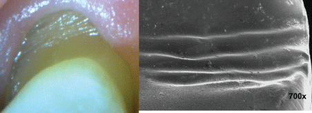
Figure 6: a-Invasion of the loss of the tooth structure to the gingival sulcus.
b-image of MEB showing the steps that indicate the presumed moments of force activity.
The emergence of steps on the basis of our hypothesis would be due to the possibility that the point of support (fulcrum) would remain unchanged where the thickness of the bone crest is greater. However, the resultant of forces does change and would be changed with the loss of periodontal support on the opposite side to the point of support (fulcrum), place where displayed the abfraction (Figure 7).
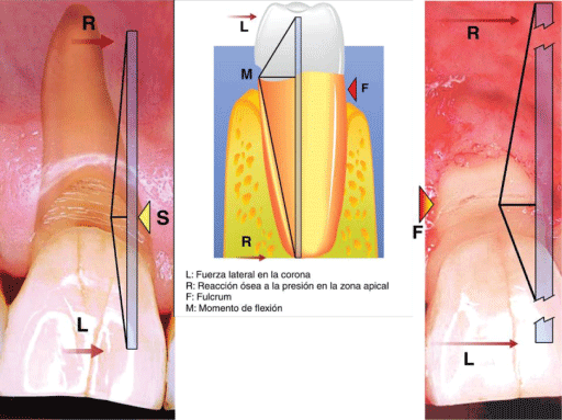
Figure 7: Scheme where seen how in the moment of the force, the fulcrum changes the position; it goes towards palatal and relies on the bone table and the tooth abfract by vestibular opposite to the place of support.
The loss of the periodontal support appears as a consequence of the original abfraction, which seeking to generate a reestablishment of the biologic periodontal width occupied by the NCCL, produces a nonbacterial inflammatory reaction resulting in bone loss of the alveolar ridge.
So, it can be stated that steps express the different periods of lesion activity caused by the occlusal overload [22].
However, it shall be proved if steps of the cementum are the result of different moments of activity, different fulcrums or cracks caused by the detachment of periodontal fibers [20,21]on the side where tension and tooth flexion occur.
Abfractions can be frequently observed in completely healthy surrounding gingivas.
In most cases, gingival margins of abfractions are evidenced at the gingival level or apical to it. Rarely, margins are supragingival.
Studies show that 32% of the abfractions have a subgingival apical limit (Figure 8).
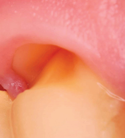
Figure 8: Invasion of the biological space creating a niche ideal to retain acid or fumes and act along with brushing causing a loss of multifactorial structure.
The margin of the subgingival abfraction invades the biological periodontal space approaching its margin at one distance less than 3 mm of bone margin. What deserves a proper restoration exhibit the same with a displaced flap apical and bone resection [20,21,23].
Thus, it can be stated that when NCCL are subgingival, they are not caused by erosion or abrasion, although it may be related to cusp flexion.
To date, researches have not been able to reproduce the role of periodontal ligaments by applying forces and dental flexion.
Schneider D, et al. [24] stated the criteria to decide if evidences of studies upon observation consider risk factors as a ground.
One of the principles is the support of experimental evidences which establishes that experimental reproduction of the disease must occur in animals (human beings when possible) exposed to risk factors.
Controlled laboratory studies, in which interventions are proved in order to prevent the disease or lesion, constitute strong evidences.
Therefore, it can be confirmed that Lilienfeld’s criteria based on experimental evidence are not fulfilled. The periodontal membrane has not been reproduced, nor its role in the application of forces regarding dental flexion as described in up-to-date published studies. So, for the time being it can be stated that it constitutes an experimental lesion based on clinical practice.
The biological space is the distance between the healthy gingival margin and crest bone. This space should be about 3 mm. The measure is approximately because can vary from tooth to tooth and even the different surfaces of the same tooth
This width can be observed in teeth with healthy periodontium.
This distance is relatively constant, in the case of being invaded there is a mechanism of bone remodelling of non-bacterial inflammatory type activated by the presence of margins subgingivals allowing periodontal tissues to recover the vital space, essentially in the form of gingival recession [25].
Recovery of biologic width is more frequent in thin bone crests; that is to say with a less than 0.5 mm thickness.
If the margin of abfraction occupies this area reducing the width, an inflammatory reaction and a resorption of the bone margin will occur. This shall be interpreted as a biologic reshaping evidenced to restore the biological distance.
Grounded on what has been previously stated and in the case of a healthy periodontium, the margin of the subgingival abfractions possibly occupies the biologic periodontal width.
Page R, et. al. [26] states that a periodontal destruction may begin and continue due to factors different to bacterial infection.
Mechanisms can be similar to periodontitis because of the production of matrix metalloproteinase and prostaglandins which can be released without the presence of bacterial plaque. These inflammatory mediators may be produced by cells which usually reside on periodontal tissues.
Fibroblasts and macrophages constitute a cellular population, predominant in healthy gingivas. These cells can activate and produce cytokines, prostaglandins and matrix metalloproteinases as a consequence of continuous or frequent aggressions like wrong oral hygiene.
Usual fibroblasts may also be taken as source of molecules that intervene in resorption of alveolar bone and destruction of connective tissue between periodontal ligament and gingiva.
When abfraction approaches its margin to the bone crest, it may activate fibroblasts and macrophages.
As the bone does not have spongy tissue but thin cortical bones, it undergoes a non-bacterial inflammatory process moving apically in an easy way.
Furthermore, many abfractions, consequence of loads, occur in canines and premolars which have a thin crest bone. Therefore, both cortical bones (periodontal and periosteal) join, almost without spongy tissue, leading to a quick loss of bone (Figure 9).
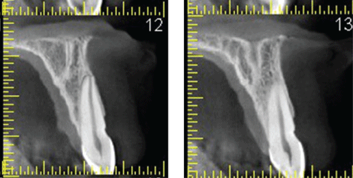
Figure 9: Acuitomo, seen the difference of tables, being the vestibular thinner with tiny spongy tissue between both cortical.
It should be recalled that, sometimes, predisposing genetic factors as bone fenestration and dehiscence may be the cause.
Bernhardt O, et al. [16], who we agree with, stated that upper premolars are the most injured teeth in 70.16% of the cases.
As a consequence, the biologic width is recovered; the gingiva joins the bone reshaping and thus, the gingival margin of the abfraction remains exposed.
So, these abfractions which show their margin at the level of the gingiva certainly undergo a reshaping process to become supragingival lesions.
If the lesion occupies the biologic width, abfraction appears before recession in the subgingival area [20,21,23]Thus, it may be confirmed that subgingival lesions cannot be caused by acid.
If in a normal depth groove neither saliva nor other mouth rinses can penetrate, it is unreasonable to think that acid or a toothbrush may do so. Saliva does not penetrate into deep grooves or into pockets as demonstrated in studies which determine the need to use irrigations of subgingival antiseptics substituting mouth rinses.
Taking into account an average volume of gingival fluid of 0.5 ml in usual groove, and a flow of 20 ml per hour [27], substances penetrating into this groove shall be diluted in a minute.
Fluid is renewed 33 times per hour in a healthy gingiva, once every two minutes, and 224 times when gingivitis appears, that is to say 4 times every minute [27].
It is unlikely that saliva penetrates into the gingival groove since fluid acts preventing this access or counteracting it by a simple cleansing action (Figure 10).
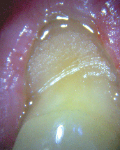
Figure 10: Presence of gingival fluid which would prevent the entry of acid to the inside.
Therefore, it is impossible that acids penetrate into the gingival groove as they do when the margin of the abfraction is subgingival. So, acid only acts at a supragingival level.
The percentage of calcium in gingival fluid is extremely low, so it is not enough to compensate the loss of dental structure caused by abfraction. Besides, saliva cannot act as a protector element when subgingival abfraction occurs.
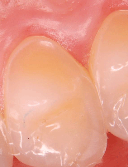
Figure 11: Patient with nutritional disorder. It is observed as in the gingival area the enamel remains intact.
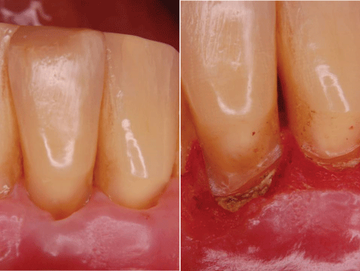
Figure 12: a-Injury covered by gum indicating a loss of subgingival structure.
b-Uncovered the loss of tissue through a periodontal surgery. Is observed the presence of calculus on the steps, therefore the loss do not correspond to the presence of acid.
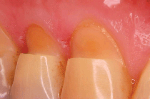
Figure 13: Abratcion tends to commit one or a few dental pieces. Can be seen in the cavo incisal edge its roughness and the inclination towards incisal of the injury.
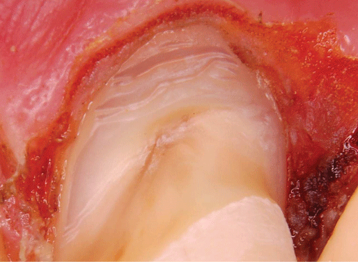
Figure 14: Periodontal Surgery: Surgical intervention that allows seeing the edge of the lost of dental structure, penetrating in the gingival sulcus, simultaneously that discovers entirely injury to restore it completely.
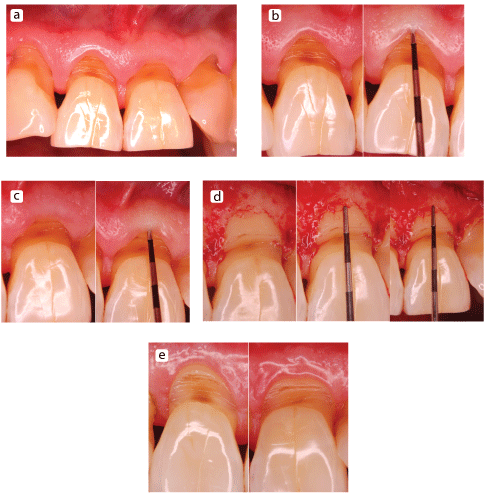
Figure 15: Flap displaced apical with bone resection.
a-clinical case, preoperative;
b and c-Probing the shallow depth of the gingival sulcus.
d-High flap. It is observed as the biological space has been invaded. With ostectomy it is recovered.
e-Postoperative. Margins of non-carious cervical lesion are exposed creating a suitable situation to isolate and to restore.
In order to take abfraction as pathology itself it is necessary to perform further investigations (particularly experimental) since it only comprises clinically and biomechanically proved lesions.
Before accepting or rejecting the hypothesis of the dental flexion as it causes of the abfraction, must be able to reproduce the periodontium, in such a way that experiences on models or asymmetric finite elements are more consistent with the real biology.
The important thing is not the occlusion but what the patient does with it.
To Dr Ricardo Macchi by it’s advising on the preparation of this work.
Download Provisional PDF Here
Article Type: CASE REPORT
Citation: Cuniberti N, Rossi G (2019) Abfraction-Myth or Reality? Why Some Wedge-shaped Cervical Lesions are not Caused by Acid Erosion? Int J Dent Oral Health 6(1): dx.doi.org/10.16966/2378-7090.309
Copyright: © 2019 Cuniberti N, et al. This is an open-access article distributed under the terms of the Creative Commons Attribution License, which permits unrestricted use, distribution, and reproduction in any medium, provided the original author and source are credited.
Publication history:
All Sci Forschen Journals are Open Access