Table 1: Age distribution of sample population


Amr M Bayoumi* Razan M Baabdullah Alaa F Bokhari Mohammed Nadershah
Department of Oral and Maxillofacial Surgery, Faculty of Dentistry, King Abdulaziz, Kingdom of Saudi Arabia*Corresponding author: Amr M Bayoumi, Department of Oral and Maxillofacial Surgery, Faculty of Dentistry, King Abdulaziz, Kingdom of Saudi Arabia, Tel: +966126400000; Fax: +9666403316; E-mail: Amrbayoumi@hotmail.com
Objective: The objective of this study was to determine the prevalence rate and pattern of third molar impaction and its association with age, gender, local, and racial factors.
Method and Materials: This was a retrospective cross-sectional study of patients treated at King Abdulaziz University Dental Hospital from January 2011 to September 2013. The inclusion criteria were; 1) age at least 18 years 2) complete third molars root formation 3) no history of third molar extraction 4) third molars are not congenitally missing 5) complete records with good quality orthopantomogram (OPG) 6) absence of any pathological dento-alveolar condition or craniofacial syndrome. Descriptive and bivariate statistics were computed and the P-value was set at 0.05
Results: The charts of 1866 patients were reviewed and only 580 patients were included. At least one impacted third molar was present in 77.1% of the included patients and there was no significant difference between different genders and nationality groups. Younger subjects showed higher prevalence rate. There was a statistical significant association between gender and angulation pattern. The predilection for mandibular impactions was higher than the maxilla, but not statistically significant.
Conclusion: The results of this study suggest that the prevalence rate of third molar impaction in Jeddah population is high. Future studies should evaluate the availability of resources and trained dental professionals to treat such a prevalent problem.
Third molar; Impaction; Prevalence; Impacted tooth
A tooth is considered impacted when it is unable to fully erupt into the oral cavity within its expected developmental time frame. It is a pathological condition in which a tooth or teeth cannot or will not erupt to its proper normal functioning position[1]. Third molar impaction is the most common among all teeth impactions [2]. The etiology of third molar impaction is multifactorial including genetic and local factors. Insufficient outgrowth of the retro molar space and the lack of space between the ramus and the second molar, differential growth between the mesial and distal roots are among the major causes of mandibular impactions [3-5].
Multiple classification systems exist for describing third molar impactions to estimate the difficulty of surgical extraction, aid in developing an optimal treatment plan, and minimize complications during surgery. The most commonly used classification systems are Winter’s [6] and Pell and Gregory’s [7] classifications. They classified the inclinations and positions of the third molars based on the relation among three factors; the dental longitudinal axis, occlusal plane, and the relationship to the anterior border of the ascending mandibular ramus [6,7]. Winter classified the impaction based on the angulation of the impacted third molar in relation to the long axis of the second molar [6].
Impactions of third molars have been associated with several pathological changes including pericoronitis, root resorption, periodontitis, and damage of the adjacent teeth, cystic lesions, and neoplasms [8]. Meisami et al. [9] reported that third molar impactions weakens the angle of the mandible and makes it more prone to fracture. It is also involved in the etiology of temporomandibular joint disorders, lower arch crowding, neuralgias, and vague orofacial pain [10,11].Chu et al. [12] reported that periodontal loss is more than 5 mm on the distal surface of the second molar adjacent to impacted mandibular third molar as well as caries (7%) among 2115 Chinese subjects. Moreover, daily and routine activities may also be affected in acute conditions attaining general health importance [13].
There is rarity in the literature reporting the prevalence of third molar impactions in Saudi [14-16]. The objective of this study was to estimate the prevalence rate, pattern, and distribution of third molar impaction among Jeddah population. Moreover, evaluate its association with age, gender, and nationality factors.
This cross-sectional retrospective study was approved by the Ethical Research Committee at King Abdulaziz University Dental Hospital in Jeddah, Saudi Arabia. It included review of 1866 digital orthopantomograms (OPG) of patients who attended the dental hospital from January 2011 to September 2013. The inclusion criteria were: 1) at least 18 years old 2)complete third molars root formation 3) no history of third molar extraction 4) third molars are not congenitally missing 5) complete records with good quality OPG 6) absence of any pathological dento-alveolar condition or craniofacial syndrome. Five hundred eighty digital OPG of patients aged at least 18 years were selected from these records including their demographic information related to age, gender, and nationality. Patient identifying data were not collected.
A third molar with fully formed roots and a level of eruption below the occlusal plane on OPG was considered impacted. Next, the depth of the impacted tooth in relation to the height of the adjacent second molar was evaluated. The impacted tooth was assigned to one of the three levels A, B, and C [13]. If the occlusal surface (OS) of the impacted tooth was at the level of the OS of the second molar, above it in the mandible, or below it in the maxilla, it is level A. Level B impaction is when the OS of the impacted tooth is between the OS and the cervical line of the second molar. If the OS of the impacted tooth was below the cervical line of the second molar in the mandible or above it in the maxilla, level C was assigned.
The width between the distal surface of the second molar and the anterior border of the ramus was also evaluated. Pell & Gregory classification included 3 classes; class 1: the mesiodistal diameter of the crown is completely anterior to the anterior border of the ascending ramus, class 2: the impacted tooth is positioned posterior so that approximately one half is covered by bone of the ascending ramus, and class 3: is when the impacted tooth located completely within the ascending ramus.
Furthermore, angulation type dependent upon Winter’s classification system was assessed measuring the angle formed between the long axis of impacted third molar to that of the second molar as described by Quek et al. [17] Two examiners conducted all radiographic assessments. Each of these examiners assessed twenty OPGs and the inter-rater reliability was tested using the Kappa reliability test.
IBM Statistical Package for the Social Sciences (Version 20; IBM SPSS) was used for statistical data analysis. Statistical tests used for the demographics were frequency distribution and simple descriptive statistics. To establish relationship between categorical variables, Chisquare Test was used as well as independent t-test to check the statistical significance difference in impaction between age groups means.
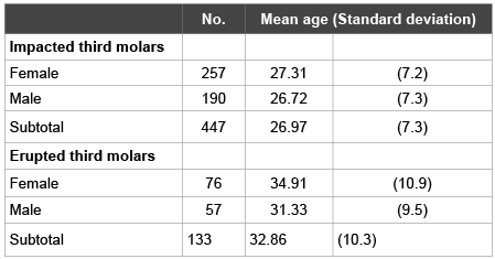
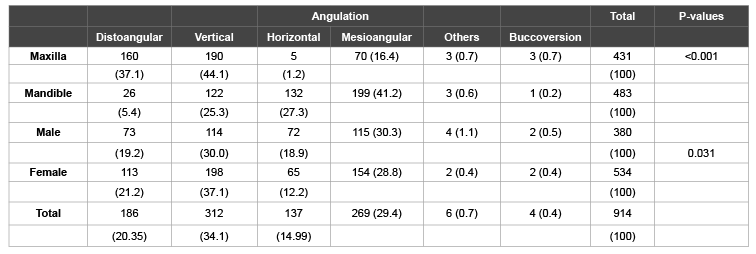
Inter-rater Kappa reliability test for radiological evaluation was 0.790 (p<0.001). Among 580 patients, a total of 447 (77.1%) presented with at least one impacted third molar. The mean age of subjects who presented with impaction was 26.97 years old 9 (Table 1). There was no significant difference between males (190; 42.5%) and females (257; 57.5%) (P=0.943), in the prevalence rate of third molar impaction, but they represented a significantly different results in the angulation as listed in table 2 (P=0.031). Likewise, there was no pivotal difference between Saudis and other nationalities in the prevalence rate of third molar impaction and in the pattern of impaction (Table 3). However, younger subjects had higher prevalence rate of third molar impaction with the mean age of 26.97 ± 7.2 year-old patients being highest, but this decreased with increasing age (mean age, 32.9 ± 10.3 years).

Table 1: Age distribution of sample population

Table 2: Distribution (%) of angulation of third molar impaction by arch and gender

Table 3: Distribution (%) of third molar impaction by gender and nationality
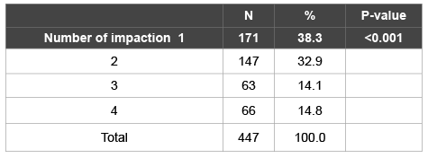
Of the 914 impacted third molars, the proportion of mandibular and maxillary third molar impactions was 52.8% and 47.2% respectively. Although mandibular impaction was more prevalent than maxillary impactions, there was no significant difference statistically (P=0.085). There was also no significant difference between right and left sides within each arch (p>0.429) (Table 4). The most frequent number of impacted third molar per subject was one (38.3%) and the least frequent numbers were four (14.8%) and three (14.1%) respectively (Table 5).

Table 4: Distribution (%) of third molar impaction by side

Table 5: Distribution (%) of third molar impaction by number of impactions
Vertical angulation was the most common angulation among all impacted teeth (34.1%). Angulation of impacted mandibular third molars was most commonly mesioangular (41.2%) followed by horizontal impaction (27.3%). Angulation of impacted maxillary third molar was most commonly vertical (44.1%) followed by distoangular impaction (37.1%) (Table 2). Level C and B impactions were the most common in the maxilla (53.6% and 40.8% respectively) while level A and B impactions were the most common in the mandible (47.6% and 42.2% respectively) (Table 6). On the other hand, class 1 and 2 were the most common, 45.3% and 45.5% respectively (Table 7).

Table 6: Distribution (%) of third molar impaction by level of impaction

Table 7: Distribution (%) of mandibular third molar impaction in relation to the ramus
Prevalence rate of third molar impaction in the present study was high (77.1%) when compared to the previous studies conducted in Saudi Arabia and other countries.
There are only few studies in Saudi Arabia reporting the prevalence of third molars impaction. Haidar and Shalhoub evaluated 1000 OPGs in the central region of Saudi Arabia and reported a prevalence rate of 31.9% for third molar impaction [14]. In 2010, Hassan conducted a similar study in Jeddah and showed a prevalence rate of 40.5% of the 1039 OPGs evaluated [15]. In 2013, Syed et al. [16] revelaed a prevalence rate of 18.8% in 3800 subjects in Asir region. The increased prevalence rate in the westren region could be related to the high ethnic and racial diversity residing in this region more than other areas of Saudi Arabia.
Racial factors were suggested to play a role in the prevalence of third molar impaction [14]. However, there was no statistical significant difference between Saudis and other nationals residing in Jeddah. This may be explained by the fact that Saudis in this area are of mixed race and ethnic backgrounds of Saudi nationals. Our results showed higher prevalence of third molar impaction compared to multiple studies in other countries [13-18].
A study conducted in Nigeria [19] found that the prevalence rate of third molar impaction in countries with high standards of living ranges from 9.5 to 25%. The prevalence rate in this study is close to what was reported in a study that examined 1000 OPG of a Singapore Chinese population (68.6%) [17] and to another study that showed impactions in 65.6% of 500 patients in USA population [20].
In this study, there was no gender predilection to the prevalence rate of third molar impaction similar to multiple studies in the literature [14,15,16,20]. However, there was a statistical significant difference in the angulation pattern of third molar impaction between males and females in our study. The gender-based distribution of third molar impaction was of statistical significance also in several other studies [13,17] not only in the prevalence rate of impaction, but also in the angulation patterns [18, 21,22] . One of the inclusion criterion in this research was being at least 18 years of age since most of growth is completed by age of 17 [23]. However, in a longitudinal study [24] over a period of 12 years, it was found that a change in the angulation of third molar is possible up to but not limited to 32 years old.
It was observed that the impaction prevalence rate decreases with increasing age. This finding is in agreement with Olasoji & Odusanya [19], Reddy [13], Garcia and Chauncey [25], and Bokhari et al. [16]. Many factors are associated with third molar impaction. Favorable path of eruption – depending on the tooth bud – affects third molars eruption [26]. Extraction therapy of premolars for orthodontic reasons improves the angulation of the third molar [27].
Other factors include the length of the mandible and the space available, the size of the third molar, insufficient eruption force, and skeletal class II [24,28-33]. The mandibular third molars are the most frequently impacted teeth [12,13,15,16,17,18]. Dachi and Howell [34] however, reported that maxillary impaction (21.9%) was more than mandibular impaction (17.5%) in a study included 1685 subjects at the University of Oregon. Kramer and Williams [35] examined 3745 OPGs and also agreed with this finding in which maxillary impactions represented 62.57% in comparison to mandibular impactions (47.4%) in an African American population. Although mandibular impaction (52.8%) in the present study was higher than maxillary impaction (47.2%), the difference was not statistically significant.
Since classification systems of angulation differ among different studies, there is no denying that it is difficult to compare angulations between studies. Vertical angulation was found to be the highest (34.1%) among all impacted teeth, which is in accordance with previous studies [13,14,36]. However, this is not coinciding with those of Hashemipour et al. [18] who reported that mesioangular impactions were the most common.
The most common angulation in the mandible was mesioangular (41.2%). This agrees with findings of Hassan [15], Quek et al. [17], Bokhari K et al. [16], Kramer and Williams [35], and Hashemipour et al. [18] Other study conducted by Bataineh et al. [37], however, reported that vertical impaction was the most common angulation impaction in the mandible.
Different method to classify the angulation was used by them; hence this could be the reason behind that. The present study shows that the most common angulation in the maxilla is vertical (44.1%) which is in agreement with other authors [15, 16, 17] but Kruger et al. [38] observed that mesioangular impaction was the most common among maxillary third molar impactions [38].
The C level impactions (53.6%) are most common seen impactions in the maxilla followed by B level (40.8%) as observed in this study. Quek et al. [17] reported that B level was the most common in both maxilla and mandible. However, they observed that level C impactions were more common in the maxilla than in the mandible and the difference was statistically significant, an interesting finding that was observed by Kanneppady [39] as well. Contrary to the level of impaction in the maxilla, level A impactions (47.6%) are most common seen impactions in the mandible followed by level B (42.2%) and this is in accordance with Hashemipour et al. [18] who observed that level A impaction was the most common. In 2014, Pillai and colleagues [40] found that level A impaction was most common in mandible and level C impaction was most common in maxilla.
Younger people had higher prevalence rate of third molar impaction than older people. Third molar impactions were observed more in the mandible than in the maxilla but the difference was not statistically significant. Vertical angulation of impaction was the most common angulation pattern. Level C impaction was the most frequent in the maxilla while level A impaction was the most frequent in the mandible. Further studies are in demand to evaluate the availability of resources and trained dental professionals to treat such a prevalent problem.
Download Provisional PDF Here
Article Type: Research Article
Citation: Bayoumi AM, Baabdullah R, Bokhari AF and Nadershah M (2016) The Prevalence Rate of Third Molar Impaction among Jeddah Population. Int J Dent Oral Health 2(4) doi http://dx.doi. org/10.16966/2378-7090.194
Copyright: © 2016 Bayoumi AM, et al. This is an open-access article distributed under the terms of the Creative Commons Attribution License, which permits unrestricted use, distribution, and reproduction in any medium, provided the original author and source are credited.
Publication history:
All Sci Forschen Journals are Open Access