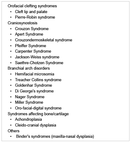
Table 1: A List of Craniofacial Anomalies reported in the Literature

Ahmad M Al-Tarawneh1* Raed H Alrbata1 Kholoud F Alazmi2 Khalid Hadaddin3
1M Clin Dent, JB Orth Royal Medical services Amman, Jordan*Corresponding author: Ahmad M Al-Tarawneh, 1M Clin Dent, JB Orth Royal Medical services Amman, Jordan, Tel: 009625713394; E-mail: tarawnehahmad@hotmail.com
A craniofacial malformation is an anomaly of embryonic development that results in a serious impairment of the normal anatomy of the head area for affected patients. A multidisciplinary, medical team approach is needed to successfully deal with such patients. Orthodontists play a major role in management of the craniofacial defects from the initial birth stages until skeletal growth maturation.
In this article, the most commonly encountered craniofacial anomalies related to the field of orthodontics will be discussed in groups with more focus will be given to the role of the orthodontists in the management of these anomalies.
Craniofacial; Anomaly; Orthodontics
A craniofacial malformation is an anomaly of embryonic development that results in a serious impairment of the normal anatomy of skull, jaws and adjacent soft tissues. Most of the malformations diagnosed at birth fall in the category “craniofacial” [1].
Children with craniofacial anomalies require a very detailed and unique medical support. Therefore, geneticists, surgeons, pediatricians, neurosurgeons, ENTs, orthodontists, ophthalmologists, speech therapists and many others who will take care of these patients should all have a very specific expertise in the field, because the problems of these patients often differ substantially from those of normal patients.
The orthodontist as one of the many specialists within the craniofacial team plays a major role in stabilization and optimization of the craniofacial defects from the initial birth stages until skeletal growth maturation. Systematic approaches of orthodontic management protocols, depending on the exact nature of the anomaly are needed to be followed to support skeletal, dental and soft tissue components. This was found to significantly improve the psychological status of the patients who normally suffer from, due to the presence of facial defects [2].
In this article, some of the most important craniofacial anomalies related to the field of orthodontics will be discussed shortly in groups. Greater emphasis will be given to the role of the orthodontists in the management of these anomalies.
A wide variety of craniofacial anomalies is reported in the literature with extensive lists of facial dysmorphology types (Table 1). The most common facial malformations are cleft lip and cleft palate. Less frequent are the syndromes of the I and II branchial arches and the forms more accurately called “craniofacial”, that primarily involve the midface and the skull; craniofacial synostosis.

Table 1: A List of Craniofacial Anomalies reported in the Literature
In this article, only the three main craniofacial anomalies groups will be presented in terms of facial and occlusal features accompanied with and the role of the orthodontist in the management as a member of the craniofacial medical team. Such management guidelines could be usefully applied for other anomalies, however, the exact pathology of the affected facial skeleton is needed to choose between the suitable and valid orthodontic treatment options.
Facial clefts may present as isolated cleft palate (CP), cleft lip (CL) or combination of both (CLP). These symptoms may be found unilateral, bilateral, isolated or as a part of complex conditions. Around 400 reported syndromes in which one of the symptoms is represented by clefting. Examples include; Van der Woude syndrome, Stickler syndrome, Treacher Collins syndrome and Pierre-Robin syndrome. The general orthodontic treatment of patients with CLP isdivided into four phases (Table 2).

Table 2: Orthodontic Treatment Phases for Clp Patients
For patients with unilateral CLP: Since the introduction of passive infant orthopaedics with acrylic plates by McNeil in 50s [3], through the 70s in which the Hotz plate [4] as a passive plate in hard acrylic was introduced, the use of passive plates with nasal stents [5] with or without tape, or the use of active plates, until now, an extremely controversial literature is encountered concerning the efficacy of these modalities.
Kozelj [6] in a retrospective study has demonstrated that the maxilla of CLP children subjected to infant orthopaedics may reach the same dimensions of five-year-old normal children, and that the plate allowed for a reduced deformity of nasal septum. On the contrary, Chan et al. [7] found no significant differences in the occlusal relationship in patients with orthopaedics or without.
A randomized comparative prospective clinical study by Parhl et al. [8] found that only differences in speech and palatal dimensions were found at age of 2.5 years for patients with orthopaedics. While either in feeding or in labial esthetics there was no difference at this age. At age of six years, no differences either in maxillary arch dimensions or in maxillary growth were found [9]. Also in terms of speech, although the burden to benefit ratio seems to be favorable to the use of orthopaedic plates, no differences any longer exist at age of 6 years [10].
For patients with bilateral CLP: The need for some presurgical orthopaedics in most bilateral CLP cases is universally accepted [11]. Severe protrusion of the maxilla which is usually accompanied with patients born with bilateral CLP is an important target for orthodontics treatment. The simplest technique is the use of a passive plate and an adhesive tape. However, as the premaxilla is slowly and physiologically retracted, tip of the nose is also retracted which might lead to flattening of the nose with a very short columella.
For this, Grayson and Cutting in 1996 [12] introduced naso-alveolar molding (NAM) protocol for primary columella lengthening along with gradual retraction of the premaxilla (two nasal stents supporting nostrils). Such protocol produces normal columella and a better nose projection, however, wider nose width and nasolabial angle is also accompanied and nasal anatomy is still not ideal [13,14].
Overall, presurgical infants orthopaedics using NAM protocol are of great benefits for the patients with bilateral CLP. However, for those with unilateral CLP, the use of such protocol is not mandatory. Supportive psychological therapy is needed for parents at this stage [1].
At this stage, only cross bite cases with mandibular shift should be targeted by simple measures like grinding of the premature contacts which caused the shift. Otherwise, waiting till the mixed dentition stage is preferred.
At this stage, correction of transverse discrepancies of the maxillary arch is often needed to create space for the alignment or eruption of permanent teeth or to prepare for secondary bone grafting if needed. Removable acrylic appliances might be not suitable and preferably be avoided because they may interfere with speech therapy the patients need. A Hyrax expander is the simplest method for parallel expansion of the two segments. If more anterior expansion is needed than posterior, a rigid “fan” expander is indicated. A quad helix expander may also be used as slow expansion. Such expansion of the palate should be normally performed as bone grafting is scheduled shortly.
An anterior crossbite related to an inadequate development in a postero-anterior direction of the maxillary bone is very frequent in CLP cases. The use of Face Mask as an external device for the maxillary protraction is one of the most traditional treatment methods. However, no scientific proof of the long term effect of this device on the growth of the maxilla. Nevertheless, such treatment option may be indicated in cases of mild skeletal severity, when there is an occlusal trauma generally at the level of central incisors, or for psychological purposes.
Management of dental anomalies especially those related to problems with number and shape of teeth is dependent on multiple factors. Anatomical factors, presence and severity of crowding, esthetic factors and financial aspects should be considered.
In summary, the main purpose of patients’ treatment at this stage is to prepare for alveolar bone grafting. Palatal arch expansion, preferably by Hyrax expander is the most effective protocol along with simple orthodontic mechanics to align erupting permanent teeth not only for functional reasons but also for the esthetic and psychological aspects for both patient and parents [1].
At this stage, orthodontic treatment may be definitive or in preparation for subsequent orthognathic surgery. CLP patients with severe skeletal discrepancies might get benefit from orthognathic surgery option. A patient with CLP, given the reduced nasal support and the insufficient thickness of the upper lip, is always more in need for extra maxillary support by maxillary advancement procedure utilizing Le Fort I osteotomy.
Some patients may present with severe palatal and labial scarring which may increase the risk of post-orthognathic surgical relapse [15]. In these patients distraction osteogenesis, especially in growing children, is needed which found to greatly improve the esthetic results.
Craniosynostosis is defined as a condition where there is premature fusion of one or more cranial sutures. Over 90 syndromes are known to be associated with this defect; roughly half of these have a mendelian modality of inheritance, with an autosomal transmission. For many of these syndromes, such as Crouzon, Apert, Pfeiffer and Saethre-Chotzen the genetic bases are well identified.
Crouzon’s Syndrome makes up approximately 4.8% of all cases of craniosynostosis, making it the most common syndrome of the more than 100 within the craniosynostosis group [16]. Facial and occlusal findings include midface hypoplasia, relative mandibular prognathism, class III malocclusion, narrow/high-arched palate, posterior bilateral crossbite and crowding of teeth [16,17].
Apert’s Syndrome is characterized by irregular craniosynostosis, midface hypoplasia, cleft palate, maxillary hypoplasia, high arched palate with bulbous palatal swellings,delayederuption,congenitally missing teeth, severe crowding and anterior open bite which is more challenging than that in the Crouzon type.
Compared to Crouzon’s and Apert’s syndromes, in Pfeiffer Syndrome, with synostosis of coronal sutures which leads to brachycephaly, the midface is usually more severely affected. Carpenter syndrome is the rarest, with only occasional patients seen, also share the facial and occlusal features with other CSF syndromes such as underdeveloped maxilla and/ or mandible and highly arched and narrow palate which make speech a very difficult skill to master.
Orthopaedic and orthodontic treatment of patients with craniofacial synostosis (CFS): In this group of craniofacial anomalies, sutural growth of the cranial base and maxillary- zygomatic complex is severely impaired [18,19] and there is mainly pathological appositional growth which leads to a significant vertical dento-alveolar growth. Consequently, maxillary orthopaedic treatment may not be suitably approached as in normal patients.
No data in the literature regarding the precise indications of when it is possible to expand the palate in a child affected by CFS. Ferraro et al. [20] suggested not performing rapid palatal expansion on patients with CFS. Schuster, [21] reported that such procedure might be only performed for patients younger than 5 years of age, only performing 2-3 mm expansion and then checking the actual expansion obtained with an occlusal X-ray of the palate. If expansion seems to be only dental, expansion appliance should be removed to avoid severe mobilization and early loss of primary teeth. Surgically assisted rapid palatal expansion may be then considered early.
The use of any device to stimulate the growth of the maxilla should be absolutely avoided in patients with CSF, as early congenital fusion of the cranial base and malar sutures is found in these patients. However, for patients received distraction osteogenesis of the midface with the use of an external device and for a reason or another, the distraction device was early removed; in these cases a face mask might be useful in the retention phase.
An important objective of the orthodontic treatment in CFS patients rather than improvement of dental aesthetics are to prepare the patient for future surgical steps required such as the need for Le Fort III and rigid external fixation, as close contact to surgeon is always required.
The vast majority of CSF patients have very severe skeletal open bite which necessitates orthognathic surgery at a time to posterior impact the maxilla with a clockwise rotation. Such procedure produces retroclination of maxillary incisors. For this, presurgical orthodontics should target the inclination of these teeth to be more proclined.
Crowding is generally such, that an extraction of permanent teeth is mostly needed. The need for surgical uncovering of teeth is very frequent and should be kept in mind.
Syndromes such as Hemifacial microsomia (HFM), Goldenhar syndrome and Treacher-Collins syndrome are related to deformities that derive from the first and second branchial arches. HFM affects the development of the lower half of the face, most commonly the ears, mouth and mandible, though it may also involve the eye, cheek, neck and other parts of the skull, as well as nerves and soft tissue. It is characterized by midface hypoplasia (usually asymmetric), mandibular hypoplasia, TMJ ankylosis, macrostomia and CL and/or CP.
Goldenhar Syndrome also known as Oculo-Auriculo-Vertebral syndrome is a rare congenital defect characterized by incomplete development of the ear, nose, soft palate, lip, and mandible. Other findings include V-shaped palate, severe class II malocclusion, increased mandibular plane angle, mandibular retrognathism and CL/CP.
In Treacher Collins Syndrome, malar and mandibular defects, convex facial profile, macrostomia due to lateral clefting and CP with or without CL and class II anterior open bite malocclusion are characteristically found.
Orthopaedic and orthodontic treatment of patients with 1st and 2nd branchial arch syndromes: As anomalies of this craniofacial group almost share the facial and occlusal features, orthodontic treatment options will be presented for HFM patients in the following section which could be systematically approached for other anomalies.
Orthopaedic treatment for patients with HFM: A great controversy in literature is found for this issue as it is the case for the mandibular orthopaedic treatment for otherwise normal growing patients with deficient mandible. According to the American association of orthodontics in 2005, no scientific proof that a functional appliance is able to modify mandibular growth [22].
A number of case reports regarding the response of HFM patients to functional simulation. Most of these cases are “Pseudo-HFM”, are misdiagnosed HFM patients with severe non-congenital mandibular asymmetries probably secondary to very early trauma [23,24].
According to Vargervik, [25] the true response of the patients with HFM to functional treatment is usually quite moderate and limited in time. In mild cases it is possible to solve an asymmetry with orthopaedic treatment obtaining mainly dentoalveolar compensation, accepting some degree of skeletal asymmetry. Some authors suggest application of asymmetrical or hybrid functional application which help maintain a less oblique occlusal plane and might stimulate the musculature on the affected side, whereby obtaining a better symmetry of the face.
Pre and post-surgical functional orthopaedics has been also suggested in order to increase stability of the surgical result when costo-chondral grafting is neededor pre and post distraction osteogenesis [26]. However, this procedure found to be not able to maintain the postsurgical mandibular skeletal symmetry at the long term [27].
Orthodontic treatment for patients with Hemifacial microsomia (HFM): Patients with HFM may present with maxillary crowding and constriction on the affected side. A rapid palatal expansion might be useful, considering the right geometry needed. The position of the midlines should be discussed with the surgeon who will perform the future osteotomies, in order to reduce the burden of care of the child and avoiding round tripping of teeth. In adults, orthodontic treatment is usually a preparation for orthognathic surgery and follows the same principles of presurgical orthodontics in asymmetries.
Orthodontic management for patients with craniofacial anomalies tends to be more complex, takes more time and clinical resources and should be based on a precise coordination with multiple dental, surgical and medical providers to achieve the best long-term esthetic and functional results.
As the orthodontic management is commonly needed prior to most surgical procedures associated with craniofacial anomalies, management protocols should be based on a precise understanding of the exact nature of the anomalies as certain mechanics may be provided efficiently, safely and with acceptable durability, while at the same time, other techniques might be not effectively applied with some complications.
Download Provisional PDF Here
Article Type: Review Article
Citation: Al-Tarawneh AM, Alrbata RH, Alazmi KF, Hadaddin K (2016) Orthodontic Management of Craniofacial Anomalies, a Summary of the Available Options. Int J Dent Oral Health 2(3): doi http://dx.doi. org/10.16966/2378-7090.184
Copyright: © 2016 Al-Tarawneh AM, et al. This is an open-access article distributed under the terms of the Creative Commons Attribution License, which permits unrestricted use, distribution, and reproduction in any medium, provided the original author and source are credited.
Publication history:
All Sci Forschen Journals are Open Access