Table 1: Age and gender wise distribution of subjects Values in parenthesis indicate percentage

Ravi Teja Chitturi* Kiran Kumar Kattapagar M Indira G Sridhar Reddy Chandrasekhar Poosarla Venkat Ramana Reddy Baddam
Department of Oral Pathology, SIBAR Institute of Dental Sciences, Guntur, India*Corresponding author: Dr. Ravi Teja Chitturi, Senior Lecturer, Dept of Oral Pathology, SIBAR institute of Dental Sciences, Guntur – 522509, India, Tel : +91-9676767387; E-mail: dr.raviteja@gmail.com
Background: In forensics estimation of age using dental findings has played a very important role over the years but its utilisation with regard to adult-juvenile conflict in court of law is very limited. This study has been done to estimate the age in this particular group using third molar radiographs in an Indian population.
Aim: To estimate the age of an individual using third molar radiographs in a South Indian population.
Materials and methods: The study was performed by using 140 digital orthopantamograms (OPG’s) of 70 males and 70 females of South Indian origin. The developmental stages of third molars according to Demirijan and modified by Kasper was used and the data was recorded. It was statistically analysed to derive a regression formula to estimate age and calculate the probability of an individual to be over various age groups.
Results: It was observed that in the south Indian population there was no significant difference between development of third molars in maxillary and mandibular arches as well as in the right and left sides.
Conclusion: Third molar radiographs can be considered a useful tool to estimate age of an individual especially in the court of law where there are conflicts of an individual being a major or not. Though conclusive results cannot be given using a single study, this data can be used as a reference for South Indian population belonging to the 18-25 years age group.
Forensic odontology; Age estimation; Third molar radiographs
Assessment of age is very important in several aspects as far as forensic medicine is concerned [1]. In this aspect estimation of age using dental findings and the branch of forensic odontology has played a very important role over the years [2]. Forensic odontologists all over the world have employed several methods to estimate age using several methods like changes that occur in the human skull and dentition. As far as the teeth are concerned several techniques have been employed such alteration in the morphology that are assessed by tooth sections, studying the developmental changes by radiographs and estimation of age by evaluating the teeth biochemically [3-5]. Studying the developmental stages by radiographs was introduced as early as 1973 by Demirjian et al. [4]. Since then various authors have successfully employed this technique to estimate age and third molar has been a precise and reliable indicator to asses age in unidentified cadavers and human remains and most importantly in living persons too to differentiate between adult and juvenile status in the court of law [6,7].
The court of law expects a very reliable method to estimate age and differentiate between an adult and juvenile. Various studies across the globe have shown that the third molar genesis is considerably varied among various ethnic groups. Hence, data from each country and ethnic group has to be recorded for forensic purposes [8-11]. The present study has been performed to estimate age using third molar radiographs. In this study we have attempted to estimate the age of an individual by using OPG’s and also record the probability of a south Indian individual to be over the age of 14, 16, 18 and 20 which is very important in the court of law.
The study was performed by using 140 digital orthopantamographs (OPG) collected from various private practitioners and diagnostic centres in Guntur, Andhra Pradesh, India. The OPG’s were of 70 male and 70 female patients who were native to Guntur district and aged between 14-23 years (Mean=19.12 ± 3.01) and for whom OPG’s were taken as a pre treatment radiographs for orthodontic purposes. Radiographs of patients for whom all the four wisdom teeth i.e 18, 28, 38 and 48 were present without any pathologies were alone included in the study. The four teeth were designated with letters for ease of use (18=UR, 28=UL, 38=LL, 48=LR). A detailed age and sex distribution of the patients has been presented in Table 1.

Table 1: Age and gender wise distribution of subjects Values in parenthesis indicate percentage
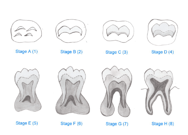
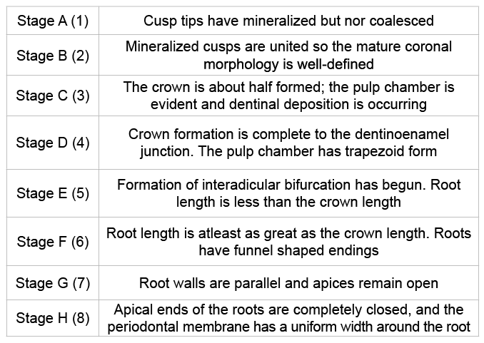
The developmental stages of all the third molar teeth were assessed by a modified Demirjian’s chart as proposed by Kasper et al. [11]. The stages were assigned from 1 to 8 (corresponding to A to H) as depicted in the schematic figure for convenience (Figure 1) and the development chart corresponding to each stage used to assess in this study has been given in Table 2. The scores were recorded by two observers independently who previously did not establish agreement regarding the classification of stages radio graphically. Intra observer differences were also recorded to determine the individual’s reliability by scoring the stages in a time interval of two months. Inter- and intra-observer agreements were analysed using weighted kappa and the scores were classified as a substantial agreement.

Figure 1: Schematic picture showing radiographic stages of tooth development given by Demirijian and modified by Kasper et al.

Table 2: Development chart of third molar development (as modified by Kasper et al.) [11]
The mean age for each developmental stage was calculated for all the wisdom teeth separately. Comparison between the development of upper and lower teeth were done using Mann-Whitney U test and the same statistical analysis was used to compare males and females too. Multiple regression formulas were obtained to calculate the age of both the sexes using the developmental stages of third molar teeth and also the probabilities of an individual being over the ages of 14, 16, 18 and 20 were also computed. All the statistics were performed using SPSS software (IBM, USA) for Windows 7 (Microsoft, USA).
The mean age of males and females in the study were 19.17 ± 3.02 and 19.07 ± 3.03 respectively. The mean age of the individual developmental stages of 18, 28, 38 and 48 for both males and females have been shown in Table 3. The formation process of all the four teeth were examined in both the sexes and the results have been computed in Table 4. There was no significant difference between the development of upper and lower teeth in both the sexes.
Also no significant differences were found when the development of each wisdom tooth was calculated between the males and females (Table 5). The probability of an individual being over the age of 14, 16, 18 and 20 was calculated based on the stage of development (Table 6). The probability of male being over the age 18 whose maxillary and mandibular third molar was in the developmental stage G was 28% and 33% respectively and for the females the probability was 33% and 40%. If the developmental stage was H the probability of males and females being over 18 was 74% and 86% for maxillary molars and 86% and 78% respectively for mandibular molars. Linear regression analysis was done to calculate multiple regression formulas to estimate age using the developmental score of the maxillary and mandibular teeth (Table 7).

Table 3: Mean ages (with 95% confidence intervals) of Demirjian’s stages UR- Maxillary right third molar, UL – Maxillary left third molar, LL – Mandibular left third molar, LR- Mandibular right third molar

Table 4: Comparison of third molar development in upper and lower jaws (Mann-Whitney U test) UR- Maxillary right third molar, UL – Maxillary left third molar, LL – Mandibular left third molar, LR- Mandibular right third molar

Table 5:Comparison of male and females with respect to third molar development in upper and lower jaws (Mann-Whitney U test) UR- Maxillary right third molar, UL – Maxillary left third molar, LL – Mandibular left third molar, LR- Mandibular right third molar

Table 6: Probability of an individual being older than 14, 16, 18, and 20 years estimated by third molar radiological examination Max – Maxillary, Mand - Mandibular
Estimation of age is one of the important aspects in forensics especially in the field of legal matters. Identification of a suspect or an accused being a juvenile or adult (i.e under or above 18 yrs) is very important in the court as punishment as well various other proceedings of the court varies for both [12,13]. Various methods have been employed to identify this particular age section such as skeletal and dental markers. Some of the skeletal markers that have been used are the use of length of the long bones, fusion of epiphyseal plate and spheno-occipital synchondrosis. Also use of bio chemical examination of bones has been used to estimate age. Some major disadvantages of these methods include environmental and genetic factors influencing the skeletal markers and inability to perform bio chemical and direct examination of bones in live individuals. The effect of genetic factors such as an individual’s normal growth pattern and environmental factors such as nutritional factors and chronic illness’ affects the skeletal markers more than the dental ones. Teeth are preserved longer than the bones and are highly resistant to chemical and thermal insults and thus providing it a reliable tool for age estimation [14-16].
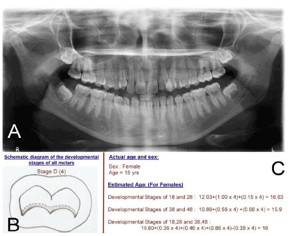
Of the dental markers, estimation of age using third molars is widely employed because of the well known fact that it is the only tooth that starts to calcify after the age of 14 and continues till after 18 [17-19]. Radiographic examination of developing third molars is a relatively easy technique to estimate age of an individual. In this study we used the Demirjian’s chart as modified by Kasper et al. (Figure 1) to study the developmental stages of third molar [11]. It is a simple, practical and widely employed method that comprises of clearly defined changes in shape that do not require speculative estimation. It has been shown to perform well in terms of both observer agreement and correlation between estimated and true age [10]. In our study, the difference between the estimated age and the actual age showed a standard error between 2.62 and 2.90 which is comparatively closer to the actual age as compared with other studies [20-22]. An example on how the regression formula was used to estimate age has been shown in Figure 2.
The present study was performed because estimation of age in various ethnic groups and populations is important for forensic purposes. The mean age of start development of third molars in our study population was similar to that seen in Japaneese and Turkish populations [8,10]. Also in our study there was no significant difference between the development of wisdom teeth well as the maxillary and mandibular teeth and right and left sides as seen in few studies done on Spanish population [20,21]. In the study done by Arany et al. and Orhan et al. there are significant differences between the development of maxillary and mandibular third molars and the study done on Spanish, Turkish and Japaneese individuals show that the development of molars in females takes place earlier than in the males [8,10,21]. Our study showed that there is no significant difference between development of third molars between males and females. Another important difference that was observed in our study is that the probability of an individual being over 18 when the developmental stage was H in the Indian population was only 74–86 % whereas in the Turkish population it was 97-99% , thus indicating the completion of third molar development occurred later than that of Turkish population. Thus, our study indicates that the third molars in a South Indian population are similar to people from Turkey and Japan as far as mean age of start of development is concerned and similar to Spanish population with regard to development in the jaws and each quadrant is concerned. However it differs from all the above populations with regard to development in sexes is concerned.

Table 7: Multiple regression formulas to estimate age using developmental stage of maxillary and mandibular third molars UR – Developmental stage of maxillary right third molar, UL- Developmental stage of maxillary left third molar, LR- Developmental stage of mandibular right third molar, LL – Developmental stage of mandibular left third molar

Figure 2: A- shows the OPG of patient with all developing third molars, B – shows the schematic diagram of the developmental stage all the third molars are in and actualage and sex of the patient, C – shows the calculation of estimated age of this particular patient using the developmental scores of upper third molars alone, lower third molars alone and upper and lower third molars combined.
As far as the studies from within Indian populations in considered, it was found that the there was a difference in the development of third molars between males and females in north India [22]. This is contrary to our study where we have not found any difference. This can be because of genetic or environmental reasons. Although large samples and examination of third molar radiographs of various geographic populations in India is required to say anything conclusively, it can be said that examination of third molar radiographs is a simple, easy and practical way of assessing a individual is legally a major or not.
Forensic odontology plays a significant role in the assessment of age and dental maturity factors especially third molar radiographs play a very important role in assessment of age in the juvenile-adult conflict in the court of law. Considering the fact that very limited studies are available regarding estimation of age using wisdom teeth radiographs in an Indian population in this age group, this study can be used as a data for a South Indian population as a reference. However, very limited conclusions can be made with a single study. Hence studies with large sample sizes and from various parts of the country have to be done and suitable comparisons with different populations and ethnic groups all over the world has to be done to provide a good data base for forensic purposes.
Download Provisional PDF Here
Aritcle Type: Research Article
Citation: Ravi Teja C, Kiran Kumar K, Indira M, Sridhar Reddy G, Chandrasekhar P, et al. (2015) A Study to Estimate Age Using Third Molar Development in a South Indian Population. Int J Dent Oral Health 1(6): doi http://dx.doi.org/10.16966/2378- 7090.161
Copyright: © 2015 Ravi Teja C, et al. This is an open-access article distributed under the terms of the Creative Commons Attribution License, which permits unrestricted use, distribution, and reproduction in any medium, provided the original author and source are credited.
Publication history:
All Sci Forschen Journals are Open Access