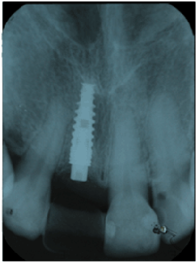
Figure 1: Control periapical radiograph

Piero Rocha Zanardi* Raquel Laia Rocha Zanardi Bruno Costa Marcio Katsuyoshi Mukai Dalva Cruz Laganá
Department of Prosthodontics, School of Dentistry, University of São Paulo, SP, Brazil*Corresponding author: Piero Rocha Zanardi, Department of Prosthodontics, School of Dentistry, University of São Paulo, SP, Brazil, Tel: +55 11 3091-7885; E-mail: pieroznd@gmail.com.br
Background: Morse taper implant connection involves the determination transmucosal height, especially because of its 1 or 2 mm position below the alveolar crest. Intraosseous position promotes a better bone stability and assures soft tissue stability.
Purpose: The aim of this article is to provide practical and objective information for prosthetic component selection in a digital form.
Case Report: The patient MVP needed the replacement of the right, central incisor. A control periapical radiograph was taken. A frontal view photograph was used for a digital aesthetic analysis of the situation. The Pink Esthetic Score (PES), White Esthetic Score (WES) and Digital Smile Design (DSD) were applied to evaluate the individual dentogingival. In planning of rehabilitation, it was chosen a solid abutment for a cemented single crown. Radiograph control of the implant was transposed to the diagnostic photograph and calibrated. With the overlap of the images, the measurement of the abutment collar could be achieved. In addition to this measurement, the 6 mm abutment height could be selected, compatible with the interocclusal space and the length of the crown since it does not invade the crown translucency area which would be compromised due to the lack of sufficient space for the prosthetic restoration.
Conclusion : Since diagnosis is the most important tool in several treatment modalities, some digital resources were used to improve the visualization of the aesthetic limitations of the patient. The use of the suggested method can assist the dentist in planning the rehabilitation while waiting for osseointegration of the implants.
Dental implant; Dental prosthesis; Digital planning
Differently from the internal/external hexagon implant, the selection of the correct prosthetic component for the Morse taper connection implant involves the determination of a correct collar height as well as other factors that are crucial for the best choice [1]. Primarily, the use of prosthetic components with a Morse taper connection involves the correct choice of transmucosal height, especially because of its1 or 2 mm preferred position below the alveolar crest. This intraosseous position seems to promote a better bone stability and in consequence, assure soft tissue stability [2,3]. The selection of the prosthetic component involves several factors that are crucial for the correct choice [4]. With an initial impression, the shape of the implant platform can be determined before treatment planning. The obtained model will also provide important information as to the angle as to depth of the implant. In addition, the type of prosthesis to be made will influence the component choice. For a unitary prosthesis, the emergence or temporary profile and the angle of the implant are especially important while in a fixed prosthesis the passive fit is critical. The purpose of this article is to provide practical and objective information for prosthetic component selection in a digital form.
The patient MVP needed the replacement of the right, central incisor. After the correct three dimensional implant installations [5] and a four month interval, a control periapical radiograph was taken (Figure 1).
A frontal view photograph was used for a digital aesthetic analysis of the situation. This type of record is very opportune to determine the procedures for the best aesthetic resolution. The Pink Esthetic Score (PES) [6], White Esthetic Score (WES) [7] Digital Smile Design (DSD) [8] were applied to evaluate the individual dentogingival alterations and, to certain point, to predict which corrections would be necessary to achieve an optimal aesthetic results. For planning the rehabilitation, we chose a solid abutment (without a passing screw) for a cemented single crown. At this point, with the intention of placing the temporary crown at the same time as the implant access is made, photographs and radiographs were used to define the height of the collar and the cementing area of the abutment with the virtual planning.
The initial assessment with PES (Figure 2), it was showed an 8 value and the WES was 6 (Figure 3). This type of diagnosis has great value when used prior to treatment as it provides enough information to plan the periodontal surgery and achieve a better aesthetic result. The value obtained with the PES/WES evaluation represents a partially compromised aesthetic condition. Those clinical cases where PES ≤ 7 / WES ≤ 5 characterize a compromised aesthetic condition; on the other hand, PES ≥ 12 / WES ≥ 9 denote values for highly appropriate aesthetics or an almost perfect rehabilitation [9,10]. Since the WES alone is not enough for planning the final rehabilitation, this was supplemented by some steps from DSD [8]. Figure 4 illustrates the phases of the digital planning of the rehabilitation. After taking a diagnostic picture (A), a line is drawn parallel to the bi-pupillary plane (B- blue line) as well as the contour of the teeth and the gum. These reference lines are transferred separately to a monochromatic background as to highlight the discrepancies between teeth (C). After the diagnosis of dentogingival discrepancies are made, the corrected plane is projected on the side that has the highest discrepancies. Thus, a red dashed line designed on teeth 11 and 12, in accordance with a symmetrical gingival height on the opposite side (D). The same projection was made from the tooth 21 to the 11. In the overlapping mirror contour it can be seen that 11 is slightly larger than the width of 21. In addition, it can also be noticed that the height of the 11 is larger than that of its counterpart.

Figure 1: Control periapical radiograph
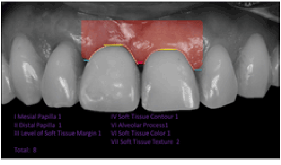
Figure 2: Pink Esthetic Score for the illustrated case
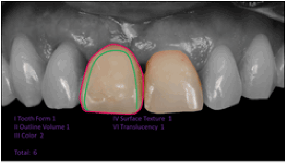
Figure 3: White Esthetic Score for the illustrated case
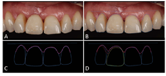
Figure 4: Digital diagnosis of dentogingival discrepancies: ADiagnosis photography; B –bipupillary line, contour of the teeth and gum; C - Projection of diagnostic traits on a monochrome background; D - Mirroring the tooth gingival contour of 21 on the 11.
Once established the parameters that need to be improved to optimize the aesthetic result, the selection of prosthetic abutment was initiated. First of all, the radiograph control of the assisted implant in the evaluation and selection of the abutment. Certain criteria must be met to ensure a satisfactory prosthesis, such as the angulation of the implant, the interocclusal space and the height of the collar [4]. One way to overlap the radiographic image of the implant on to the diagnostic photograph is calibrate both measurements. For this purpose, a digital scale is positioned on the radiograph (Figure 5) matching the size of the radiograph to the measure on the ruler (35 mm, at its greatest length). With this reference, it was possible to measure the anatomical crown of the 21 (red line) and transfer to the clinical picture, adjusting the dimension so that the radiographic image and the picture were equivalent in size. When the crown-implant ratio is suitable the position of the implant is transported to the photograph (Figure 6A). With the overlap of the images, the measurement of the abutment collar can now be initiated (Figure 6B). The green line represents the distance from the implant platform (with a 2 mm healing cap since its position was 1 mm bellow the bone crest) to the gingival zenith. In this case the distance was 3 mm according to the condition of the crown. In addition to this measurement, the abutment height was compared to the length of the tooth (Figure 6C). In this case, a 4 mm abutment height seemed too short for the crown length so a 6 mm was positioned (Figure 6D). The latter presented a height compatible with the interocclusal space and the length of the crown. This set up also allowed the assessment as to the abutment does not invade the area of crown translucency which would be compromised due to the lack of sufficient space for the prosthetic restoration.
The diagnosis is the most important tool in several treatment modalities. After an accurate diagnosis, one can proceed to proper planning and thorough execution. In the case presented, some digital resources were used to aid the visualization of the patient aesthetic limitations. Although the periapical radiograph present some distortions, the use of the suggested method can assist the dentist in the planning of rehabilitation while waiting for osseointegration of the implants.
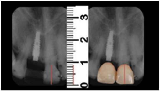
Figure 5: Image size adequacy by radiography scale
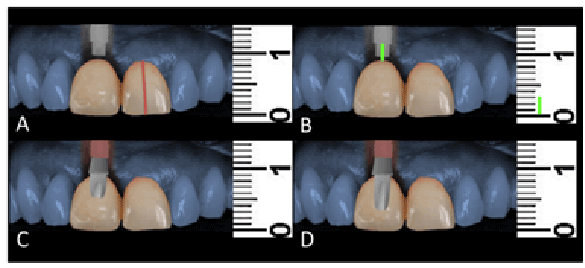
Figure 6: A- adequacy of the radiographic image diagnosis on the photo; B - representative measurement of transmucosal height; C - Overlay a digital pillar 4 mm high; D - overlapping a digital pillar 6 mm long.
Download Provisional PDF Here
Aritcle Type: Research Article
Citation: Rocha Zanardi P, Rocha Zanardi RL, Costa B, Mukai MK, Laganá DC (2015) Digital workflow for selecting the best prosthetic component for a morse taper implant. Int J Dent Oral Health 1(5): doi http:// dx.doi.org/10.16966/2378-7090.142
Copyright: © 2015 Rocha Zanardi P, et al. This is an open-access article distributed under the terms of the Creative Commons Attribution License, which permits unrestricted use, distribution, and reproduction in any medium, provided the original author and source are credited.
Publication history:
All Sci Forschen Journals are Open Access