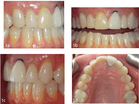
Figure 1: a) Preoperative right lateral view b): Preoperative frontal view c): Preoperative left lateral view d): Preoperative maxillary occlusal view

Suhasini Mandiga Denise Estafan*
Esthetic Dentistry, Department of General Dentistry, Division of Reconstructive and Comprehensive Care, New York University College of Dentistry, USA*Corresponding author: Denise Estafan, Associate Professor and Directo r, Esthetic Dentistry, Department of General Dentistry, Division of Reconstructive and Comprehensive Care, New York University College of Dentistry, USA, E-mail: de1@nyu.edu
Restorative dentistry is the art and science of replacing human tooth structure. It has been said that individually, enamel and dentin are lowstrength materials but when combined, have a unique bond that can last a lifetime [1]. Finding the perfect dental material to mimic nature’s bioengineering through the years has emerged from a monolithic metal design to a bi-layered metal-ceramic or polycrystalline-ceramic design and then to monolithic ceramic design.1 The success of an esthetic rehabilitation not only depends on the material selection or the lab utilized but more importantly, understanding how to work with the unique characteristics of different restorative materials to achieve a predictable, functional and beautiful outcome for each individual case.
The purpose of this case report is to describe a treatment modality utilizing two different restorative materials in the smile zone to achieve an acceptable esthetic outcome. Monolithic pressed lithium disilicate and porcelain fused to high noble metal were the restorative materials utilized.
A 48-year-old Hispanic male presents to the dental clinic with a chief complaint of discoloration and cracks existing on his 16-year-old veneers. Examination revealed 5 discolored veneers with several areas of wear and crack propagation and an existing crown with recurrent decay. Recession of his anterior teeth displayed the margins of his existing restorations. He was unhappy with his smile and wanted a smile makeover that would appear whiter than his existing veneers and appear as natural as possible.
Restoration of a pleasant smile by successfully matching two different materials in the smile zone was achieved to the patent’s satisfaction.
Matching two central incisors with different underlying substructures poses a great restorative challenge for the clinician as well as the laboratory technician especially in esthetically driven demanding patients. For some patients, the use of two different materials is warranted and in such a case, a basic understanding of the material’s characteristics becomes very important to take advantage of its pros and cons. A classification system used by Stefano Gracis et al. [2] divides dental ceramics and ceramic like materials into 1) Glass-matrix ceramics 2) Polycrystalline ceramics 3) Resin-matrix ceramics gives clinicians a better understanding of the materials that they are using.
The two materials chosen for this case report to replace and restore six maxillary anterior teeth are monolithic pressed lithium disilicate and porcelain fused to metal.
Monolithic refers to a material that is homogenous in its construction. The ability of ceramics to be made in monolithic form is advantageous because its mechanical properties remain superior throughout the restoration when compared to bi-layered restorative material that is made by fusing two different homogenous materials together creating a weak link in between them. Monolithic lithium disilicate is a glass ceramic composed of 70% small interlocking prismatic lithium-disilicate crystals randomly dispersed in a glassy matrix [3]. This crystal size and orientation account for an increased flexural strength up to 400 MPa [4] without compromising optical properties in thin restorations allowing for conservative tooth preparation even in patients with a history of bruxism. Monolithic pressed lithium disilicate has proven to have great potential in terms of utilization, esthetics, strength and good wear compatibility to opposing teeth [5,6,7].
Porcelain fused to metal (PFM) crown is preferred by many clinicians because of their high structural performance and esthetic capability and has been dependable for more than five decades. A study conducted by Behr et al., shows that PFM crowns showed 96.4% survival rate over 5 years with 98.2% free of chipping over 10 years for anterior teeth [8]. The substructure of the crown is made of high noble alloy (over 60% noble metal i.e. gold, palladium, platinum of which 40% must be gold), noble alloy (over 25% noble metal content) or non-noble alloy (less than 25% noble metal content). Layers of feldspathic porcelain are then allowed to fuse to the metal substructure in a high heat oven in order to make it more esthetically pleasing. Though this is a bi-layered restorative material, it is still popularly used because it has good marginal finish, can mask any stump color, has good wear compatibility to opposing teeth and has long well documented history of providing lasting service [8,9].
A 48-year-old Hispanic male presents to the dental clinic with a chief complaint of discoloration and cracks that developed on his 16-year-old veneers. He was unhappy with his smile complaining of yellow and black margins around the gum line and wanted a smile makeover that would get him a whiter, brighter smile appearing as natural as possible. The patient reported awareness to clenching and grinding his teeth during periods of stress at work for the past five years. The patient had no other medical contraindications or allergies and exhibited good oral hygiene.
Clinical and radiographic examination revealed 5 discolored veneers (Teeth #6,7,8,10,11) with several areas of wear and crack propagation and an existing crown (Tooth #9) with recurrent decay. Dental-Facial analysis shows a facial midline coinciding with the maxillary dental midline. Occlusal analysis revealed a class 1 occlusion with acceptable overbite and over jet. Photographs, articulation of study models and a diagnostic wax up were used to discuss and evaluate the patient’s concerns, goals and expectations before, during and after treatment. Smile evaluation showed teeth angulations and proportions to be within normal limits. Gingival zeniths of the upper anterior teeth showed good symmetry but uniform recession displayed the margins of his existing restorations and underlying tooth structure. Anterior incisal embrasures were less than normal giving a worn appearance. (Figures 1a-1d) The lip line is normal displaying 80- 100% of tooth structure with 0-2 mm of gingiva in a full smile and the smile line is straight, not following the anatomy of the lower lip.
The treatment plan included removal of existing restorations and decay #6-11, evaluate condition of underlying post and core #9 and restore using pressed monolithic lithium disilicate veneers #6,7,8,10,11 and porcelain fused to metal #9. A metal lingual was chosen for #9 (Figure 3d) instead of porcelain due to the patient’s history of bruxism.
Existing veneers were removed from teeth #6,7,8,10,11 using diamond burs and were prepped following the contour of the tooth surface trying to preserve as much as enamel as possible. Cervical margins were extended to the crest of the gingiva and finished with a shoulder finish line paralleling the entire gingival margin. This finish line was continued inter-proximally and was kept flush with the adjacent tooth. The veneer prep was wrapped around the incisal edge towards the lingual. Any sharp line angles were rounded off using a fine diamond bur (Figures 2a-2c).
Figure 1: a) Preoperative right lateral view b): Preoperative frontal view c): Preoperative left lateral view d): Preoperative maxillary occlusal view
After removal of existing crown on tooth # 9, an underlying metal cast post and core with recurrent decay was present. Caries was removed with a round bur. Clinical judgment was to leave the post and core intact as the previous endodontic treatment was asymptomatic and refine the margin to a 90-degree shoulder that extended subgingivally (Figures 2b-c).
A buccal index was used to verify sufficient tooth reduction on the prepared teeth. Shade selection was done to the patient’s satisfaction in a natural light source. Prepared teeth were cleaned using pumice slurry. #000 cord soaked in hemodent packed around Tooth # 9 and a final impression was taken using medium and light bodied vinyl poly siloxane. One piece provisional restoration was fabricated with Luxatemp utilizing the putty matrix made from the diagnostic wax up. Excess flash material was removed and occlusion was checked and relieved to avoid excess pressure. Glaze was applied and cured on the provisional for added temporary esthetics. Minor adjustments were made and a diagnostic impression of the satisfied provisional restorations was sent to the laboratory for better communication of the desired changes.
Five veneers and one crown were inspected on the model for proper fit and color consistency after returning back from the lab. Provisional restorations were removed and the teeth were cleaned using pumice slurry to remove any debris. Permanent restorations were tried in and checked for proper fit, marginal accuracy, contacts, shade and overall esthetics. The internal surfaces of the final restorations #6-11 were pretreated with 35% phosphoric acid for 20 sec, washed thoroughly, dried and then silanated for 60 sec and allowed to dry. Teeth #6-11 were well isolated and treated with 35% phosphoric acid for 15 sec, rinsed and lightly dried. Isolation was redone. Two bottles adhesive (All-bond) was mixed and applied and thinned out with air. Monolithic lithium disilicate veneers #6,7,8,10,11 were cemented using resin cement (Choice 2) and porcelain fused to high noble metal crown #9 was cemented using resin modified glass ionomer cement (FujiCem) (Figsures 3a-3d). Excess cement was removed and contacts were rechecked. Occlusion was checked for proper canine guidance and final photographs were taken. In the next visit, a lab fabricated occlusal guard was delivered after adjustment was made to ensure proper canine guidance in all excursive movements.
Matching two central incisors with dissimilar substructures can be a challenge. Though superior metal free restorative materials are available in the market, certain clinical situations still demand the use of traditional porcelain fused to metal restorations. In any instance, a clinician’s well understanding of material properties enable for effective color selection in a desired case to achieve an acceptable result. A general comparison among the common esthetic materials can be seen in Figure 5. While there is an increase in durability of ceramics towards the right, there is a decrease in its optical properties and vice versa. All factors and variable must be taken into account while weighing the pros and cons of restorative materials in any individual case to achieve an esthetic and functional balance and overall treatment success.
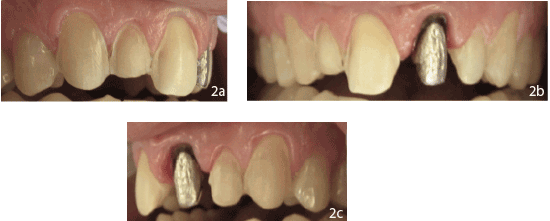
Figure 2: a) Postoperative right lateral view b): Postoperative frontal view c): Postoperative left lateral view d): Postoperative maxillary view
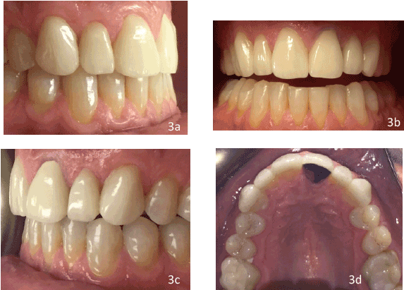
Figure 3: a) Postoperative right lateral view b): Postoperative frontal view c): Postoperative left lateral view d): Postoperative maxillary view
Factors to consider when choosing the restorative materials include:
Though ceramic materials were contraindicated in the past for patients with parafunctional habits, recent advances indicate otherwise. Studies show that monolithic materials such as lithium disilicate are two times more fracture resistant when compared to layered porcelain on another substructure and therefore are preferred in patients that clench or grind teeth [10,11].
A tooth present in the posterior region of the mouth would require a material that is stronger than an anterior tooth. Similarly fixed partial denture frameworks or implant abutments would require a restorative material with superior mechanical properties. Square faces tend to have stronger muscles of mastication and a chewing pattern that is more harmful than oval faces impacting the teeth.
Increased demand for metal free tooth colored restorations for teeth that are subjected to more force led to the development of high strength alumina and zirconia copings. Still certain occlusions where there is insufficient inter-occlusal distance or a deep overbite, all ceramic restorations are not indicated.
Consideration of the opposing tooth material e.g. natural tooth, resin composite, ceramic, metal is also important. Many studies have reported that ceramic substrates to produce more wear on opposing tooth structure than enamel with higher rates for zirconia [12-15].

Figure 4: Postoperative smile view
Margins of the preparation depends primarily on the final restorative material chosen Conservative tooth preparation is the standard of treatment. Preservation of enamel to allow for enamel bonding is one of the most significant achievements in dentistry [16].
Variables affecting optical properties include: stump shade selection, final shade, and amount of tooth reduction necessary to achieve the final shade selection, color and thickness of luting agent, translucency of the restorative material.
An untrained eye can easily detect slight differences in value where as hue and chroma cannot. Masking a dark stump shade with a material that is more translucent (less value) requires more tooth preparation. It is said that masking should be carried out in the substructure of the restoration rather than the build up because the technician has better control of value in the deeper layers of the restoration [17].
Certain clinical situations may require greater tooth reduction on the facial surface so that the laboratory technician can manage the final shade matching during ceramic buildup of the restoration.18 e.g. one stump shade is darker than adjacent stump shades.
Alternatively, by choosing a restorative material with an opaque substructure, unnecessary tooth preparation can be avoided. According to Stephen Chu, [19] teeth that are low in translucency are best served with metal ceramic restorations or CAD-CAM based ceramics and teeth that are high in translucency are best served with all-ceramics.
In this case report, monolithic pressed lithium disilicate was chosen because its monolithic characteristic offered better mechanical properties in patients with bruxism while having superior life like aesthetics. The pros and cons of using all-ceramic or porcelain-fused-to-metal for tooth #9 were weighed keeping in mind, the amount of reduction required to block out the metal post, color preference, the choice of surrounding restorative materials, functional demand, patient history and cost.
There is no one material that is ideal for every case as advantages and disadvantages exist for every material. In this case, restoration of a pleasant smile (Figure 4) utilizing two different materials to restore six maxillary anterior teeth in the esthetic zone was achieved to the patient and clinician’s satisfaction.
This case report was previously presented as a poster at NYU College of Dentistry and won:
Best Case Study Presentation by senior student in the pre-doctoral clinics for the 2015 Clinical and Educational Scholarship Showcase awarded by NYU Academy of Distinguished Educators DSG Americus Award for the 2015 NYU Aesthetic Poster Competition awarded by DSG Americus Dental Lab
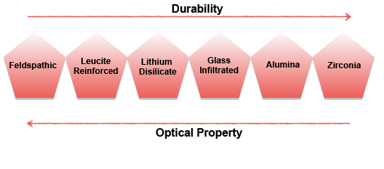
Figure 5: Relative comparison among common ceramics used in dentistry
Download Provisional PDF Here
Aritcle Type: Case Report
Citation: Mandiga S, Estafan D (2015) An Esthetic Challenge: Case Report Utilizing a Combination of Monolithic Ceramic Veneers and Porcelain Fused to Metal Crown. Int J Dent Oral Health 1(5): doi http:// dx.doi.org/10.16966/2378-7090.128
Copyright: © 2015 Mandiga S, et al. This is an open-access article distributed under the terms of the Creative Commons Attribution License, which permits unrestricted use, distribution, and reproduction in any medium, provided the original author and source are credited.
Publication history:
All Sci Forschen Journals are Open Access