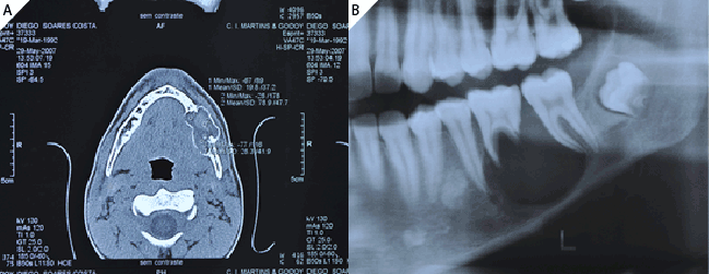
Figure 1a: Radiographic feature showing a large radiolucency lesion and radicular resorption of the first lower left molar (36)
Figure 1b: Axial computed tomography (CT) image showing the expansive mandibular lesion

Belini Freire Maia Élton Gonçalves Zenóbio* Hayder Egg Gomes Roberta Paula Colen Bustamante Martinho C. R. Horta Jamil Awad Shibli Mauricio Greco Cosso
Dental Research Division, Department of Dentistry, PUCMINAS, Brazil*Corresponding author: Prof. Élton Gonçalves Zenóbio, DDS, MS, PhD, Department of Dentistry PUCMINAS Dental Research Division - Implantology and Periodontology, Brazil, Tel: +55 (31) 33194414; E-mail: zenobio@pucminas.br
Ameloblastoma is a slow-growing polymorphic neoplasm that presents a follicular or plexiform pattern in the fibrous stromal. Although considered a benign tumor, it is aggressive and recidivistic, reappearing even after surgical removal due to its ability to infiltrate the trabecular bone [1-3].
Ameloblastoma is the most common neoplasia among the odontogenic tumors, with 11% prevalence. Approximately, 80% of the cases of ameloblastoma occur in the mandible, with the mandibular ramus and angle being the most affected areas [4].
The diagnosis is usually made between the fourth and fifth decade of an individual’s life, with the exception of the unicystic type, which is commonly diagnosed in patients from 20 to 30 years of age.
In most cases, these are virtually asymptomatic, except for the occurrence of facial asymmetry. This tumor can present as a radiolucent area in radiographic exams [5].
The treatment modality decision is based on the type of ameloblastoma (solid, multicystic, unicystic, or peripheral), location, size, and the patient’s age. It is believed that the unicystic type is the least aggressive and responds most favorably to conservative surgery [5,6]. Furthermore, the aesthetic and functional rehabilitation of patients that undergo respective surgery presents an enormous challenge to oral surgeons. Immediate bone reconstruction and implant placement are options for treatment therapy in such patients [7, 8].
This paper presents a case report of unicystic ameloblastoma in a 15-year-old patient who was rehabilitated by using extra-short implants after the removal of the lesion with six years of preservation.
A 15-year-old male was referred to the Department of Maxillofacial Surgery and Traumatology, at the Pontifical Catholic University of Minas Gerais - School of Dentistry, presenting an asymptomatic edema in the posterior region, left side of the mandible.
Radiographic examination revealed the presence of an extensive unilocular radiolucent image involving the mandibular angle and body with evidence of root resorption of tooth 36 (Figures 1a and 1b). The clinical periodontal examination (bleeding or altered depths on probing, periodontal clinical insertion loss, and good dental plaque index) indicated the good periodontal health of the tooth. The clinical and radiological exams suggested an ameloblastic lesion.
The patient underwent removal and curettage of the lesion and extraction of teeth 36, 37, and 38 under general anesthesia. Histological diagnosis established unicystic ameloblastoma with mural invasion (Figures 2a and 2b).
Twenty-three months after the surgery to remove the ameloblastoma lesion, three implants (RN standard plus, Straumann, Switzerland - 4.1 mm/6 mm) were installed in the remaining bone (Figure 3). Five months after the implant surgery, metallic-ceramic prosthesis was installed (Figure 4). Radiographic and clinical exams were used to monitor the patient, at 24 and 36 months, after the installation of the prosthesis and at 72 months after ameloblastoma resection surgery (Figure 5).
The invasive and destructive behavior displayed by ameloblastoma often leads to the choice of ressective treatment of the affected area, which can significantly affect the oral rehabilitation of individuals with this disease [7, 8].
Therefore, the multidisciplinary approach is extremely important in the patient’s rehabilitation [3]. Cases of recurrences may be considered in the prognosis, and in the literature they might be usually detected at 32 months on average following initial treatment; and surgically treated by means of marginal mandibulectomies and segmental mandibulectomies with bone reconstruction, performed primarily with cortical iliac crest grafts, cancellous iliac crest bone graft, proximal tibia bone graft and fibula free flap. The basic reconstruction may involve the use of nonvascularized bone grafts together with restoration of lost teeth by means of dental implants and implant-supported prostheses.
In such cases, the statistics balances of cost savings versus risk of recurrence are favorable to the dental rehabilitation with dental implants because no further recurrences were observed after the second operation.
Immediate reconstruction of the bone defect with placement of dental implants and rehabilitation with implant-supported prostheses in a second stage can improve jaw function and facial harmony of the patient once dental implants allow the restoration of masticator function, phonetics and aesthetics in maxillofacial rehabilitation, thus contributing to an increase in the patient’s quality of life [9,10].
However, respective surgery for the removal of these tumors may result in a decreased bone stump, making it impractical to place regular implants. Thus, the short implants could be a viable alternative treatment [11].
Studies have shown that the success rate of short implants placed in the posterior areas of the jaw is similar to that of conventional implants [12-14]. However, reports in the literature are scarce regarding the installation of short implants in patients who have undergone unicystic ameloblastoma curettage. Data from some studies show that conventional length implants installed in the areas subjected to tumor resection and immediate reconstructions with bone grafts have presented high success rates [15-17]. Additionally, the success rate of prostheses installed on short implants is similar to that obtained with prostheses supported by conventional size implants [18,19].
Figure 1a: Radiographic feature showing a large radiolucency lesion and radicular resorption of the first lower left molar (36)
Figure 1b: Axial computed tomography (CT) image showing the expansive mandibular lesion
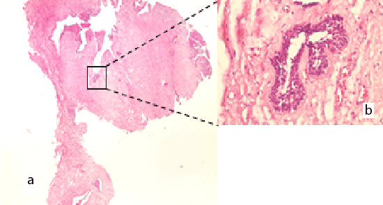
Figure 2a: 20X increasing – Lesion with cystic cavity
Figure 2b: 400X increasing – Areas showing mural infiltration mass.
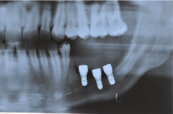
Figure 3: 18 months follow-up – implants were placed after surgical lesion removal.
The use of short implants in areas with trauma and severe bone resorption, or in sites subjected to the removal of tumors, has become a feasible option. In this case, the choice of installing this type of implant resulted in less morbidity for the patient because he had undergone bone graft surgery, in addition to representing a reduction in treatment cost and time. The tree-year post rehabilitation preservation has revealed no failures in the implants or the prostheses.
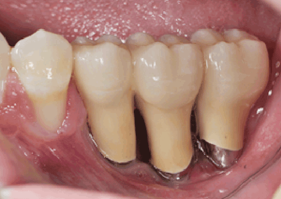
Figure 4: dental metal-ceramic prosthesis after occlusal adjustment.
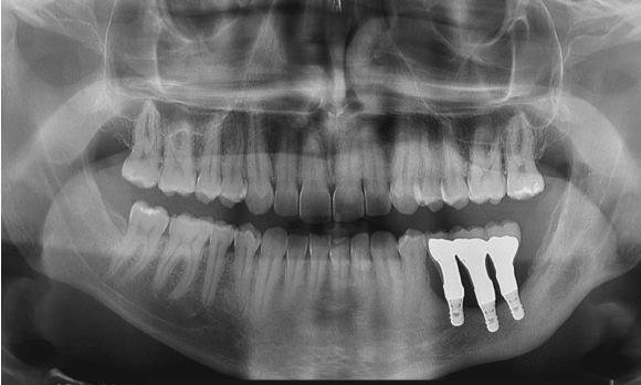
Figure 5: Radiography of the installed prosthesis, taken 24 months later
The treatment of unicystic ameloblastoma involves a multidisciplinary approach that includes surgery for tumor removal, pathologic analysis, aesthetic and functional rehabilitation of the patient by the use of implantsupported prostheses; and the installation of short implants in low height bone remaining tissue may well be a viable and less aggressive option of rehabilitation.
No potential conflict of interest relevant to this article was reported.
Download Provisional PDF Here
Article Type: Case Report
Citation: Freire-Maia B, Zenóbio EG, Gomes HG, Bustamante RP, Horta MC, et al. (2016) Rehabilitation through Short Implants Following Conservative Surgery for a Unicystic Ameloblastoma: Case Report. Clin Res Open Access 2(1): doi http://dx.doi.org/10.16966/2469-6714.109
Copyright: © 2016 Freire-Maia B, et al. This is an open-access article distributed under the terms of the Creative Commons Attribution License, which permits unrestricted use, distribution, and reproduction in any medium, provided the original author and source are credited.
Publication history:
All Sci Forschen Journals are Open Access