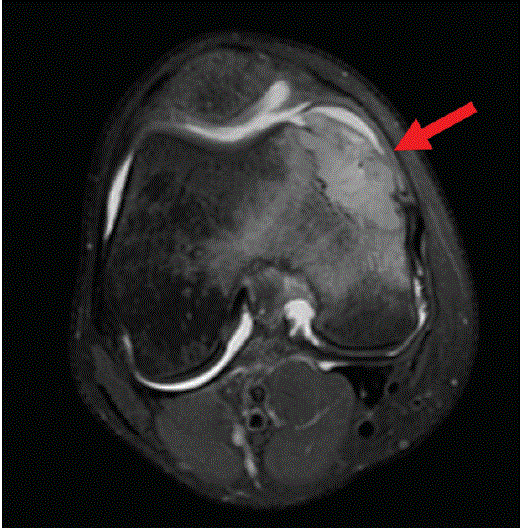
Figure 1: MRI of the right knee (axial, fat suppressed) showing a mass arising from the right medial femoral condyle (arrow).

Nida Ashraf1 Rachel T Weihe2* Rachael M Liesman3 Anders Meyer3 Kyle R Sweeney4 D Matthew Shoemaker2*
1Division of Infectious Diseases, Department of Internal Medicine, University of Pittsburgh Medical Center-Hamot, Erie, USA*Corresponding author: D Matthew Shoemaker, Division of Infectious Diseases, Department of Internal Medicine, University of Kansas Medical Center, 3901 Rainbow Blvd, MS 1028, Kansas City, KS-66160, USA, Tel: (913) 588-3858; Fax: (913) 588-6024; E-mail: dshoemaker2@kumc.edu
Rachel T Weihe, Division of Infectious Diseases, Department of Internal Medicine, University of Kansas Medical Center, 3901 Rainbow Blvd, MS 1028, Kansas City, KS-66160, USA, Tel: (913) 588-8708; Fax: (913) 588-6024; E-mail: rweihe@kumc.edu
Blastomyces dermatitidis is an endemic mycosis in certain regions of the United States of America and can cause disseminated infection in immunocompetent individuals. We present a case of osseous blastomycosis in a previously healthy 22-year-old male from Missouri, who developed sudden onset right knee pain in June 2019. Magnetic Resonance Imaging (MRI) demonstrated an enhancing mass that was concerning for osseous malignancy. Antigen and antibody testing for dimorphic fungi was confounding. He underwent debridement and surgical specimen cultures grew B. dermatitidis. He was treated initially with liposomal amphotericin B and transitioned to oral itraconazole to complete therapy. Blastomycosis of the bone can be mistaken for malignancy and should be included on a differential for these types of cases. Blastomycosis of the bone and joints can be successfully treated with surgical debridement and prolonged antifungal therapy.
Blastomyces dermatitidis; Osteoarticular blastomycosis; Disseminated blastomycosis
Blastomycosis is caused by Blastomyces dermatitidis, a thermally dimorphic fungus [1-3]. In nature, Blastomyces exists in the mold form and is found in woodland areas with moist soil and decomposing organic matter, commonly found near bodies of freshwater [1,4]. The yeast form is found in tissue and characterized by broadbased budding with a prominent cell wall [1]. Infection is usually acquired via inhalation of B. dermatitidis conidia, though cases of direct inoculation have been described [4-6]. The clinical spectrum of disease can include asymptomatic infection, acute or chronic pneumonia, and disseminated disease [7]. In those patients who do become symptomatic, the incubation period ranges from 30 to 45 days. Although pulmonary infection is the most common disease manifestation, the most common extra pulmonary sites include skin, genitourinary, and bone and joint infections [7]. The presentation of osteoarticular blastomycosis can mimic osseous tumors and should be considered in the differential diagnosis.
The gold standard for diagnosis of B. dermatitidis is growth in culture from a clinical specimen. Histopathological identification in clinical specimens can be supportive, and serological tests are often used as less invasive techniques, but sensitivity and specificity can be low [7]. Treatment with antifungal therapy and surgical debridement in cases of osseous lesions yields the best chance of clinical cure.
A previously healthy 22-year-old male from Missouri developed sudden onset right knee pain in June 2019. He denied any antecedent trauma. He had been on a float trip on a local river in southwest Missouri a few weeks prior to the onset of his right knee pain. He denied fevers, chills, night sweats, lymphadenopathy, or unexplained weight loss. The right knee pain was exacerbated by weight bearing and movement. Radiographs of the right knee were unremarkable. He developed worsening pain and swelling of the medial right knee which prompted him to seek evaluation in August 2019. On physical examination, he was afebrile and vital signs were normal. His right knee was swollen and non-erythematous, with a tender, palpable mass on the medial aspect overlying the right medial femoral condyle, and his range of motion was limited due to pain. Magnetic resonance imaging (MRI) demonstrated an enhancing mass with areas of central necrosis arising from the right medial femoral condyle with cortical breakthrough and extension into the joint space (Figure 1). The MRI findings were concerning for osseous malignancy. He underwent Computed Tomography (CT) guided biopsy of the medial right distal femur. Pathology revealed suppurative, granulomatous inflammation with fungal organisms on Gömörimethenamine-silver, FontanaMasson, and mucicarmine stains (Figures 2a-2c).
Figure 1: MRI of the right knee (axial, fat suppressed) showing a mass arising from the right medial femoral condyle (arrow).
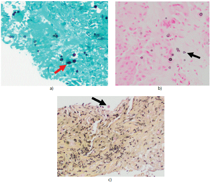
Figure 2: Histopathology of CT-guided biopsy tissue of the right knee.
a) Gömöri Methenamine-Silver Nitrate (GMS) stain, 100× magnification demonstrating yeast (arrow).
b) Fontana-Masson stain, 100× magnification demonstrating yeast-like organisms (arrow).
c) Mucicarmine stain, 100× magnification demonstrating yeast-like organisms (arrow).
Additional evaluation included a negative HIV antigen/antibody test, and routine and fungal blood cultures were both negative. The patient denied any respiratory symptoms, but due to concern for possible disseminated disease, a CT chest was obtained and revealed an irregular opacity in the right lung apex with scattered irregular, ovoid lucencies, a right middle lobe 0.3 cm nodule, and mediastinal, right axillary, and supraclavicular lymphadenopathy (Figure 3).
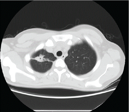
Figure 3: CT chest (axial view) showing an irregular opacity in the right lung apex.
Serum Blastomyces antigen testing was positive (below the limit of quantification). Serum Blastomyces antibody immune diffusion testing was positive. Serum and urine Histoplasma antigen testing were positive (below the limits of quantification). Serum Histoplasma antibody and serum cryptococcal antigen testing were negative.
He was started on liposomal amphotericin B 3 mg/kg intravenously per day. He underwent open arthrotomy of the right knee and debridement of the medial femur lesion. Intra-operatively he was found to have brown necrotic bone, cartilage, and soft tissue extending from the area of the lesion seen on the MRI into the suprapatellar pouch. All nonviable tissue was sharply excised using rongeurs and curettes. The intraoperative right knee tissue culture grew B. dermatitidis.
He was transitioned to oral itraconazole with therapeutic drug monitoring to guide dosing, with a plan to treat for one year from the time of debridement. He completed nine months of therapy with a good clinical response at that point in time. An interim CT chest for monitoring approximately four months into his antifungal therapy showed resolution of the right upper lobe cavitary lesion and the adjacent consolidation as well as improvement in the lymphadenopathy. Follow up plain radiographs of his right femur showed no recurrence of osteomyelitis. He was subsequently lost to follow up.
Blastomycosis was first described in 1894 by Thomas C Gilchrist. Blastomycosis is a pyogranulomatous disease caused by Blastomyces dermatitidis, thermally dimorphic fungi that is endemic to certain regions of the United States, Canada, and can be found in several countries in Africa and in India [8]. In nature, Blastomyces exists in the mold form and is found in woodland areas with moist soil and decomposing organic matter, commonly found near bodies of freshwater [1,4]. The yeast form is found in tissue and characterized by broad-based budding with a prominent cell wall [1]. Infection is usually acquired via inhalation of B. dermatitidis conidia, though cases of direct inoculation have been described [4-6]. The clinical spectrum of disease can include asymptomatic infection, acute or chronic pneumonia, and disseminated disease [7]. In those patients who do become symptomatic, the incubation period ranges from 30 to 45 days. Although pulmonary infection is the most common disease manifestation, the most common extrapulmonary sites include skin, genitourinary, and bone and joint infections [7]. The presentation of osteoarticular blastomycosis can mimic osseous tumors and should be considered in the differential diagnosis.
The clinical presentation is sometimes indistinct from infection due to other dimorphic fungi, such as Histoplasma or Coccidioides, and can also be confused with tuberculosis, non-tuberculous mycobacteria, or malignancy. Consequently, a detailed history of travel and environmental exposures is essential to establish the differential diagnosis and guide the evaluation.
B. dermatitidis is endemic to the midwestern and southern regions of the United States, particularly in the Ohio and Mississippi River valleys and the Great Lakes region [1,2,5,9]. It is twice as common in men as in women, with overall incidence rates in the hyperendemic areas of the United States of America ranging from 0.5-100/100,000 [4]. As of 2019, in the United States, cases of blastomycosis are considered reportable diseases in only 5 states, including Arizona, Louisiana, Michigan, Minnesota, and Wisconsin, however, cases have been reported in 26 of the 50 states, including Missouri [8]. The true prevalence of B. dermatitidis world-wide has historically been difficult to determine given the dearth of sensitive and specific serologic and skin tests that are available, along with the lack of mandatory public health reporting. The endemic areas may in fact be larger than previously described [4,6].
Our patient underwent surgical debridement of bone and soft tissue from the right femoral lesion, and tissue was submitted for culture and histopathological analysis, combined with serological evaluation. The diagnosis of B. dermatitidisis best achieved by culture from a clinical specimen, but can also be accomplished by direct microscopy. B. dermatitidis can be readily seen on Hematoxylin and Eosin stain (H&E), Periodic Acid-Schiff (PAS), GömörimethenamineSilver Nitrate (GMS), and Fontana-Masson stains. Mucicarmine stain is considered selective for Cryptococcus neoformans due to the strong uptake by its capsule; however, B. dermatitidis cell walls can stain weakly positive and confound the diagnosis [3]. Additional diagnostics include serum antibody and serum and urine antigen testing, but there can be some cross reactivity with other dimorphic fungi, in particular Histoplasma capsulatum, as was demonstrated in our case. Crossreactivity of B. dermatitidis antigen with Histoplasma antigen testing is present in 64% to 90% of cases [10-13]. This cross reactivity makes diagnosis difficult given that the areas of endemicity for B. dermatitidis and H. capsulatum have a significant overlap. Therefore, culture of B. dermatitidis remains the most specific method for diagnosis. Growth in culture is typically detected in 10-14 days, although some strains can be slower and can take up to 6 weeks.
Pulmonary infection is the most common presentation, followed by cutaneous involvement, and dissemination to the bones, joints, and the genitourinary tract [2,4]. Disseminated blastomycosisis not uncommon in immunocompetent individuals [14]. Blastomyces involving bone has been reported in the modern medical literature as far back as 1950 [15]. Dissemination usually occurs by lymphohematogenous spread in those with pulmonary disease [3-5,9,16]. Blastomyces septic arthritis is most commonly monoarticular [16]. Isolated cases of osteomyelitis without any pulmonary involvement have been reported; the knee being the most common site of involvement, followed by the vertebral body [2,5,9,17]. Blastomyces can cause disease regardless of host immunocompetence [14]. Our patient did not have lung tissue biopsied to determine if his CT chest findings were reflective of pulmonary blastomycosis, but the clinical findings would suggest he had disseminated disease; which was responsive to appropriate antifungal therapy. The presentation of osteoarticular blastomycosis can mimicosseous tumors and should be considered in the differential diagnosis. Depending on the site and extent of involvement, blastomycosis can be successfully treated with surgical debridement and prolonged antifungal therapy [17].
Treatment options for blastomycosis are largely dependent upon the immunocompetence of the host, the severity of disease (including the presence or absence of Central Nervous System (CNS) involvement), and toxicity of the agent [7]. In severe cases, such as with CNS involvement, liposomal amphotericin B 3-5 milligrams/kilogram/day intravenously is first initiated, followed by oral itraconazole 200 mg twice daily once clinical response is achieved [7]. In mild cases, such as those that are non-life threatening and without CNS involvement, itraconazole therapy alone is sufficient. Duration of therapy is then dependent upon disease severity and clinical response, but typically will range from 6 to 12 months [7]. Surgical debridement is warranted in cases of osteomyelitis or excision of mass osseous lesions.
None.
None.
Download Provisional PDF Here
Article Type: CASE REPORT
Citation: Ashraf N, Weihe RT, Liesman RM, Meyer A, Sweeney KR, et al. (2021) 22-Year-Old with Invasive Mass of the Right Femur. J Clin Lab Med 6(2): dx.doi.org/10.16966/2572-9578.140
Copyright: © 2021 Ashraf N, et al. This is an open-access article distributed under the terms of the Creative Commons Attribution License, which permits unrestricted use, distribution, and reproduction in any medium, provided the original author and source are credited.
Publication history:
All Sci Forschen Journals are Open Access