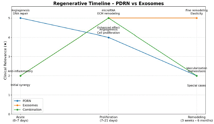
Figure 1: Clinical indications according to regeneration phase.


Denise Barcelos1,2 Cyro Hirano1,2 Tércio Elyan Azevedo Martins3 Fernanda Dutra Santiago-Bassora1,*
1Changeover Education, Brazil*Corresponding author: Fernanda Dutra Santiago-Bassora, Changeover Education, Brazil, E-mail: [email protected]
Regenerative therapies have become a relevant tool in clinical practice in dermatology, aesthetic medicine and related fields, particularly in the context of tissue repair, healing of complex wounds, management of alopecia and cutaneous aging. Among these approaches, polydeoxyribonucleotide (PDRN) and exosomes play a central role due to their ability to modulate inflammation, promote angiogenesis, stimulate fibroblasts and remodel the extracellular matrix in a coordinated manner. This narrative literature-based article is derived from a previously structured practical guide and describes the main biological mechanisms of these therapies, discusses the chronobiology of tissue regeneration and proposes a model for clinical integration based on the phases of injury, proliferation and remodeling. PDRN is emphasized as a fast-acting agent, with robust evidence in chronic ulcers and complex wounds, whereas exosomes are discussed as modulators of the tissue microenvironment, with impact on medium- and longterm remodeling. In this article, we present data from clinical trials and systematic reviews and propose practical recommendations for the rational combination of these therapies in clinical protocols.
Regenerative aesthetics; Dermatologic aesthetics; Polydeoxyribonucleotide; Exosomes
The interface between basic science, regenerative medicine, and clinical practice in aesthetics and dermatology currently constitutes one of the most dynamic fields in translational medicine. Biomolecules, acellular therapies, and extracellular vesicles have been widely investigated as tools capable of selectively modulating tissue repair, cutaneous regeneration, and extracellular matrix remodeling processes. This progress responds to the growing clinical demand for natural, predictable, and sustainable outcomes, while simultaneously challenging oversimplified approaches or promises of “miraculous effects,” often dissociated from the physiology and chronobiology of regenerative processes [1,2].
In this context, the rational use of regenerative therapies requires an in-depth understanding of the temporal dynamics of tissue repair, the main cellular targets involved-including fibroblasts, keratinocytes, endothelial cells, and immune system cells-and the level of scientific evidence available for each intervention. Inadequate modulation of these pathways may result in inefficient repair, delayed healing, or disorganized extracellular matrix remodeling, with a direct impact on final tissue quality [3].
Polydeoxyribonucleotide (PDRN) stands out as a wellcharacterized acellular regenerative therapy composed of short-chain DNA fragments, generally derived from purified animal sources. Its main mechanism of action involves activation of adenosine A2A receptors, with a consequent reduction in pro-inflammatory cytokines, increased expression of Vascular Endothelial Growth Factor (VEGF), and stimulation of fibroblast and endothelial cell proliferation. In parallel, PDRN acts as a nucleoside donor for the salvage pathway, supporting DNA synthesis in cells subjected to metabolic stress or tissue damage [4-6]. Randomized clinical trials and systematic reviews demonstrate consistent evidence of its efficacy in chronic wound healing, complex ulcers, and improvement in scar quality in post-procedural settings [7,8].
Exosomes, in turn, represent a distinct class of acellular regenerative therapies, consisting of nanosized extracellular vesicles (30-100 nm), released by different cell types and rich in membrane proteins, microRNAs, bioactive lipids, and messenger RNA fragments. Unlike agents with a predominantly proliferative mode of action, exosomes function as advanced intercellular communication systems capable of reprogramming the inflammatory microenvironment, modulating the immune response, and coordinately influencing fibroblast activity, angiogenesis, and extracellular matrix remodeling [9,10].
Preclinical, translational, and emerging clinical evidence indicates that exosomes derived from platelets, mesenchymal stem cells, and other sources exert relevant effects throughout all phases of tissue repair, promoting macrophage polarization toward the M2 phenotype, reducing persistent inflammation, enabling more organized collagen deposition, and improving tissue quality in the medium and long term [11-13]. These properties make exosomes particularly attractive in protocols aimed at improving skin quality, global rejuvenation, and the management of alopecia.
Given this scenario, it becomes evident that PDRN and exosomes should not be regarded as competing therapies, but rather as biologically complementary tools, with distinct action profiles and synergistic potential when applied in alignment with the chronobiology of tissue regeneration. Thus, this article aims to organize the content of a practical guide on regenerative therapies by describing the main biological mechanisms, the available levels of clinical evidence, and proposing rational models for the isolated and integrated use of PDRN and exosomes in dermatologic and aesthetic clinical protocols.
Tissue regeneration is a dynamic, highly orchestrated, and timedependent process in which cellular, molecular, and biomechanical events occur sequentially and partially overlap. Didactically, this process may be divided into three main phases: the injury or acute phase (0-7 days), the proliferative phase (7-21 days), and the remodeling or maturation phase (approximately 3 weeks to 6 months), although the duration and intensity of each stage vary according to tissue type, extent of injury, and the individual’s systemic conditions [14,15].
In the initial phase, hemostasis and acute inflammation predominate, with platelet activation, formation of the provisional clot, and immediate release of inflammatory mediators and growth factors such as PDGF, TGF-β, and VEGF. The coordinated recruitment of neutrophils and macrophages is essential for the removal of cellular debris and microorganisms, as well as for establishing a permissive microenvironment for initial angiogenesis and subsequent cellular activation [15,16]. In this phase, physiologic inflammation plays a reparative role; however, its persistence or exacerbation may impair the proper progression of the regenerative process.
The proliferative phase is characterized by intense metabolic and cellular activity, especially involving fibroblasts, keratinocytes, and endothelial cells. Fibroblasts migrate to the wound bed and initiate deposition of a provisional extracellular matrix rich in fibronectin, proteoglycans, and type III collagen, which provides temporary structural support to the regenerating tissue. In parallel, reepithelialization occurs, along with expansion of the vascular network through angiogenesis and vasculogenesis, as well as the initial reorganization of elastic fibers [14,15]. Communication between structural cells and immune system cells, mediated by cytokines and extracellular vesicles, is critical to the success of this stage.
In the remodeling phase, the newly formed tissue undergoes a gradual maturation process in which type III collagen is progressively replaced by type I collagen, which has greater mechanical strength, and the elastic fibers undergo spatial and functional reorganization. Simultaneously, vascular density decreases, selective apoptosis of excess cells occurs, and extracellular matrix architecture is refined, culminating in the partial or complete restoration of skin elasticity, thickness, and biomechanical function [15,16]. Alterations in this phase are directly associated with the formation of hypertrophic scars, keloids, or functionally inferior tissues.
Respect for and understanding of tissue chronobiology is therefore essential for the appropriate application of regenerative therapies. Interventions that excessively stimulate extracellular matrix synthesis in the early phases may result in disorganized fibrosis, whereas strategies that fail to provide adequate angiogenic and proliferative support may delay reepithelialization and restoration of the cutaneous barrier [14,16].
Polydeoxyribonucleotide (PDRN) is a short-chain DNA polymer, generally obtained from purified animal sources, with a molecular weight ranging from 50 to 1500 kDa. Its best-established mechanism of action involves selective activation of adenosine A2A receptors, leading to inhibition of pro-inflammatory cytokines, increased expression of angiogenic factors-particularly VEGF-and direct stimulation of fibroblast and endothelial cell proliferation [17,18].
Additionally, the DNA fragments released by PDRN act as substrates for the nucleotide salvage pathway, supporting DNA synthesis in cells subjected to hypoxia, inflammation, or metabolic damage, a condition frequently observed in chronic wounds and aged tissues [19,20]. This dual mechanism-A2A receptor-mediated signaling and cellular metabolic support-confers upon PDRN an action profile that is particularly relevant during the acute and proliferative phases of tissue regeneration.
From a clinical standpoint, PDRN may be conceptualized as a “rapid builder” of tissue repair, providing immediate signaling and raw material for regeneration. Randomized clinical trials have demonstrated its efficacy in diabetic foot ulcers, with significantly higher rates of complete healing and increased epithelialized area when compared with placebo [18]. Similar results have been observed in pressure ulcers, with a significant reduction in wound area and PUSH scores, without an increase in adverse events [8]. More recently, clinical studies have shown that early PDRN injections into surgical scars reduce excessive vascularization, erythema, pigmentation, and scar height, preventing the development of hypertrophic scars [7].
Exosomes are small extracellular vesicles, with an average diameter between 30 and 100 nm, released by different cell types and rich in membrane proteins, microRNAs, bioactive lipids, and messenger RNA fragments.
Exosomes may originate from various sources. In plant cells, they are described as nanoparticles or exosome-like extracellular vesicles derived from plants and tend to be considered more scalable, lowercost, and potentially less immunogenic alternatives, whereas vesicles of human origin offer greater biomimicry and a translational basis more directly related to human physiology; to date, however, humanderived vesicles remain the most studied and the ones with the highest level of clinical evidence, whereas plant-derived vesicles remain predominantly at the preclinical stage. From a safety standpoint, the available clinical data suggest that the use of human extracellular vesicles, both autologous and heterologous/allogeneic, is generally well tolerated in early studies, with no significant difference in serious adverse events between the two approaches, although the evidence is still limited by small and heterogeneous trials; for plant-derived vesicles, the profile appears promising in terms of biocompatibility, but greater clinical validation and long-term safety studies are still required.
In regenerative medicine, exosomes derived from platelets, mesenchymal stem cells, and other cellular sources have been widely investigated due to their ability to modulate inflammation, promote functional angiogenesis, and coordinately influence extracellular matrix remodeling [20,21].
From a practical standpoint, exosomes act as “intelligent orchestrators” of regeneration, exerting predominantly regulatory and long-term effects on the tissue microenvironment. Growing evidence indicates that these extracellular vesicles play a fundamental role in macrophage polarization toward the M2 phenotype, which is associated with inflammatory resolution and tissue repair, in addition to modulating fibroblast activity, type III collagen deposition, and neovascularization during the proliferative phase [12,22].
In the remodeling phase, exosomes contribute to a more harmonious organization of the extracellular matrix by regulating collagen turnover, elastic fiber reorganization, and reduction of hypertrophic scar formation, with a positive impact on the quality, elasticity, and uniformity of regenerated skin [23,24]. These effects make exosomes particularly relevant in protocols aimed at improving skin quality, global rejuvenation, and the management of alopecia.
Table 1 presents the main characteristics of PDRN and exosomes regarding their classification, molecule type, main function, specific receptor, initial effect, primary focus of action, and response time.
| Characteristics | PDRN | Exosomes | References (author-year) |
| Classification | Cell-free therapy based on nucleotides | Cell-free therapy based on signaling vesicles | [10,18] |
| Type of molecule | Double-stranded polymeric DNA fragments [typically 50-1500 base pairs], commonly derived from salmon/trout sperm DNA | Nanometric extracellular vesicles [commonly ~30- 150 nm] released by cells, containing proteins, lipids, mRNAs and microRNAs | [9,18] |
| Main function | Activation of cellular signaling to induce tissue repair, angiogenesis and support DNA synthesis | Transport of biological signals to modulate cellular functions and remodel the tissue microenvironment | [1,10,11,17] |
| Key receptor/ uptake | Adenosine A2A receptor → downstream signaling [cAMP/PKA] | Internalization via endocytosis and/or membrane fusion, delivering regulatory cargo [miRNAs, mRNAs, proteins] | [10,27] |
| Initial effect | Support to DNA synthesis and promotion of cell proliferation | Functional modulation of target cells [e.g., fibroblasts/keratinocytes] toward pro-regenerative responses | [5,11,17] |
| Primary focus of action | Tissue repair and angiogenesis | Immune modulation/anti-inflammatory action and extracellular matrix reorganization [remodeling] | [2,11,17] |
| Response time | Typically, more evident in early repair dynamics [days], aligned with acute/ proliferative phases | Broader and sustained effects associated with remodeling and scar modulation [weeks to months] | [2,13,26] |
Table 1: Main characteristics of PDRN and exosomes.
The therapeutic integration between polydeoxyribonucleotide (PDRN) and exosomes should be grounded in an understanding of the chronobiology of tissue regeneration and in the precise identification of the predominant phase of the reparative process, as well as the principal clinical target to be modulated. Although both approaches share regenerative properties, their mechanisms of action, biological response times, and impact on the tissue microenvironment differ substantially, which supports a phase-dependent and potentially synergistic application, as presented in figure 1.
Figure 1: Clinical indications according to regeneration phase.
Acute or injury phase (0-7 days): In the acute phase, the primary therapeutic objective is to control the initial inflammatory response, preserve tissue viability, initiate the reparative process, and promote functional angiogenesis. This period is characterized by intense inflammatory activation, local hypoxia, and elevated cellular metabolic stress, factors that impair DNA synthesis and cell proliferation if not adequately modulated [24,25].
PDRN has a clear advantage in this phase because of its rapid and well-defined mechanism of action. Activation of adenosine A2A receptors results in inhibition of pro-inflammatory cytokines (such as TNF-α and IL-6), increased VEGF expression, and direct stimulation of fibroblast and endothelial cell proliferation. Additionally, the provision of nucleosides for the salvage pathway supports DNA synthesis under conditions of hypoxia and inflammation, favoring the orderly progression of tissue repair [23-25].
Although exosomes possess anti-inflammatory and immunomodulatory properties, they do not constitute an absolute priority in this phase, since their effects are predominantly regulatory and depend on cellular internalization and gene reprogramming. Their use may be considered as supportive therapy in specific settings of exacerbated inflammation or immune dysfunction, but, in general, they do not replace the need for the immediate angiogenic and proliferative stimuli provided by PDRN [10,11].
Proliferative phase (7-21 days): The proliferative phase represents a critical period of cellular expansion, provisional extracellular matrix deposition, and reepithelialization. At this stage, fibroblasts, keratinocytes, and endothelial cells become the main therapeutic targets, and the quality of intercellular communication becomes determinant for the regenerative outcome [2].
PDRN may be maintained during this phase to sustain angiogenesis, cellular proliferation, and metabolic support, especially in tissues with compromised vascularization or those subjected to ablative procedures. However, exosomes begin to assume a central role by providing microRNAs and regulatory proteins capable of modulating fibroblast activity, stimulating the coordinated deposition of type I and type III collagens, promoting elastin synthesis, and directing macrophage polarization toward the M2 phenotype, which is associated with inflammatory resolution and efficient tissue repair [11-13].
In this context, the combination of PDRN and exosomes appears particularly attractive and biologically coherent: whereas PDRN accelerates initial structural repair and angiogenesis, exosomes act as fine modulators of the microenvironment, promoting a more organized and functional regeneration (Table 2). This complementarity reduces the risk of accelerated, yet disorganized, repair, which is often associated with isolated proliferative stimuli [5,24].
| Phase | PDRN | Exossomes | Integrated |
| Acute (0-7d) | + + + + + | + + | + + |
| Proliferation (7-21d) | + + + + | + + + + | + + + + |
| Remodeling (3wks-6mo) | + + | + + + + + | + + |
Table 2: Suggested classification of regenerative therapies according to the chronology of regeneration.
Remodeling phase (3 weeks to 6 months): In the remodeling phase, the therapeutic focus shifts from structural repair to tissue maturation, extracellular matrix reorganization, and optimization of skin quality. This period is characterized by the progressive replacement of type III collagen by type I collagen, reorganization of elastic fibers, reduction in vascular density, and refinement of dermal architecture [15,17].
Exosomes assume a central role at this stage because of their ability to regulate collagen turnover, modulate matrix metalloproteinases, influence tissue biomechanics, and reduce the formation of hypertrophic scars or disorganized fibrosis. Recent evidence demonstrates that exosomes derived from mesenchymal and platelet sources contribute to greater dermal uniformity, improved elasticity, and optimization of skin quality in the medium and long term [13,25].
The use of PDRN in this phase should be more selective, reserved for at-risk areas, regions with poor vascularization, or tissues subjected to repeated injury, where its angiogenic effect and support for DNA synthesis may still provide clinical benefit. In general, the indiscriminate maintenance of proliferative stimuli during this phase may be counterproductive, reinforcing the importance of a chronobiologically guided approach.
The higher rating assigned to PDRN in chronic wounds and postlaser/ablative procedures reflects the robustness of the current clinical evidence, supported by randomized clinical trials demonstrating significant acceleration of reepithelialization, increased angiogenic activity, and improved scar quality through A2A receptor activation and salvage pathway-mediated DNA synthesis [4,5,8].
In contrast, exosomes receive the highest classification for improvement of skin quality, treatment of alopecia, and global rejuvenation because of their ability to modulate the dermal microenvironment through the coordinated delivery of microRNAs and regulatory proteins that influence fibroblast function, melanogenesis, extracellular matrix renewal, and resolution of inflammation, as consistently reported in preclinical and translational studies [12,26,27].
Finally, both PDRN and exosomes exhibit comparable classification scores in inflammatory dermatoses, since each demonstrates immunomodulatory properties-PDRN through downregulation of pro-inflammatory cytokines via A2A receptor engagement, and exosomes through macrophage polarization toward a reparative M2 phenotype-supporting their complementary use in targeted regenerative protocols [5,12]. Figure 1 presents a summary of the clinical indications by product.
Table 3 presents an integrated, evidence-based synthesis of the clinical applications of regenerative therapies with polydeoxyribonucleotide (PDRN) and exosomes across various dermatologic indications. This phase-dependent representation is particularly useful for clinicians and researchers, as it allows a comparative assessment of the relative potential benefits of each therapeutic approach in heterogeneous conditions such as wound care, improvement of skin quality, alopecia, post-procedural recovery, global rejuvenation, and chronic inflammatory diseases.
| Clinical Indication | PDRN | Exossomes |
| Wound care | +++++ | ++++ |
| Skin Quality | +++ | +++++ |
| Alopecia | +++ | +++++ |
| Post-laser and peelings | +++++ | ++++ |
| Global Rejuvenation | ++++ | +++++ |
| Chronic Inflammatory disease | ++++ | ++++ |
Table 3: Suggested regenerative therapies according to clinical protocols.
Regenerative therapies based on polydeoxyribonucleotide (PDRN) and exosomes represent a rapidly evolving field within regenerative dermatology, offering a promising conceptual framework for the integrated management of chronic wounds, complex scars, alopecia, and cutaneous aging. Although both approaches share reparative properties, their mechanisms of action, biological kinetics, and impact on the cutaneous microenvironment differ substantially, supporting a rational, phase-dependent application aligned with the chronobiology of tissue regeneration.
PDRN stands out as a relatively fast-acting intervention, with wellcharacterized mechanisms involving activation of adenosine A2A receptors, modulation of inflammation, angiogenic stimulation, and metabolic support for DNA synthesis through the nucleotide salvage pathway. These effects justify the greater robustness of the available clinical evidence for PDRN in wound-healing contexts, particularly during the early phases of tissue repair. In contrast, exosomes act predominantly as modulators of intercellular communication, influencing immune polarization, fibroblast activity, extracellular matrix turnover, and tissue organization over the medium and long term, making them especially relevant during the proliferative and remodeling phases.
The integrated analysis of the literature suggests that combining these strategies may provide additional benefits when applied in a chronobiologically guided manner, avoiding both indiscriminate proliferative stimulation in late phases-which may potentially be associated with disorganized fibrosis-and the premature application of predominantly regulatory interventions in settings that require immediate angiogenic and metabolic stimuli. In this regard, careful selection of cellular targets, clear definition of therapeutic objectives, and a critical understanding of the available level of evidence are essential to the safety and efficacy of these therapies in clinical practice.
As a narrative review, this work has limitations inherent to its methodological design, including the heterogeneity of the available studies, variability in exosome sources and characterization methods, as well as the scarcity of directly comparative randomized clinical trials or studies evaluating combination protocols in a standardized manner. Future perspectives include the need for larger controlled clinical studies, standardization of products and production methods, definition of phase-specific therapeutic protocols, and deeper understanding of the molecular mechanisms involvedelements that are essential to consolidating the role of PDRN and exosomes as evidence-based tools in contemporary regenerative dermatology.
Download Provisional PDF Here
Aritcle Type: REVIEW ARTICLE
Citation: Barcelos D, Hirano C, Martins TEA, Santiago-Bassora FD (2026) Regenerative Therapies in Clinical Protocols: A New Approach to Isolated Protocols and Integrated Use of Polydeoxyribonucleotides (PDRN) and Exosomes. J Clin Cosmet Dermatol 10(1): dx.doi.org/10.16966/2576- 2826.190
Copyright: © 2026 Barcelos D, et al. This is an open-access article distributed under the terms of the Creative Commons Attribution License, which permits unrestricted use, distribution, and reproduction in any medium, provided the original author and source are credited.
Publication history:
All Sci Forschen Journals are Open Access