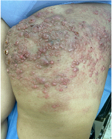
Figure 1: Multiple indurated erythematous nodules are observed on the left thigh, some confluent and some with ulceration, accompanied by firm edema 1.


Laura Vanessa Leal Guevara1* Juan Roberto Apud Gonzalez2 Leticia Martinez Perez3 Marcelo Cuevas Leal4 Roberto Perez Reyes5 Eduardo Daniel Villarreal Santos6 Laura Samantha de Leon Puga6 Luisa Engracia Sofia Galvan Vargas6 Omar Eduardo Benitez Robles6 Enrique Huerta Marquez6
1Dermatologist, Pemex Regional Hospital, Madero, Tamaulipas, Mexico*Corresponding author: Laura Vanessa Leal Guevara, Dermatologist, Pemex Regional Hospital, Madero, Tamaulipas, Mexico, E-mail: [email protected]
Introduction: Cutaneous involvement arising from colorectal adenocarcinoma is uncommon and typically indicates advanced systemic disease with a poor prognosis.
Case Presentation: A 43-year-old female was diagnosed with moderately differentiated infiltrating adenocarcinoma of the sigmoid colon. Surgical management included a sigmoidectomy with end-to-end colorectal anastomosis. Five years after the initial diagnosis, the patient presented with a dermatosis localized to the left thigh, characterized by multiple indurated nodules. Histopathological analysis confirmed metastatic colon cancer.
Discussion: Colorectal adenocarcinoma is one of the most prevalent malignancies worldwide, with well-established metastatic patterns to the liver, lungs, and peritoneum. However, cutaneous involvement is rare, even in metastatic disease, and usually manifests during the late stages of the clinical course.
Conclusion: Clinically, this case illustrates that cutaneous metastases serve as a marker of advanced disease progression rather than an initial manifestation.
Cutaneous metastases; Colon cancer
Cutaneous manifestations secondary to colorectal adenocarcinoma are infrequent and usually indicate advanced systemic disease with a poor prognosis. Early recognition is essential, as these lesions may signal disease progression or recurrence. Lesions may present as firm nodules, infiltrated plaques, ulcerations, or poorly demarcated inflammatory changes, often mimicking benign dermatoses, skin infections, or viral processes such as herpes zoster. We present the following case report.
A 43-year-old female was diagnosed in December 2017 via endoscopic biopsy with moderately differentiated infiltrating adenocarcinoma of the sigmoid colon. In February 2018, she underwent a sigmoidectomy with end-to-end colorectal anastomosis.
Histopathology: Microglandular adenocarcinoma infiltrating through the muscularis propria and serosa, extending into the peritoneal adipose tissue to within 0.5 cm of the surgical margin. Distal and proximal resection margins were negative for malignant involvement, located 2.5 cm and 1 cm from the tumor, respectively. Positive angiolymphatic invasion was noted, with mild to moderate peritumoral lymphocytic infiltrate. Three of twenty-two lymph nodes dissected from the pericolic fat were positive for metastasis, and one tumor deposit was identified. Stage III (T3 N1 M0).
In May 2018, adjuvant chemotherapy with concomitant radiotherapy was recommended; however, radiotherapy was ultimately not administered, as the tumor did not involve the rectum. Follow-up abdominal computed tomography revealed only postoperative changes, with no evidence of active disease.
In October 2018, PET-CT identified suspicious areas at the rectosigmoid junction, left adnexa, peritoneum, and left lung. Rectosigmoidoscopy revealed no endoluminal tumor. In November 2018, due to PET-CT findings, second-line treatment with capecitabine, oxaliplatin, and bevacizumab was initiated. After three months on this regimen, oxaliplatin was discontinued due to severe neuropathy.
In June 2019, an exploratory laparotomy was performed for a tumor mass and peritoneal fluid. Pathology of the resected left ovarian specimen revealed metastatic intestinal-type adenocarcinoma. Immunohistochemical studies were as follows: CK20 positive, CK7 negative, WT1 negative.
Due to the pandemic, the patient remained on capecitabine monotherapy for 16 months, followed by disease progression and elevated carcinoembryonic antigen levels (732 ng/mL).
In February 2020, due to disease progression, molecular testing was performed, revealing wild-type EGFR. Third-line treatment with 5-fluorouracil and panitumumab was initiated and discontinued after 10 months due to lack of response.
In September 2021, left thoracotomy with resection of a pulmonary nodule was performed. Pathology revealed metastasis of moderately differentiated adenocarcinoma.
In April 2022, due to documented pulmonary disease progression, fourth-line treatment with regorafenib was initiated. This regimen was discontinued in September 2022, as thoraco-abdominopelvic CT scan revealed para-aortic and subcarinal lymph node clusters with pericardial and pleural effusion. Fifth-line therapy with irinotecan was subsequently initiated.
In April 2023, the patient presented to our service with a 4-month history of dermatosis, reporting significant pain and edema of the left lower limb. The dermatosis was localized to the left thigh and characterized by multiple erythematous, indurated, and confluent nodules, ranging from 10 to 20 mm, some with ulceration and diffuse edema (Figures 1,2).
Figure 1: Multiple indurated erythematous nodules are observed on the left thigh, some confluent and some with ulceration, accompanied by firm edema 1.
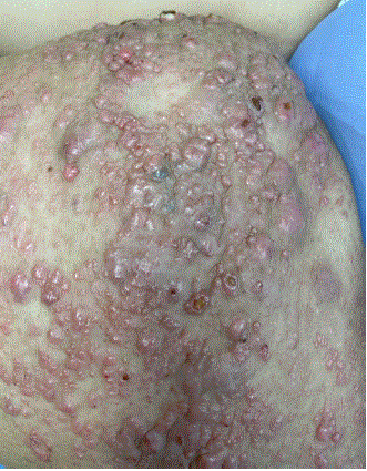
Figure 2: Multiple indurated erythematous nodules are observed on the left thigh, some confluent and some with ulceration, accompanied by firm edema 2.
A punch biopsy of a thigh nodule was performed. Histopathology revealed mild epidermal hyperkeratosis. In the papillary and reticular dermis, multiple glands were observed; cells exhibited increased nuclear size, hyperchromatism, atypical mitoses, and cyanophilic cytoplasm. Additionally, mild inflammatory infiltrate, marked desmoplasia, moderate necrosis, and perineural invasion were present, consistent with metastatic adenocarcinoma (Figures 3,4).
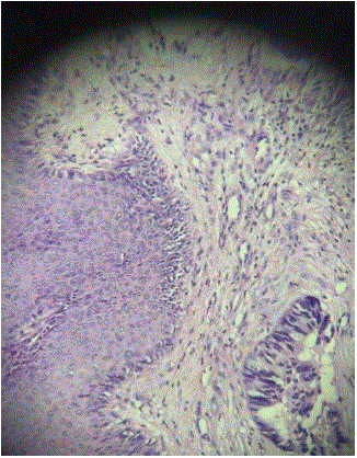
Figure 3: Histopathological examination with H&E staining showsIn the papillary and reticular dermis, glands are observed whose cells show increased nuclear size with hyperchromatism,atypical mitoses, and cyanophilic cytoplasm 1.
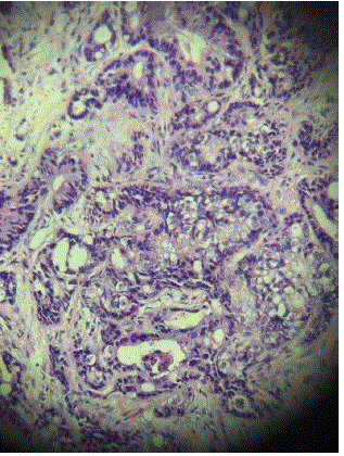
Figure 4: Histopathological examination with H&E staining showsIn the papillary and reticular dermis, glands are observed whose cells show increased nuclear size with hyperchromatism,atypical mitoses, and cyanophilic cytoplasm 2.
Due to disease progression despite five lines of treatment, in September 2023, it was decided to transition to palliative care with pain management using duloxetine, pregabalin, and buprenorphine. In November 2023, the patient passed away due to sepsis of pulmonary origin.
| Date | Procedure | Report |
| 12/2017 | Colonoscopy with biopsy | Invasive moderately differentiated infiltrating adenocarcinoma. |
| 02/2018 | Left hemicolectomy | Microglandular adenocarcinoma, which penetrates the muscularis propria, the serosa and infiltrates the peritoneal fat up to 0.5 cm, from the radial resection edge. -3 metastasis-positive nodes of 22 nodes are dissected |
| 06/2019 | Exploratory laparotomy with left ophorectomy | Metastatic intestinal adenocarcinoma. Immunohistochemistry: -ck20: positive -ck7: negative -wti: negative |
| 09/2021 | Thoracotomy with pulmonary nodule biopsy | Moderately differentiate adenocarcinoma metastasis |
| 08/2022 | Sigmoidectomy and colostomy | Adenocarcinoma of the invasive intestinal type. Moderately differentiated |
| 09/2022 | Thoracoabdominopelvic CT scan of the with report by: para-aortic and subcarinal ganglion clusters with pericardial effusion left pleural effusion | |
| 2023 | Nodule biopsy in the left thigh | Consistent with metastatic adenocarcinoma |
In clinical practice, the emergence of new cutaneous lesions in patients with advances malignancies is often erroneously attributed to inflammatory, infectious, or treatment-related causes. Such diagnostic inertia frequently leads to empirical management, delaying the consideration of a metastatic etiology-particularly when the primary malignancy is not classically associated with cutaneous dissemination f1,2]. Colorectal adevocarcinoma (CRC) typically exhibits a predictable pattern of metastatic spread, involving the liver, lungs, and peritoneum. Conversely, cutaneous metastasis remains a rare event, generally surfacing only in late-stage disease [1,3].
Although rare, the presence of cutaneous metastasis is a potent marker of high tumor burden, systemic progression, and an unfavourable prognosis [4,5]. The clinical heterogeneity of these lesions often contributes to underdignosis. Metastases may manifest as firm nodules, infiltrated plaques, ulcerated lesions, or ill-defined inflammatory changes, often masquerading as benign dermatologic conditions or herpes zoster [2,6]. This variability necessitates a high index of suspicion among clinicians managing oncological patients. While cutaneous metastases in CRC are uncommon, they are clinically significant. They often signal advanced systemic diseases, however, they may occasionally serve as the sentinel sign of tumor recurrence or occult progression, underscoring the necessity for prompt identification [7]. Morphologically, CRC cutaneous metastases typically present as firm, asymptomatic, rapidly growing nodules. While these lesions have a predilection for the abdominal wall-often appearing near prior surgical scars, which suggests possible seeding during previous interventions-their anatomical distribution is unpredictable, ehich complicates timely diagnosis [8]. Given their propensity to mimic benign entities such as epidermal cysts, lipomas, or localized infections, definitive diagnosos mandates tissue biopsy and histopathological confirmation. Immunohistochemistry is essential for diagnostic accuracy, with markers such as cytokeratin 20 (CK20) and the transcription factor CD × 2 serving as standard tools to confirm colorectal origin and differentiate these lesions from primary adnexal tumors [8]. Although the solitary nodular pattern is standard, atypical presentations exist. Zosteriform patterns, characterized by a dermatomal distribution, can misleading mimic viral reactivation, potentially appropriate oncologic intervention [9]. The pathophysiology of these metastases encompasses hematogenous spread, lymphatic dissemination, and iatrogenic implantation. These diverse mechanisms account for the morphological and anatomical variability observed in clinical practice [9]. Rarely, metastases occur in unconventional sites, such as the axilla. In such instances, molecular profiling-specifically assessing microsatellite instability (MSI-H) and BRAF V600E mutation status - provides valuable prognostic insight [10]. In highly selected cases, a multimodal therapeutic approachintegrating neoadjuvant chemotherapy and aggressive surgical resection- may achieve local control.
This case illustrates that cutaneous metastases serve as a marker of advanced disease progression rather than an initial sign of malignancy. Consistent with the literature, their appearance is associated with poor prognosis and limited therapeutic options, shifting management toward a palliative approach focused on symptom control and quality of life. Early diagnosis will be the key element in effective management, which requires careful physical examination. Once any change in the skin is noted, further evaluation should be undertaken.
The authors declare that there are no conflicts of interest regarding the publication of this manuscript.
Download Provisional PDF Here
Aritcle Type: CASE REPORT
Citation: Guevara LVL, Gonzalez JRA, Perez LM, Leal MC, Reyes RP, et al. (2026) Cutaneous Metastases Secondary to Advanced Colorectal Adenocarcinoma: Case Report and Literature Review. J Clin Cosmet Dermatol 10(1): dx.doi.org/10.16966/2576-2826.188
Copyright: © 2026 Guevara LVL, et al. This is an open-access article distributed under the terms of the Creative Commons Attribution License, which permits unrestricted use, distribution, and reproduction in any medium, provided the original author and source are credited.
Publication history:
All Sci Forschen Journals are Open Access